. 2021 Jun 11:2021:10.17912/micropub.biology.000399.
doi: 10.17912/micropub.biology.000399.

A simple one-step PCR assay for SNP detection

Affiliations

A simple one-step PCR assay for SNP detection

Jian Chen et al. MicroPubl Biol. .

Abstract

Polymerase Chain Reaction (PCR) is a powerful tool to detect natural variation or experimentally introduced variation in research and clinical settings and a widely-used method for genotyping. Single nucleotide polymorphisms (SNP) detection is challenging by PCR as the variant and wild type alleles differ by only one nucleotide. Traditional methods to detect SNPs, including Sanger sequencing and commercial kits, are usually time-consuming. Here we describe a simple primer design strategy that enables specific variant detection through regular one-step PCR. The strategy employs the differential efficiency of genomic PCR using a primer that has a single mismatch with the chromosome that contains the SNP to be detected (typically the variant allele) versus two mismatches with the corresponding alternative allele (typically the wild type allele). To date, we have successfully employed this approach to detect more than 20 SNPs. The simplicity and robustness of the approach allows rapid application to legacy mutations as well as newly discovered or generated SNPs.


Figures

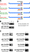
Figure 1. Novel primer design strategy to detect single nucleotide polymorphisms (SNPs) by PCR.
(A) Schematic showing the primer design strategy to discriminate between the variant and wild type alleles through PCR (compare I and II, III and IV). The SNP residue is denoted as position 0. G, in red, is the variant allele, while T, in green, is the wild type allele. The forward PCR primer was designed to specifically detect either the variant or wild type allele, with the 3’ end starting at the SNP residue (G or T at position 0). For the allele to be detected, there is no mismatch at position 0, while for the other allele, there is a mismatch (I versus II, and III versus IV). To increase the specificity for the allele to be detected by PCR, a second change (C to A in these examples, in blue) was introduced at the -2 position, two nucleotides upstream of the SNP position. For examples I and II, the variant forward primer can bind more efficiently to the variant allele (one mismatch in I) than the wild type allele (two mismatches in II), resulting in a more robust PCR amplification of the variant allele. Similarly for examples III and IV, the wild type forward primer can detect the wild type allele (shown in III) more effectively than the variant allele (shown in IV). “X” indicates mismatch, | indicates pairing. The reverse PCR primer was identical for both the variant and wild type allele (see Table 1) and has generated from 200 to 400 nucleotide amplification products. (B & C) Representative agarose gel images of single worm PCR products amplified with primers designed to specifically detect alleles of the indicated genes, either the variant or the wild type allele (Table 1). The genotype is shown above the corresponding lane, with – indicating the variant allele and + indicating the wild type allele. (B) Gel images from primers to detect the glp-1(bn18) mutant allele or the wild type allele. Single worm lysis of animals with the indicated genotype were sampled twice, and PCR was performed with variant primer (upper image) or the wild type primer (lower image). -/-, glp-1(bn18) homozygote; +/+, N2 wild type; -/+, glp-1(bn18)/ hT2::gfp[bli-4(e937) let-?(q782) qIs48]. (C) Gel images showing specific detection of the variant allele for six genes and eight variants. -/-, indicates the relevant gene-variant homozygote; +/+, N2 wild type. (D). The distribution of nucleotide changes introduced at the -2 position from a total of 24 primers. The nucleotide at -2 position was either changed to an A or a T.

References

    1. Barstead RJ, Kleiman L, Waterston RH. Cloning, sequencing, and mapping of an alpha-actinin gene from the nematode Caenorhabditis elegans. Cell Motil Cytoskeleton. 1991;20(1):69–78. doi: 10.1002/cm.970200108. - DOI - PubMed
    1. Brenner S. The genetics of Caenorhabditis elegans. Genetics. 1974 May 01;77(1):71–94. - PMC - PubMed
    1. Johnston RJ Jr, Hobert O. A novel C. elegans zinc finger transcription factor, lsy-2, required for the cell type-specific expression of the lsy-6 microRNA. Development. 2005 Nov 16;132(24):5451–5460. doi: 10.1242/dev.02163. - DOI - PubMed
    1. Kodoyianni V, Maine EM, Kimble J. Molecular basis of loss-of-function mutations in the glp-1 gene of Caenorhabditis elegans. Mol Biol Cell. 1992 Nov 01;3(11):1199–1213. doi: 10.1091/mbc.3.11.1199. - DOI - PMC - PubMed
    1. Lu X, Horvitz HR. lin-35 and lin-53, two genes that antagonize a C. elegans Ras pathway, encode proteins similar to Rb and its binding protein RbAp48. Cell. 1998 Dec 23;95(7):981–991. doi: 10.1016/s0092-8674(00)81722-5. - DOI - PubMed