

Preoperative planar lymphoscintigraphy allows for sentinel lymph node detection in 51 dogs improving staging accuracy: Feasibility and pitfalls
- PMID: 34131982
- PMCID: PMC8518895
- DOI: 10.1111/vru.12995
Preoperative planar lymphoscintigraphy allows for sentinel lymph node detection in 51 dogs improving staging accuracy: Feasibility and pitfalls
Abstract
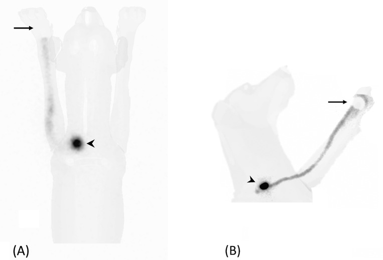
Sentinel lymph node (SLN) mapping is the current gold standard for the oncological staging of solid malignancies in humans. This prospective observational study describes the feasibility and the limits of preoperative lymphoscintigraphy for SLN detection in dogs with spontaneous malignancies and the improvements in staging accuracy. Client-owned dogs with confirmed malignant neoplasia and absence of distant metastasis were prospectively enrolled. Lymphoscintigraphy was performed after the peritumoral injection of Technetium-99m labeled nanocolloids. Regional dynamic and static images were acquired, with and without masking of the injection site with a lead shield. The dogs were then subjected to surgery for tumor excision and SLN extirpation. Intraoperative SLN detection was performed by combining methylene blue dye and a dedicated gamma probe. Overall, 51 dogs with a total of 60 solid malignant tumors were enrolled. Lymphoscintigraphy identified at least one SLN in 57 of 60 cases (95%). The SLN did not always correspond to the regional lymph node (35/57, 61.4%). The use of a lead shield, masking the injection site, markedly improved the SLN visibility. The median time of SLN appearance was 11.4 ± 9.3 min. No side effects were observed. Preoperative lymphoscintigraphy allows for SLN detection in dogs and can improve staging accuracy by either identifying the SLN in a different lymphosome than clinically expected or discriminating the draining node in uncertain cases. The combined use of preoperative and intraoperative techniques is recommended to increase the SLN detection rate.
Keywords: 99mTc; lymphatic metastasis; lymphoscintigraphy; sentinel lymph node; tumor staging.
© 2021 The Authors. Veterinary Radiology & Ultrasound published by Wiley Periodicals LLC on behalf of American College of Veterinary Radiology.
Conflict of interest statement
The authors declare no conflict of interest.
Figures

References
-
- Cabanas, RM. An approach for the treatment of penile carcinoma. Cancer. 1977;39(2):456‐466. - PubMed
-
- Ferrari, R , Marconato, L , Buracco, P , et al. The impact of extirpation of non‐palpable/normal‐sized regional lymph nodes on staging of canine cutaneous mast cell tumours: a multicentric retrospective study. Vet Comp Oncol. 2018;16:505‐510 - PubMed
-
- Balogh, L , Thuróczy, J , Andócs, G , et al. Sentinel lymph node detection in canine oncological patients. Nucl Med Rev Cent East Eur. 2002;5:139‐144. - PubMed
-
- Langenbach, A , Mcmanus, PM , Hendrick, MJ , Shofer, FS , Sorenmo, KU. Sensitivity and specificity of methods of assessing the regional lymph nodes for evidence of metastasis in dogs and cats with solid tumours. JAVMA. 2001;218:1424‐1428. - PubMed
