Review
. 2021 Sep;22(9):1462-1474.
doi: 10.3348/kjr.2020.1479. Epub 2021 May 26.

Key Imaging Findings for the Prospective Diagnosis of Rare Diseases of the Gallbladder and Cystic Duct

Affiliations
Review

Key Imaging Findings for the Prospective Diagnosis of Rare Diseases of the Gallbladder and Cystic Duct

Shintaro Ichikawa et al. Korean J Radiol. 2021 Sep.

Abstract

There are various diseases of the gallbladder and cystic duct, and imaging diagnosis is challenging for the rare among them. However, some rare diseases show characteristic imaging findings or patient history; therefore, familiarity with the imaging presentation of rare diseases may improve diagnostic accuracy and patient management. The purpose of this article is to describe the imaging findings of rare diseases of the gallbladder and cystic duct and identify their pathological correlations with these diseases.

Keywords: Granular cell tumor; Intracystic papillary neoplasm; Mucinous carcinoma; Multiseptate gallbladder; Tubular adenoma.


Conflict of interest statement

The authors have no potential conflicts of interest to disclose.

Figures

Fig. 1
Fig. 1. Multiseptate gallbladder in an asymptomatic 77-year-old male.
Gallbladder wall thickening incidentally found on contrast-enhanced computed tomography. Laboratory test results including tumor markers were negative. A. Contrast-enhanced computed tomography demonstrates multiple linear structures with a honeycomb pattern in the gallbladder lumen (arrows). B. A grape-like cluster in the gallbladder with heterogeneous signal intensity is observed on magnetic resonance cholangiopancreatography (dotted arrow). C. Gross pathology after laparoscopic cholecystectomy reveals multiple edematous septa dividing the gallbladder lumen into many compartments.
Fig. 2
Fig. 2. Xanthogranulomatous cholecystitis in an asymptomatic 71-year-old male.
Gallbladder wall thickening found on ultrasonography during an annual medical checkup. Serum C reactive protein was slightly elevated (6.2 mg/L), while other laboratory test results, including tumor markers, were negative. A–D. Precontract (A), arterial phase (B), portal venous phase (C), and delayed phase contrast-enhanced computed tomography (D) reveal diffuse gallbladder wall thickening, continuous mucosal line (arrows), and intra-mural hypoattenuating nodules (arrowheads). Macroscopic hepatic invasion and intrahepatic bile duct dilatation are not observed. Open cholecystectomy was performed, and xanthogranulomatous cholecystitis was diagnosed.
Fig. 3
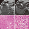
Fig. 3. Xanthogranulomatous cholecystitis in a 77-year-old male with recurrent right upper quadrant pain.
Serum C reactive protein was slightly elevated (6.2 mg/L), while other laboratory test results including tumor markers were negative. A, B. In-phase (A) and opposed-phase (B) T1-weighted images show diffuse thickening of the gallbladder wall. A signal drop on the opposed-phase image, compared with the in-phase image, reflecting lipid-laden macrophages is observed (arrows). C. Microscopic pathology (hematoxylin and eosin stain, × 100) after open cholecystectomy shows severe inflammatory cell infiltration, hemorrhage, and the accumulation of foamy histiocytes (dotted arrows). D. Magnified image (hematoxylin and eosin stain, × 400) of foamy histiocytes (arrowheads).
Fig. 4
Fig. 4. Intracystic papillary neoplasm (low grade) in an asymptomatic 75-year-old female.
Nodular lesions in the gallbladder found on ultrasonography during an annual medical checkup. Serum carcinoembryonic antigen was slightly elevated (8.4 µg/L), while other laboratory test results were negative. A. Contrast-enhanced coronal computed tomography reveals two enhanced nodular lesions protruding into the gallbladder (arrows). B. Microscopic pathology (hematoxylin and eosin stain, × 200) after laparoscopic cholecystectomy shows papillary proliferation with cellular atypia characteristic of low-grade dysplasia.
Fig. 5
Fig. 5. Intracystic papillary neoplasm (high grade) in an asymptomatic 76-year-old female.
A mass lesion in the gallbladder found on ultrasonography during an annual medical checkup. Laboratory test results, including tumor markers, were negative. A. Arterial phase contrast-enhanced magnetic resonance imaging shows a heterogeneously enhanced mass, measuring 18 mm in diameter, in the body of the gallbladder (arrow). B. Fat-saturated T2-weighted imaging shows a mass with peripheral nodular hyperintense areas, which could be intratumoral mucin (dotted arrows). C. Gross pathology after laparoscopic cholecystectomy shows a papillary mass measuring 20 mm at its maximum diameter (open arrow). D, E. Microscopic pathology (hematoxylin and eosin stain, × 2 (D) and × 200 (E)) shows papillary or tubular proliferation of atypical glandular ducts with a gastric phenotype (arrowheads) as well as cellular atypia characteristic of high-grade dysplasia (arrows).
Fig. 6
Fig. 6. Pyloric gland adenoma (low grade) in an asymptomatic 37-year-old female.
A mass lesion in the gallbladder found on ultrasonography during an annual medical checkup. Laboratory test results, including tumor markers, were negative. A. Endoscopic ultrasonography reveals a well-defined mass, measuring 12 mm in diameter, in the gallbladder. Small cystic spots are observed within the lesion (arrows). B. Fat-saturated coronal T2-weighted imaging shows a mass with hyperintense spots (dotted arrows). These findings may reflect enlarged intratumoral glandular ducts. C. Microscopic pathology (hematoxylin and eosin stain, × 200) after laparoscopic cholecystectomy shows papillary or tubular epithelium with mild atypia. Enlarged glandular ducts are also observed (arrowheads).
Fig. 7
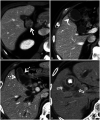
Fig. 7. Typical images of gallbladder cancer in an asymptomatic 52-year-old male (A), an asymptomatic 72-year-old male (B), a 55-year-old male (C) with abdominal pain and jaundice, and an 83-year-old male (D) with fever and appetite loss.
A. Portal venous phase CT demonstrates polypoid mass within the gallbladder lumen (arrow). B. Portal venous phase CT reveals focal thickening of the gallbladder wall (arrowhead). C. Portal venous phase CT demonstrates diffuse thickening of the gallbladder wall (dotted arrows). D. Portal venous phase CT reveals a mass in the gallbladder fossa with severe hepatic invasion (open arrows). CT = computed tomography
Fig. 8
Fig. 8. Poorly differentiated mucinous carcinoma in an asymptomatic 78-year-old female.
Papillary mass in the gallbladder was incidentally found on contrast-enhanced computed tomography. Laboratory test results, including tumor markers, were negative. A, B. Arterial-phase (A) and delayed-phase (B) contrast-enhanced computed tomography show a papillary mass with delayed enhancement in the enlarged gallbladder (arrows). C. T2-weighted images show a heterogeneous signal by the contents of the gallbladder, representing abundant mucin (dotted arrows). D. Extended cholecystectomy was performed and microscopic pathology (hematoxylin and eosin stain, × 200) reveals that the mass contains abundant mucin. Several signet ring cells are also noted.
Fig. 9
Fig. 9. Adenosquamous carcinoma in an 82-year-old male with a high fever.
Serum inflammatory markers (white blood cell count 22560/µL and C reactive protein 137 mg/L) were markedly elevated, while tumor markers were negative. A, B. Portal venous phase contrast-enhanced computed tomography (A) and axial T2-weighted imaging (B) show a large hypovascular mass in the gallbladder fossa (arrows). It is difficult to differentiate the mass from a hepatic tumor because this mass shows severe hepatic invasion. Lymph node metastasis (arrowheads) and numerous liver metastases are also noted (dotted arrows). The disease progressed rapidly, and he died two months later. An autopsy was performed, and the gallbladder lesion was diagnosed as adenosquamous carcinoma.
Fig. 10
Fig. 10. Gallbladder metastasis from RCC in an asymptomatic 74-year-old female.
A mass lesion in the gallbladder was found on ultrasonography during an annual medical checkup. Laboratory test results, including tumor markers, were negative. The female had undergone right radical nephrectomy for RCC (clear cell, pT3b, 45 × 38 mm, G1 > 2, v(+)) 6 years prior. A, B. Arterial phase (A) and portal venous phase (B) contrast-enhanced computed tomography showing a polypoid lesion measuring 15 mm in diameter in the body of the gallbladder (arrows). The mass shows marked contrast enhancement in the arterial phase and iso-attenuation to the liver in the portal venous phase. C. Microscopic pathology (hematoxylin and eosin stain, × 200) revealed that the mass was composed of large nests and sheets of moderately sized polygonal cells with abundant clear nonpapillary cytoplasm, typical of clear cell carcinomas. RCC = renal cell carcinoma
Fig. 11
Fig. 11. Lymphoma in a 46-year-old male with left back pain.
Serum interleukin-2 receptor 2540 U/mL, lactate dehydrogenase 328 U/L, and C reactive protein 50.5 mg/L were elevated. A, B. Contrast-enhanced CT shows several nodules in the gallbladder (arrows). Retroperitoneal mass is also noted (arrowheads). C. Non-contrast CT after four weeks of (A) and (B) shows the rapid growth of the polypoid lesions (open arrow) and the large retroperitoneal mass (curved arrows). CT-guided biopsy of the retroperitoneal mass was performed, and the diagnosis of diffuse large B-cell lymphoma was made. D. Non-contrast CT after chemotherapy shows that the polypoid lesions in the gallbladder and retroperitoneal mass have almost disappeared (dotted arrows). CT = computed tomography
Fig. 12
Fig. 12. Cystic duct carcinoma in a 67-year-old male with abdominal pain.
A mass lesion near the gallbladder was found on ultrasonography during an annual medical checkup. Laboratory test results, including tumor markers, were negative. Contrast-enhanced computed tomography demonstrates enhanced circumferential wall thickening in the cystic duct (arrow) with gallbladder enlargement. Open cholecystectomy was performed, and the lesion was diagnosed as cystic duct adenocarcinoma.
Fig. 13
Fig. 13. Granular cell tumor in an asymptomatic 58-year-old female.
A mass lesion in the cystic duct was found on ultrasonography during an annual medical checkup. Laboratory test results, including tumor markers, were negative. A. Portal venous phase computed tomography shows a thickened cystic duct wall with prolonged enhancement (arrow). B. Open cholecystectomy was performed, and microscopic pathology (hematoxylin and eosin stain, × 200) reveals that the mass contains dense proliferation of polygonal atypical cells with eosinophilic granules (arrowheads). These cells are weakly positive for the Periodic acid-Schiff stain and broadly positive for the S100 stain (not shown).
Fig. 14
Fig. 14. Amputation neuroma in an asymptomatic 84-year-old female with a history of cholecystectomy.
Enhancing soft tissue at the cystic stump was incidentally found on contrast-enhanced computed tomography. Laboratory test results, including tumor markers, were negative. Contrast-enhanced coronal computed tomography reveals an enhanced mass in the cystic duct stump (arrow). This lesion did not change in size for more than four years during follow-up.

Similar articles

Cited by

References

    1. Whittle C, Skoknic V, Maldonado I, Schiappacasse G, Pose G. Multimodality imaging of congenital variants in the gallbladder: pictorial essay. Ultrasound Q. 2019;35:195–199. - PubMed
    1. Nakazawa T, Ohara H, Sano H, Kobayashi S, Nomura T, Joh T, et al. Multiseptate gallbladder: diagnostic value of MR cholangiography and ultrasonography. Abdom Imaging. 2004;29:691–693. - PubMed
    1. Singh VP, Rajesh S, Bihari C, Desai SN, Pargewar SS, Arora A. Xanthogranulomatous cholecystitis: what every radiologist should know. World J Radiol. 2016;8:183–191. - PMC - PubMed
    1. Goshima S, Chang S, Wang JH, Kanematsu M, Bae KT, Federle MP. Xanthogranulomatous cholecystitis: diagnostic performance of CT to differentiate from gallbladder cancer. Eur J Radiol. 2010;74:e79–e83. - PubMed
    1. Kang TW, Kim SH, Park HJ, Lim S, Jang KM, Choi D, et al. Differentiating xanthogranulomatous cholecystitis from wall-thickening type of gallbladder cancer: added value of diffusion-weighted MRI. Clin Radiol. 2013;68:992–1001. - PubMed