Review
. 2021 Aug;48(2):149.
doi: 10.3892/ijmm.2021.4982. Epub 2021 Jun 16.

Recent advances on the mechanisms of kidney stone formation (Review)

Affiliations
Review

Recent advances on the mechanisms of kidney stone formation (Review)

Zhu Wang et al. Int J Mol Med. 2021 Aug.

Abstract

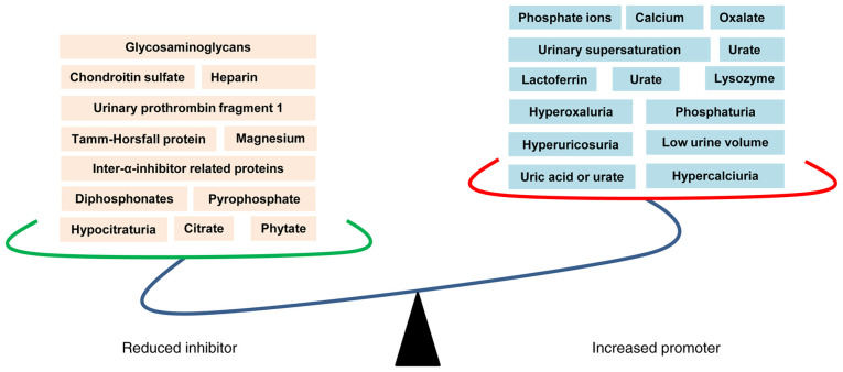
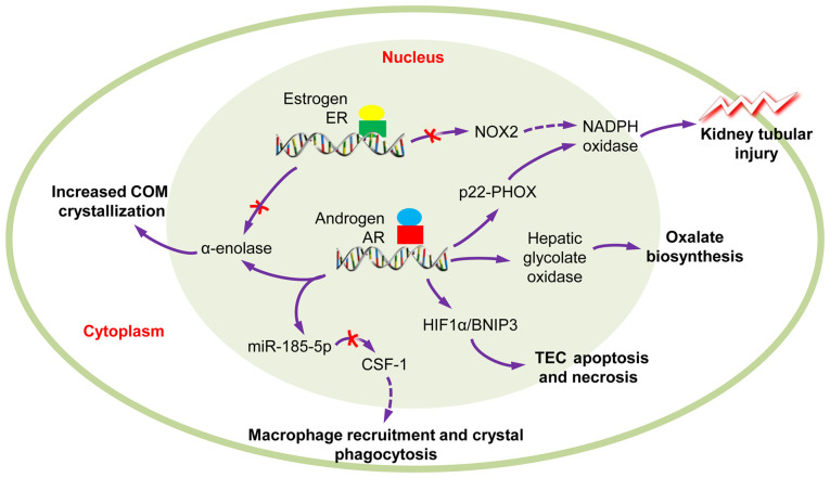
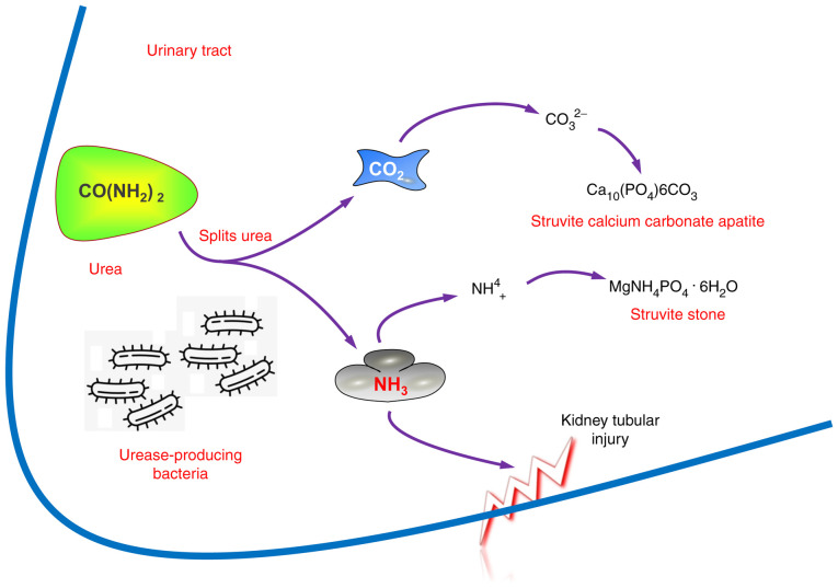
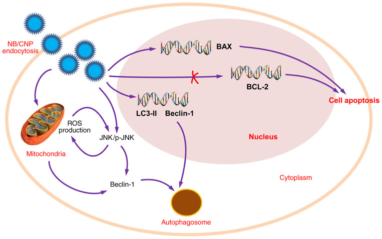
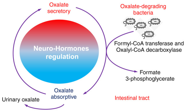
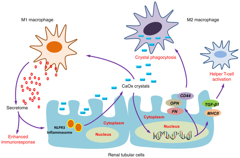
Kidney stone disease is one of the oldest diseases known to medicine; however, the mechanisms of stone formation and development remain largely unclear. Over the past decades, a variety of theories and strategies have been developed and utilized in the surgical management of kidney stones, as a result of recent technological advances. Observations from the authors and other research groups suggest that there are five entirely different main mechanisms for kidney stone formation. Urinary supersaturation and crystallization are the driving force for intrarenal crystal precipitation. Randall's plaques are recognized as the origin of calcium oxalate stone formation. Sex hormones may be key players in the development of nephrolithiasis and may thus be potential targets for new drugs to suppress kidney stone formation. The microbiome, including urease‑producing bacteria, nanobacteria and intestinal microbiota, is likely to have a profound effect on urological health, both positive and negative, owing to its metabolic output and other contributions. Lastly, the immune response, and particularly macrophage differentiation, play crucial roles in renal calcium oxalate crystal formation. In the present study, the current knowledge for each of these five aspects of kidney stone formation is reviewed. This knowledge may be used to explore novel research opportunities and improve the understanding of the initiation and development of kidney stones for urologists, nephrologists and primary care.

Keywords: kidney stone; mechanism; microbiome; nanobacteria; urolithiasis.


Conflict of interest statement

The authors declare that they have no competing interests.

Figures

Figure 1
Figure 1
Physicochemical mechanisms of kidney stone formation. The reduced inhibitors (left panel) and increased promoters (right panel) are suggested to play critical roles in kidney stone formation.
Figure 2
Figure 2
Role of sex hormones in calcium oxalate nephrolithiasis. The AR signaling could induce TECs apoptosis and necrosis and kidney tubular injury, promote COM crystallization and oxalate biosynthesis; however, macrophage recruitment and crystal phagocytosis are inhibited. Conversely, ER signaling can reduce ROS-mediated kidney tubular injury and COM crystallization. COM, calcium oxalate monohydrate; AR, androgen receptor; ER, estrogen receptor; ROS, reactive oxygen species.
Figure 3
Figure 3
Role of urease-producing bacteria in stone formation. The urease-producing bacteria split urea and promote the formation of ammonia and carbon dioxide, leading to kidney tubular injury and urine alkalinization and subsequent formation of phosphate salts.
Figure 4
Figure 4
Role of nanobacteria in stone formation. The nanobacteria may induce ROS production through the JNK/p-JNK signaling induction, may decrease mitochondrial membrane potential and promote cell apoptosis through the downregulation of Bcl-2 expression and the upregulation of Bax expression. Additionally, nanobacteria may lead to autophagy through the upregulation of LC3-II and Beclin-1 expression. ROS, reactive oxygen species; LC3-II, microtubule-associated proteins 1A/1B light chain 3B.
Figure 5
Figure 5
Role of oxalate-degrading bacteria in stone formation. Oxalate-degrading bacteria use oxalate as a carbon energy source and thrive in the presence of the oxalate anion, reduce urinary oxalate level and exhibit growth inhibition in the calcium oxalate crystallization in the kidney.
Figure 6
Figure 6
Immune response to urinary crystals. Macrophage accumulation and macrophage-related inflammation or anti-inflammation is the main immune response alteration observed as a result of kidney stone formation. M1 macrophages are important effectors of CaOx stone formation, while M2 macrophages could prevent CaOx inflammatory damage through crystal phagocytosis. CaOx, calcium oxalate.

References

    1. Romero V, Akpinar H, Assimos DG. Kidney stones: A global picture of prevalence, incidence, and associated risk factors. Rev Urol. 2010;12:e86–e96. - PMC - PubMed
    1. Morgan MS, Pearle MS. Medical management of renal stones. BMJ. 2016;352:i52. doi: 10.1136/bmj.i52. - DOI - PubMed
    1. Zeng G, Mai Z, Xia S, Wang Z, Zhang K, Wang L, Long Y, Ma J, Li Y, Wan SP, et al. Prevalence of kidney stones in China: An ultrasonography based cross-sectional study. BJU Int. 2017;120:109–116. doi: 10.1111/bju.13828. - DOI - PubMed
    1. Ziemba JB, Matlaga BR. Epidemiology and economics of nephrolithiasis. Investig Clin Urol. 2017;58:299–306. doi: 10.4111/icu.2017.58.5.299. - DOI - PMC - PubMed
    1. Eisner BH, Goldfarb DS. A nomogram for the prediction of kidney stone recurrence. J Am Soc Nephrol. 2014;25:2685–2687. doi: 10.1681/ASN.2014060631. - DOI - PMC - PubMed