. 2021 Aug 10;95(17):e0026421.
doi: 10.1128/JVI.00264-21. Epub 2021 Aug 10.

Identification of the Viral Determinant of Hypovirulence and Host Range in Sclerotiniaceae of a Genomovirus Reconstructed from the Plant Metagenome

Affiliations

Identification of the Viral Determinant of Hypovirulence and Host Range in Sclerotiniaceae of a Genomovirus Reconstructed from the Plant Metagenome

Chenchen Feng et al. J Virol. .

Abstract

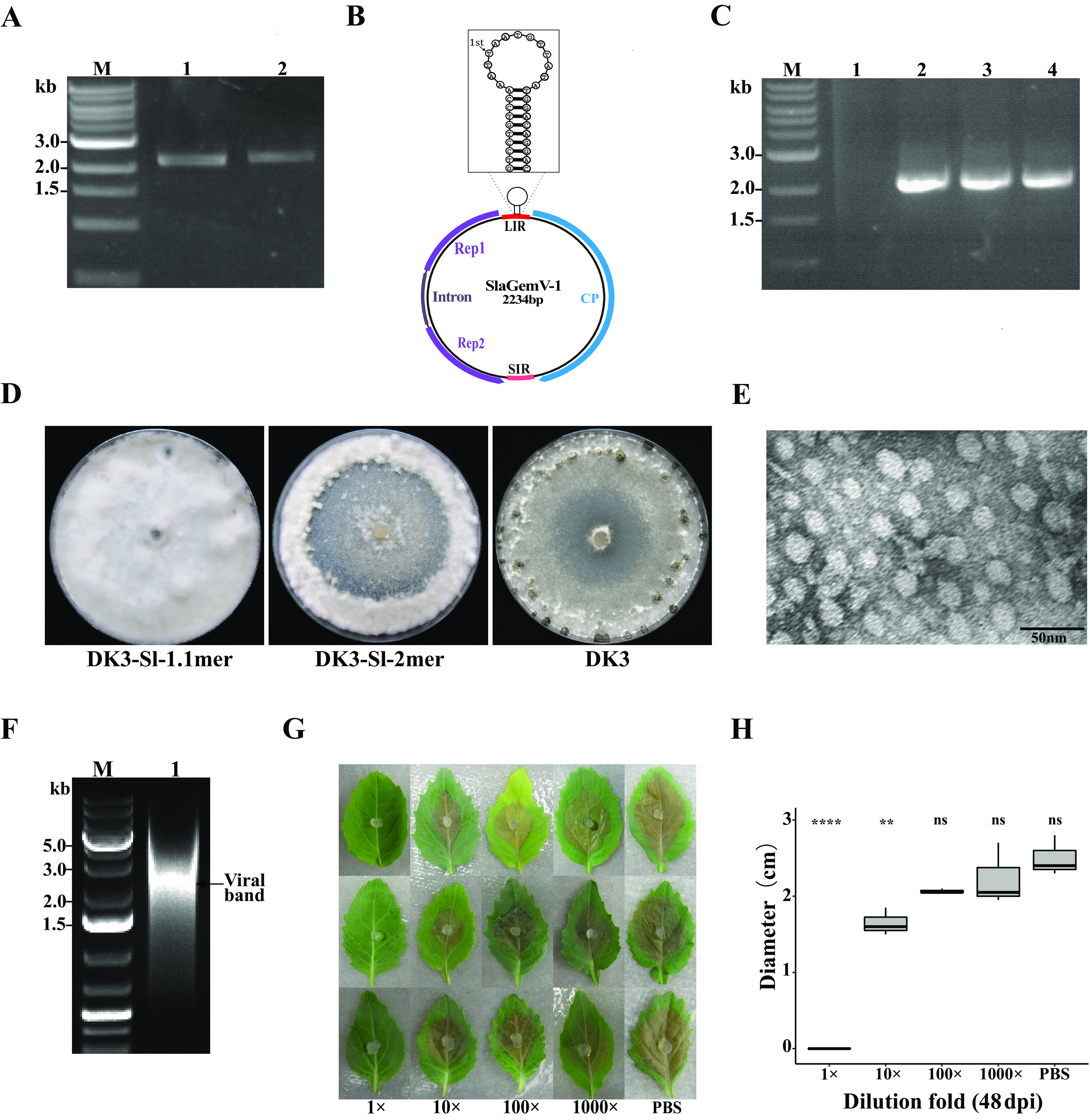
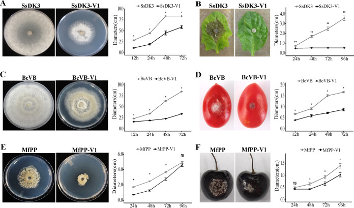
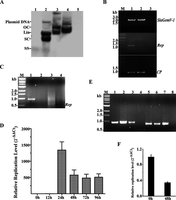
Uncharacterized viral genomes that encode circular replication-associated proteins of single-stranded DNA viruses have been discovered by metagenomics/metatranscriptomics approaches. Some of these novel viruses are classified in the newly formed family Genomoviridae. Here, we determined the host range of a novel genomovirus, SlaGemV-1, through the transfection of Sclerotinia sclerotiorum with infectious clones. Inoculating with the rescued virions, we further transfected Botrytis cinerea and Monilinia fructicola, two economically important members of the family Sclerotiniaceae, and Fusarium oxysporum. SlaGemV-1 causes hypovirulence in S. sclerotiorum, B. cinerea, and M. fructicola. SlaGemV-1 also replicates in Spodoptera frugiperda insect cells but not in Caenorhabditis elegans or plants. By expressing viral genes separately through site-specific integration, the replication protein alone was sufficient to cause debilitation. Our study is the first to demonstrate the reconstruction of a metagenomically discovered genomovirus without known hosts with the potential of inducing hypovirulence, and the infectious clone allows for studying mechanisms of genomovirus-host interactions that are conserved across genera. IMPORTANCE Little is known about the exact host range of widespread genomoviruses. The genome of soybean leaf-associated gemygorvirus-1 (SlaGemV-1) was originally assembled from a metagenomic/metatranscriptomic study without known hosts. Here, we rescued SlaGemV-1 and found that it could infect three important plant-pathogenic fungi and fall armyworm (S. frugiperda Sf9) insect cells but not a model nematode, C. elegans, or model plant species. Most importantly, SlaGemV-1 shows promise for inducing hypovirulence of the tested fungal species in the family Sclerotiniaceae, including Sclerotinia sclerotiorum, Botrytis cinerea, and Monilinia fructicola. The viral determinant of hypovirulence was further identified as replication initiation protein. As a proof of concept, we demonstrate that viromes discovered in plant metagenomes can be a valuable genetic resource when novel viruses are rescued and characterized for their host range.

Keywords: Botrytis cinerea; Sclerotinia sclerotiorum; biocontrol; gemygorvirus; genomovirus; mycovirus; virulence determinants.


Figures

FIG 1
FIG 1
SlaGemV-1 replication in Sclerotinia sclerotiorum after transfection with infectious clones was confirmed. (A) PCR amplification from extracted mycelial DNA of SlaGemV-1-transfected S. sclerotiorum using primers pJetR and 3′R. Lanes: M, 1-kb ladder; 1, 1.1-mer transfection; 2, 2-mer transfection. (B) SlaGemV-1 genome organization with the nonanucleotide structure specified. (C) PCR amplification with primers pJetR and 3′R and with the rolling circle amplification product from the transfected S. sclerotiorum mycelial DNA as the template. The resulting PCR products were subjected to Sanger sequencing to confirm their identities. Lanes: M, 1-kb ladder; 1, RCA product with a smear; 2, PCR product using the RCA product from the 1.1-mer-transfected culture as the template; 3, PCR product of RCA product from 2-mer-transfected culture; 4, positive control from PCR product without RCA from the transfected culture. (D) Comparison of virus-transfected S. sclerotiorum (strain DK3) grown on PDA for 21 days after transfection with pJet-SlaGemV-1 1.1-mer and 2-mer constructs. (Left) pJet-SlaGemV-1 1.1-mer transfection; (middle) pJet-SlaGemV-1-2-mer transfection; (right) original DK3 strain, used for comparison. (E) Transmission electron microscopy image of purified SlaGemV-1 particles. (F) Agarose gel (1%) electrophoresis of DNA extracted from 25% sucrose gradient fractions containing virus particles. (G) Viral particles (1 mg/ml) extracted from infected S. sclerotiorum mycelia were diluted to different concentrations (1 mg/ml, 0.1 mg/ml, 0.01 mg/ml, and 0.001 mg/ml; PBS, phosphate-buffered saline control) used as inoculants (10 µl) on virus free agar plug to inhibit fungal growth. As the viral particles were resuspended in PBS, all dilution used PBS. Each row represents one replication for three replications shown here. (H) Dose-response relationship of undiluted and diluted treatments of viral particles compared to the PBS control. Two-way analysis of variance and Bonferroni post hoc test. **, P < 0.01; ****, P <0.0001.
FIG 2
FIG 2
The extracellular transmission of SlaGemV-1 and its induced hypovirulence in fungi in the family Sclerotiniaceae, including Sclerotinia sclerotiorum, Botrytis cinerea, and Monilinia fructicola, were confirmed. Effects of SlaGemV-1 on (A) fungal growth on PDA at 3 days old and (B) virulence at 4 days postinoculation (dpi) of S. sclerotiorum. Effects of SlaGemV-1 on (C) fungal growth on PDA at 4 days old and (D) virulence at 4 dpi of B. cinerea. Effects of SlaGemV-1 on (E) fungal growth on PDA at 3 days old and (F) virulence at 4 dpi of M. fructicola. All resulted in significantly reduced virulence and growth (*, P < 0.05; **, P < 0.01; ns, not significant).
FIG 3
FIG 3
The high infectivity of SlaGemV-1 virions was confirmed. (A) Regardless of Sclerotinia sclerotiorum strain, individual protoplasts as single cells were largely infected by the virion inoculation. Three strains were tested: DK3, 1980, and 274. The whole viral genome was amplified by PCR after the regenerated virus-treated fungal cultures were transferred several times. Protoplast suspensions (100 μl, 107 protoplasts/ml) from different strains were mixed gently with viral particles (20 μl, 1 mg/ml). At the same time, the viral genomic DNA was mixed with protoplasts as a control. The mixture was incubated on ice for 40 min and held at room temperature for an additional 30 min; then, 2 ml of regeneration medium was added and shaken gently for 2 h. The virus-treated protoplasts were regenerated at room temperature for 2 to 3 days. The whole SlaGemV-1 genome was amplified and run on a gel for >10 cells for each strain. (Top) Lanes 1 to 10, virus-treated DK3; 11, viral-DNA-treated DK3. (Middle) Lanes 1 to 10, virus-treated strain 274; 11, viral-DNA-treated strain 274. (Bottom) Lanes 1 to 12, virus-treated strain 1980; 13, viral-DNA-treated strain 1980. (B) Phenotypic changes in the virus-infected (left) cultures compared to virus-free (right) cultures of S. sclerotiorum strains on PDA plates. (C) The infectivity of SlaGemV-1 virions was confirmed by PCR on alternative fungal hosts. The whole viral genome (2.2 kb) was amplified from inoculated B. cinerea and M. fructicola. Lanes: M, ladder; 1, virus-infected B. cinerea; 2, virus-infected M. fructicola; 3, virus-free strain of B. cinerea as a negative control; 4, virus-free strain of M. fructicola as a negative control.
FIG 4
FIG 4
The crude extracts of SlaGemV-1 virions protected the tomato plants from S. sclerotiorum infection. (A) Fungal homogenate was prepared by blending fungal hyphae in PDB and using low-speed centrifugation to separate the hyphal fragments as a method of extracting viral particles, diluted to OD600 values of 4.0, 3.0, and 2.0, and sprayed on plants which were inoculated with hyphal agar plugs applied to two leaves per plant. The control was sprayed with double-distilled water. The fungal-pathogen-infected plants were kept in an incubator with 100% humidity at 22°C, and images of the lesions were taken at 6 dpi. (B) Protective activity of SlaGemV-1 against white mold disease. The crude preparation efficiently protected intact plants from infection with S. sclerotiorum when the OD600 of the viral particles was greater than or equal to 2.0. The images of whole plants were taken at 16 dpi. (C) The disease severity was rated at 16 dpi (*, P < 0.05; ****, P < 0.0001). The rating was performed based on a scale of 0 to 5: 0, no disease; 1, restricted lesion on leaf; 2, expanded lesion on leaf; 3, leaf with expanded lesion and start of stem colonization; 4, severe stem colonization; 5, plant completely dead.
FIG 5
FIG 5
Replication of the SlaGemV-1 genome in bacteria and insect cells. (A) Southern hybridization to confirm different conformations of the replicative forms of SlaGemV-1 in bacteria. Lanes were loaded with total DNA: 1, virus-infected fungal mycelia; 2, E. coli carrying SlaGemV-1 1.1-mer clone in pJet1.2; 3, E. coli carrying SlaGemV-1 2-mer clone in pJet1.2; 4, E. coli carrying 2-mer clone in pOET-1; 5, E. coli culture without any plasmids. OC, open circular double stranded; Lin, linear; SC, supercoiled double stranded; SS, single-stranded forms. (B) PCR detection of SlaGemV-1 in total DNA extracted from transfected Sf9 insect cells. Lanes: M, ladder, 1, Rep/CP genes amplified from transfected S. sclerotiorum as the positive control for PCR; 2, Rep/CP genes amplified from transfected Sf9 cells; 3, virus-free Sf9 cells as the negative control for PCR. (C) RT-PCR detection of SlaGemV-1 CP/Rep gene expression in total RNA extracted from the supernatant of transfected Sf9 insect cells. Lanes: M, ladder; 1, CP transcript amplified from the supernatant of infected cells; 2, negative amplification of CP transcript from virus-free Sf9 cells; 3, Rep transcript amplified without the intron (650 bp) from the supernatant of transfected Sf9 cells; 4, negative amplification of Rep transcript from the virus-free Sf9 cells. (D) Viral replication kinetics measured at different time points by qPCR in transfected Sf9 cells. (E) Confirmation of positive SlaGemV-1 replication in Sf9 cells. Viral amplification of the cellular and supernatant DNA extracted from the second passage of the infected insect cells. Lanes: 1 and 2, viral CP gene amplification in cellular DNA; 3, viral CP gene amplification in supernatant DNA of virus-infected Sf9 cells; 4, virus-free Sf9 cells; 5 and 6, viral Rep gene amplification in cellular DNA; 7, viral Rep gene amplification in supernatant DNA of virus-infected Sf9 cells; 8, virus-free Sf9 cells. (F) Confirmation by RT-qPCR of the negative replication of SlaGemV-1 in Arabidopsis thaliana mesophyll protoplasts.
FIG 6
FIG 6
Site-specific homologous expression of SlaGemV-1 CP or REP in Botrytis cinerea. (A) Construction diagram of displacement of bcniaD by the SlaGemV-1 CP and Rep gene fragments. (B) Comparison of growth on PDA and lesion diameters on tomatoes and grapes. BcVB, wild-type B. cinerea; BcVB-OGG, BcVB transformed by the empty OGG vector; BcVB-CP-Sl, CP transformant; BcBV-V1, SlaGemV-1-infected BcBV; BcVB-Rep-Sl, Rep transformant. (C) Viral Rep and CP gene expression was confirmed from site-directed expression transformants using RT-PCR with specific primers (CP-F/CP-R, Rep-242F/Rep-1072R). Lanes: M, ladder; 1, BcVB-Rep-Sl without intron; 2, BcVB-CP-Sl; 3, BcVB-OGG as the negative control. (D) Mycelial growth on PDA measured by diameters. (E) Lesion diameters on tomatoes. (F) Lesion diameters on grapes.
FIG 7
FIG 7
Confirmation of site-directed expression of Rep and CP in B. cinerea. (A) Homologous recombination at the 5′- and 3′-flanking regions was validated for transformants BcVB-CP-Sl and BcVB-Rep-Sl using the diagnostic PCR primers (bcniaD-hi5F/TgluchiF, hph-hiF/bcniaD-hi3R) (35). (Left) Confirmation of the size of the 5′ region of homologous integration transformants; (right) confirmation of the 3′ region. Lanes: M, ladder; 1, BcVB-CP-Sl transformant; 2, BcVB-Rep-Sl transformant; 3, BcVB-OGG transformant. (B) Sequencing was conducted to confirm the intron (red bases) removal of the BcVB-Rep-Sl transformant in B. cinerea. (C) Confirmation of virulence determinant of SlaGemV-1 by BcVB-CP-Sl and BcVB-Rep-Sl transformants on grapes. Lesion diameters were compared on green grapes inoculated with different transformants. BcVB, wild-type B. cinerea; BcVB-OGG, BcVB transformed by empty OGG vector; BcVBCP-Sl, CP transformant; BcBV-VI, SlaGemV-1-infected BcBV; BcVB-Rep-Sl, Rep transformant. (D) Average lesion diameters measured on 8 grapes after 24, 48, 72, and 96 h after inoculation with the strains from one of the three trials. All three trials showed reduced virulence caused by BcVB-Rep-Sl.

Similar articles

Cited by

References

    1. Marzano SYL, and, Domier LL. 2016. Novel mycoviruses discovered from metatranscriptomics survey of soybean phyllosphere phytobiomes. Virus Res 213:332–342. doi:10.1016/j.virusres.2015.11.002. - DOI - PubMed
    1. Neupane A, Feng C, Feng J, Kafle A, Bücking H, Lee Marzano S-Y. 2018. Metatranscriptomic analysis and in silico approach identified mycoviruses in the arbuscular mycorrhizal fungus Rhizophagus spp. Viruses 10:707. doi:10.3390/v10120707. - DOI - PMC - PubMed
    1. Márquez LM, Redman RS, Rodriguez RJ, Roossinck MJ. 2007. A virus in a fungus in a plant: three-way symbiosis required for thermal tolerance. Science 315:513–515. doi:10.1126/science.1136237. - DOI - PubMed
    1. Bao X, Roossinck MJ. 2013. Multiplexed interactions: viruses of endophytic fungi. Adv Virus Res 86:37–58. doi:10.1016/B978-0-12-394315-6.00002-7. - DOI - PubMed
    1. Li C, and, Samulski RJ. 2020. Engineering adeno-associated virus vectors for gene therapy. Nat Rev Genet 21:255–272. doi:10.1038/s41576-019-0205-4. - DOI - PubMed

MeSH terms