Review
. 2021 Apr;79(4):321-333.
doi: 10.1590/0004-282X-ANP-2020-0105.

Arteriopathy in pediatric stroke: an underestimated clinical entity

Affiliations
Review

Arteriopathy in pediatric stroke: an underestimated clinical entity

Ronaldo Pizzatto et al. Arq Neuropsiquiatr. 2021 Apr.

Abstract

Background: Pediatric arterial ischemic stroke (AIS), which was thought to be a rare disorder, is being increasingly recognized as an important cause of neurological morbidity, thanks to new advances in neuroimaging.

Objective: The aim of this study was to review the main etiologies of stroke due to arteriopathy in children.

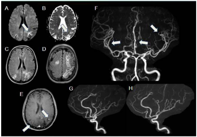
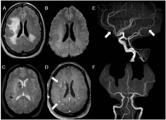
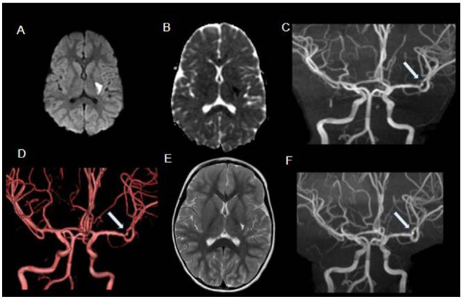
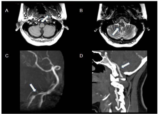
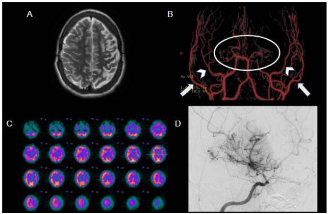
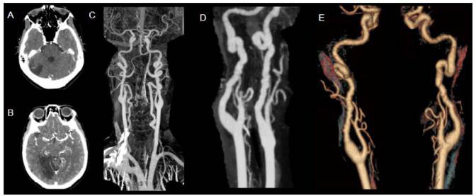
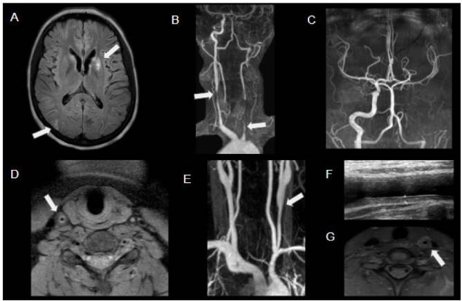
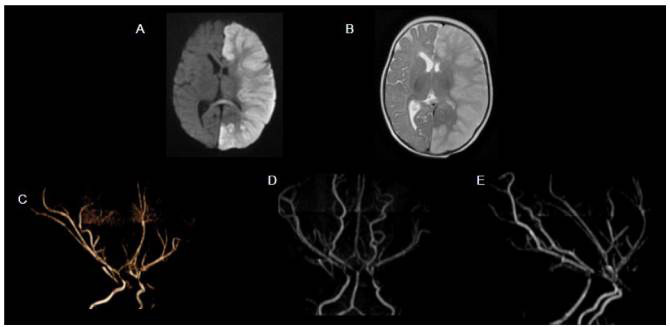
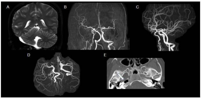
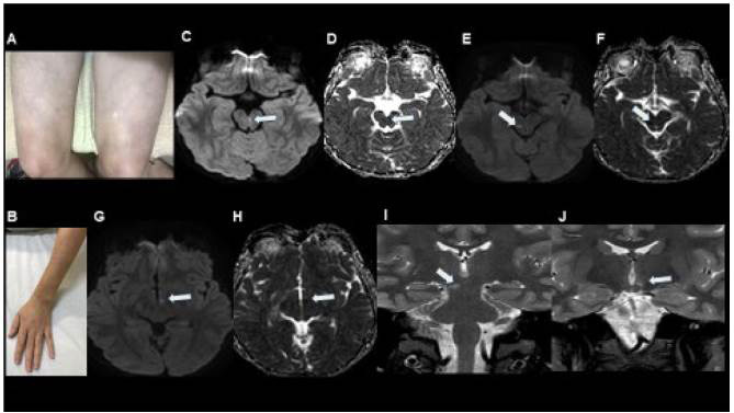
Methods: Using a series of cases from our institution, we addressed its epidemiological aspects, physiopathology, imaging findings from CT, MR angiography, MR conventional sequences and MR DWI, and nuclear medicine findings.

Results: Through discussion of the most recent classification for childhood AIS (Childhood AIS Standardized Classification and Diagnostic Evaluation, CASCADE), we propose a modified classification based on the anatomical site of disease, which includes vasculitis, varicella, arterial dissection, moyamoya, fibromuscular dysplasia, Takayasu's arteritis and genetic causes (such as ACTA-2 mutation, PHACE syndrome and ADA-2 deficiency). We have detailed each of these separately. Conclusions: Prompt recognition of AIS and thorough investigation for potential risk factors are crucial for a better outcome. In this scenario, neurovascular imaging plays an important role in diagnosing AIS and identifying children at high risk of recurrent stroke.

Introdução:: O acidente vascular cerebral (AVC) pediátrico, considerado um distúrbio raro, está sendo cada vez mais reconhecido como importante causa de morbidade neurológica, graças aos novos avanços na neuroimagem.

Objetivo:: Revisar as principais etiologias do AVC por arteriopatia em crianças.

Métodos:: Utilizando-se de uma série de casos de nossa instituição, abordamos seus aspectos epidemiológicos, fisiopatológicos e de imagem na angiotomografia computadorizada e angiorressonância magnética, sequências convencionais e avançadas de ressonância magnética e medicina nuclear.

Resultados:: Com base na classificação mais recente de AVC na infância (Classificação Padronizada e Avaliação Diagnóstica do AVC na Infância - CASCADE) propusemos uma classificação modificada com base no local anatômico da doença, que inclui vasculite, varicela, dissecção arterial, Moyamoya, displasia fibromuscular, arterite de Takayasu e causas genéticas (como mutação ACTA-2, síndrome PHACE e deficiência de ADA-2), detalhando cada uma separadamente.

Conclusões:: O reconhecimento imediato do AVC na infância e a investigação minuciosa de possíveis fatores de risco são cruciais para um melhor resultado. Nesse cenário, a imagem neurovascular desempenha papel importante no diagnóstico de AVC e na identificação de crianças com alto risco de recorrência.


Conflict of interest statement

Conflicts of interest: There is no conflict of interest to declare.

Figures

Figure 1.
Figure 1.. Systemic lupus erythematosus vasculitis. Multiple infarctions affecting different vascular regions, in various stages of healing in a 14-year-old female patient presenting with right lower limb weakness. Hyperintensity areas on DWI (A) with corresponding low-signal ADC map (B) show restricted diffusion in the left parietal region, in the anterior border of a previous infarct, compatible with acute stroke (arrows). Axial FLAIR images (C and D) show multiple cortical-subcortical areas of infarction in different vascular regions of both cerebral hemispheres, predominantly on the left. Axial T1 C+ FS (E) shows cortical areas of contrast enhancement in parietal regions, compatible with subacute / chronic infarctions (arrows). MRA images (F, G and H) depict multiple irregularities and stenosis of multiple distal artery branches (arrows), compatible with vasculitis.
Figure 2.
Figure 2.. Primary angiitis of the central nervous system, in 17-year-old female with progressive unexplained neurological deficits. Axial FLAIR image (A) shows an extensive area of abnormal signal in the right frontoparietal white matter and left peritrigonal white matter, without restricted diffusion (B). SWI (C) depicts foci of magnetic susceptibility in the right caudate and subcortical white matter, compatible with microhemorrhages. After administration of gadolinium (D), multiple bilateral parenchymal nodular foci of enhancement are evident in the areas of abnormal FLAIR signal, with perivascular distribution (arrows). MRA images (E and F) show irregularities in multiple distal arterial branches (arrows), consistent with vasculitis.
Figure 3.
Figure 3.. Varicella. Acute stroke in a 3-year-old female patient presenting with lower-limb weakness, weeks after chickenpox. DWI (A) and corresponding ADC map (B) show restricted diffusion in the posterior left putamen and internal capsule. MRA (C and D) depicts irregularities in the M1-M2 segments of the left middle cerebral artery (arrows). Follow-up MRI after 5 months demonstrates residual findings (E, T2WI) and partial resolution of the vascular irregularities (F, MRA, arrow).
Figure 4.
Figure 4.. Arterial dissection in 16-year-old female with acute onset of Wallenberg syndrome. Axial T1WI (A) shows high-signal eccentric thickening in the right vertebral artery, compatible with mural hematoma (arrow). T2WI (B) depicts infarcted area in the right dorsolateral portion of the medulla oblongata (arrow). MRA (C) highlights tapering of the V4 segment of the right vertebral artery (arrow), suggestive of dissection. Follow-up CTA (D) shows right V4 dissecting a pseudoaneurysm (arrow).
Figure 5.
Figure 5.. Moyamoya syndrome secondary to sickle cell disease. 18-year-old male patient with sickle cell disease and history of multiple cerebrovascular events, who underwent bilateral encephalo-duro-arterial synangiosis (EDAS). Axial T2WI (A) shows a large left frontoparietal area of encephalomalacia. MRA (B) depicts progressive tapering of the distal internal carotid arteries, culminating in distal subocclusion/occlusion (circle), with extensive collateral vessel network deriving from external carotid arteries; the dilated branches of these arteries, especially the medial meningeal arteries (arrowheads) and superficial temporal arteries (arrows), are related to EDAS. SPECT (C) demonstrates reduced perfusion in the left cerebral hemisphere, while the right hemisphere is relatively preserved, possibly due to EDAS. Digital subtraction angiography (D) on another sickle cell patient (13 years old) outlines the classical angiographic pattern of moyamoya syndrome (internal carotid artery stenosis with compensatory hypertrophy of lenticulostriate arteries).
Figure 6.
Figure 6.. Fibromuscular dysplasia in 60-year-old patient under investigation regarding acute reduction of visual acuity. Axial CT after contrast administration shows infarcted areas in posterior circulation (A and B). CTA with MIP (C and D) and 3D reconstruction (E) show bilateral irregularity and tortuosity of the internal carotid arteries, forming vascular loops in a classic string-of-beads fashion. This classical imaging presentation is not commonly seen in children, who usually have less specific arterial changes that are described as focal or segmental stenosis or occlusions.
Figure 7.
Figure 7.. Takayasu’s arteritis in a patient with acute stroke in the left lenticulostriate region. Axial FLAIR (A) shows infarcted areas in the left nucleocapsular region and in the right posterior border zone (arrows). MRA imaging shows stenosis of the right common carotid artery, occlusion of the left common carotid artery (arrows), and also from both vertebral arteries, with distal filling by collaterals (B and C). Axial T1WI (D) shows circumferential thickening of the common right carotid artery (arrow). MRA (E) on another female teenager with Takayasu’s arteritis showing left common carotid stenosis (arrow). There was corresponding wall thickening on grayscale ultrasound (calipers in F) and marked wall thickening and enhancement on MR T1WI C+ FS (arrow in G).
Figure 8.
Figure 8.. ACTA-2 mutation in 10-month-old female infant presenting with extensive left cerebral hemisphere infarct. DWI (A) shows a large hemispherical area of restricted diffusion. Axial T2WI (B) shows corresponding diffuse high signal intensity and mass effect. MRA with 3D rendering (C, D and E) demonstrates rectification of the intracranial arteries (loss of normal curvature) and tapering of distal internal carotid arteries.
Figure 9.
Figure 9.. PHACE syndrome in 12-year-old female with a right-face hemangioma. Coronal T2WI (A) shows asymmetry of cerebellar hemispheres with volumetric reduction of the right hemisphere and hypoplastic vermis. MRA (B, C and D) shows thinning of the right internal carotid artery and multiple tortuous and irregular ipsilateral vessels (rete mirabile). CT of the skull base (E) depicts narrowing of the right internal carotid canal (arrow), thus signaling hypoplasia.
Figure 10.
Figure 10.. ADA-2 deficiency in 13-year-old boy with livedo reticularis (A, B) and sudden onset of left ophthalmoplegia. DWI and ADC mapping (C, D) shows restricted diffusion in the left paramedian portion of the mesencephalon in the projection of the left III cranial nerve fibers (arrows), compatible with acute stroke. One year later, this boy presented with another acute symptom, this time consisting of paresthesia and loss of taste in the left portion of the tongue. DWI and ADC mapping (E, F, G, H) showed restricted diffusion in the right paramedian portion of the mesencephalon and in the left hypothalamus (arrows), both compatible with acute stroke. Coronal T2-weighted images (I, J) confirmed these findings (arrows). Whole-exome sequencing was performed and showed heterozygosity for missense mutations in CECR1, thus encoding ADA-2.

Similar articles

Cited by

References

    1. 1. DeVeber G, Roach ES, Riela AR, Winznizer M. Stroke in children: recognition, treatment, and future directions. Semin Pediatr Neurol. 2000 Dec;7(4):309-17. https://doi.org/10.1053/spen.2000.20074 - PubMed
    1. 2. Ganesan V, Hogan A, Shack N, Gordon A, Isaacs E, Kirkham FJ. Outcome after ischaemic stroke in childhood. Dev Med Child Neurol. 2000 Jul;42(7):455-61. https://doi.org/10.1017/s0012162200000852 - PubMed
    1. 3. Fullerton HJ, Wu YW, Sidney S, Johnston SC. Risk of recurrent childhood arterial ischemic stroke in a population-based cohort: the importance of cerebrovascular imaging. Pediatrics. 2007 Mar;119(3):495-501. https://doi.org/10.1542/peds.2006-2791 - PubMed
    1. 4. Fullerton H, Wintermark M, Hills N, Dowling MM, Tan M, Rafay MF, et al. Risk of recurrent arterial ischemic stroke in childhood: a prospective international study. Stroke. 2016 Jan;47(1):53-9. https://doi.org/10.1161/STROKEAHA.115.011173 - PMC - PubMed
    1. 5. Mallick AA, Ganesan V, Kirkham FJ, Fallon P, Hedderly T, McShane T, et al. Childhood arterial ischaemic stroke incidence, presenting features, and risk factors: a prospective population-based study. Lancet Neurol. 2014 Jan;13(1):35-43. https://doi.org/10.1016/S1474-4422(13)70290-4 - PubMed