. 2021 Jun 24;184(13):3467-3473.e11.
doi: 10.1016/j.cell.2021.05.040. Epub 2021 Jun 1.

Low-dose Ad26.COV2.S protection against SARS-CoV-2 challenge in rhesus macaques

Affiliations

Low-dose Ad26.COV2.S protection against SARS-CoV-2 challenge in rhesus macaques

Xuan He et al. Cell. .

Abstract

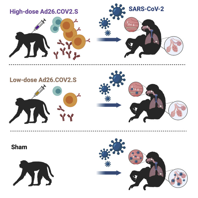
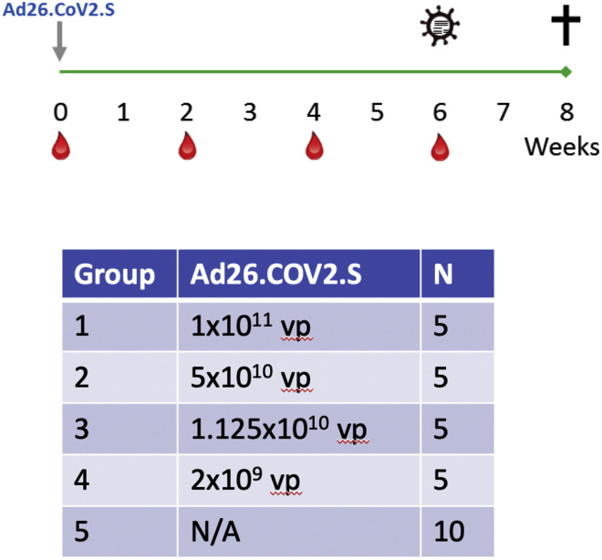
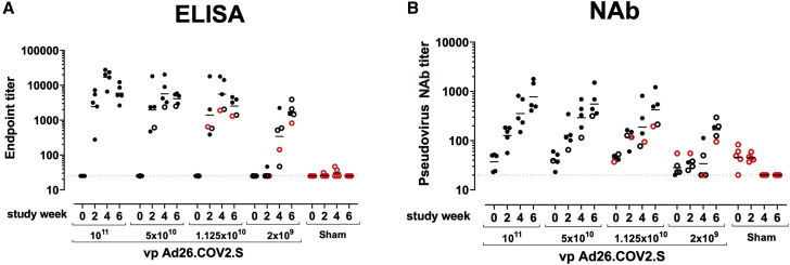
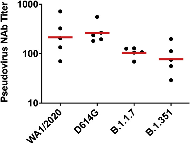
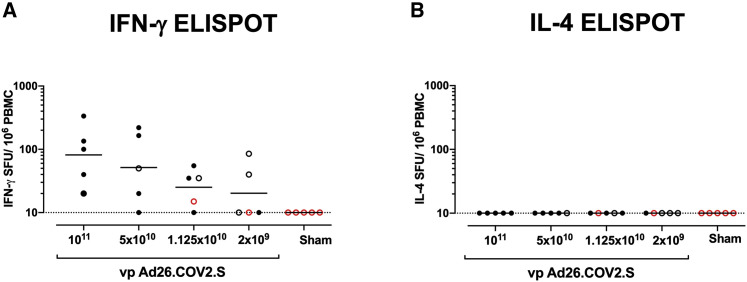
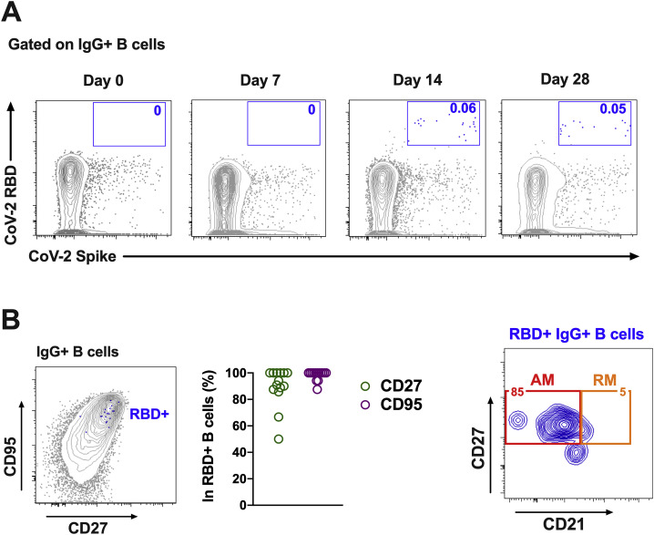
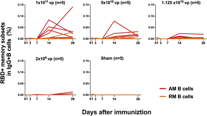
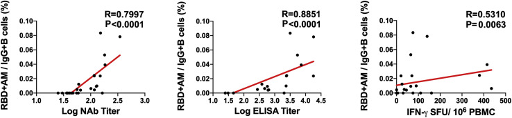
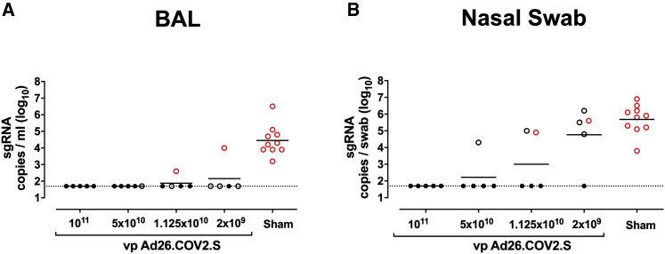
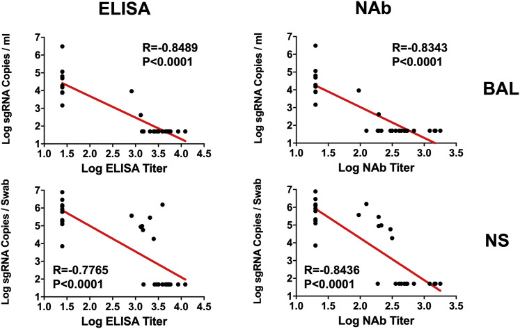
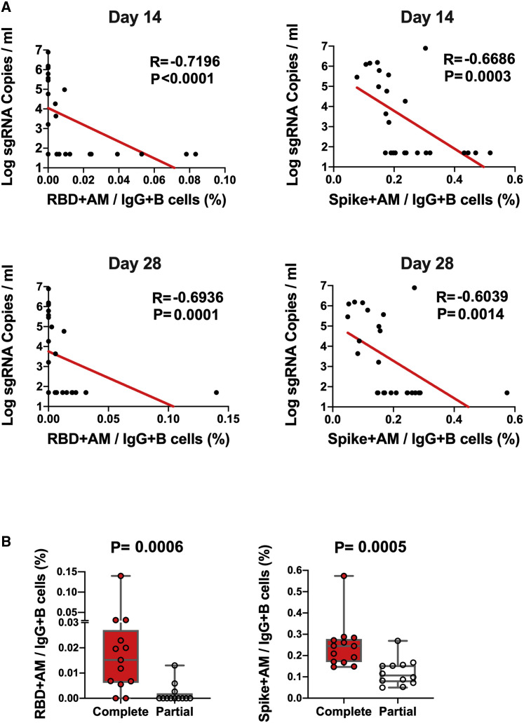
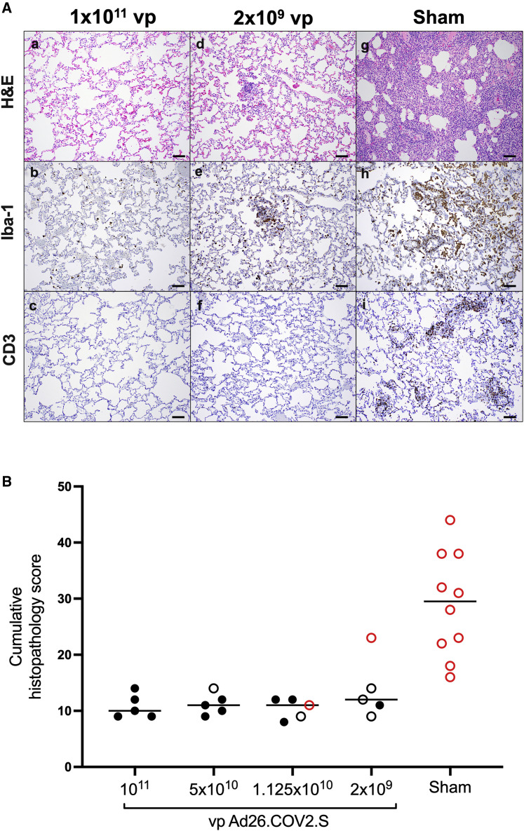
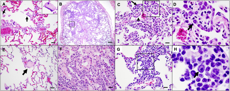
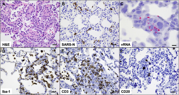
We previously reported that a single immunization with an adenovirus serotype 26 (Ad26)-vector-based vaccine expressing an optimized SARS-CoV-2 spike (Ad26.COV2.S) protected rhesus macaques against SARS-CoV-2 challenge. To evaluate reduced doses of Ad26.COV2.S, 30 rhesus macaques were immunized once with 1 × 10¹¹, 5 × 10¹⁰, 1.125 × 10¹⁰, or 2 × 10⁹ viral particles (vp) Ad26.COV2.S or sham and were challenged with SARS-CoV-2. Vaccine doses as low as 2 × 10⁹ vp provided robust protection in bronchoalveolar lavage, whereas doses of 1.125 × 10¹⁰ vp were required for protection in nasal swabs. Activated memory B cells and binding or neutralizing antibody titers following vaccination correlated with protective efficacy. At suboptimal vaccine doses, viral breakthrough was observed but did not show enhancement of disease. These data demonstrate that a single immunization with relatively low dose of Ad26.COV2.S effectively protected against SARS-CoV-2 challenge in rhesus macaques, although a higher vaccine dose may be required for protection in the upper respiratory tract.

Keywords: Ad26.COV2.S; COVID-19; SARS-CoV-2; humoral immunity; immunology; memory B cells; non-human primates; protection; vaccination.


Conflict of interest statement

Declaration of interests D.H.B., R.Z., F.W., and H.S are co-inventors on provisional vaccine patents (63/121,482; 63/133,969; 63/135,182). R.Z., F.W., S.R.H., M.v.H., L.v.d.F., and H.S. are employees of Janssen Vaccines & Prevention BV and may hold stock in Johnson & Johnson.

Figures

None
Graphical abstract
Figure S1
Figure S1
Study schema, related to Figure 1
Figure 1
Figure 1
Humoral immune responses in vaccinated rhesus macaques (A and B) Humoral immune responses were assessed at weeks 0, 2, 4, and 6 by (A) RBD-specific binding antibody ELISA and (B) pseudovirus neutralizing antibody (NAb) assays elicited by a single immunization of 1 × 1011, 5 × 1010, 1.125 × 1010, or 2 × 109 vp Ad26.COV2.S (n = 5/group) or sham negative controls (n = 10). Horizontal bars reflect geometric mean responses. Dotted lines reflect assay limit of quantitation. Solid black circles indicate animals that showed no virus in bronchoalveolar lavage (BAL) and nasal swabs (NSs) following challenge; open black circles indicate animals that showed virus in NSs, but not BAL, following challenge; and open red circles indicate animals that show virus in both BAL and NS following challenge. See also Figures S1 and S2.
Figure S2
Figure S2
NAb responses against SARS-CoV-2 variants, related to Figure 1 Pseudovirus neutralizing antibody (NAb) assays elicited by 1.125x10¹⁰ Ad26.COV2.S (n = 5) against the WA1/2020, D614G, B.1.1.7, and B.1.351 SARS-CoV-2 variants. Horizontal bars reflect geometric mean responses.
Figure 2
Figure 2
T cell responses in vaccinated rhesus macaques (A and B) Cellular immune responses were assessed at week 4 following immunization by (A) IFN-γ and (B) IL-4 ELISpot assays in response to pooled S peptides. Horizontal bars reflect geometric mean responses. Dotted lines reflect assay limit of quantitation. Solid black circles indicate animals that showed no virus in BAL and NSs following challenge; open black circles indicate animals that showed virus in NSs, but not BAL, following challenge; and open red circles indicate animals that show virus in both BAL and NSs following challenge.
Figure S3
Figure S3
B cell responses in vaccinated rhesus macaques, related to Figure 3 Percentages of RBD-specific B cells in total IgG+ B cell populations following Ad26.COV2.S immunization. (A) Representative flow cytometry of PBMCs from one monkey in the 1x10¹¹ vp dose group at days 0, 7, 14, and 28 after vaccination, gated on class-switched IgG+ B cells. (B) Expression level of CD27 and CD95 on RBD-specific B cells. (C) Flow cytometry showing activated memory (AM) and resting memory (RM) B cells, gated on RBD-specific IgG+ B cells.
Figure 3
Figure 3
B cell responses in vaccinated rhesus macaques Frequencies of RBD-specific CD27+CD95+ activated memory B cells (red) and resting memory B cells (orange) in total IgG+ B cell populations on days 0, 1, 3, 7, 14, and 28 following Ad26.COV2.S or sham immunization. See also Figures S3 and S4.
Figure S4
Figure S4
Correlations of B cell responses with antibody and T cell responses, related to Figure 3 Correlations of RBD-specific activated memory B cell frequencies with log NAb, log ELISA, and ELISpot responses in vaccinated rhesus macaques. Red lines reflect the best linear fit relationship between these variables. P and R values reflect two-sided Spearman rank-correlation tests. n = 25 biologically independent animals.
Figure 4
Figure 4
Protective efficacy following SARS-CoV-2 challenge Rhesus macaques were challenged by the intranasal and intratracheal routes with 1.0 × 105 TCID50 SARS-CoV-2. (A) Peak log10 sgRNA copies/mL (limit of quantification 50 copies/mL) were assessed in BAL following challenge. (B) Peak log10 sgRNA copies/swab (limit of quantification 50 copies/swab) were assessed in NSs following challenge. Horizontal lines reflect geometric mean values. Solid black circles indicate animals that showed no virus in BAL and NS following challenge; open black circles indicate animals that showed virus in NSs, but not BAL, following challenge; and open red circles indicate animals that show virus in both BAL and NSs following challenge.
Figure 5
Figure 5
Antibody correlates of protection in BAL and NS Correlations of log ELISA titers and log NAb titers at week 6 following vaccination with log peak sgRNA copies/mL in BAL and NSs following challenge. Red lines reflect the best linear fit relationship between these variables. p and R values reflect two-sided Spearman rank-correlation tests. n = 30 biologically independent animals.
Figure 6
Figure 6
B cell correlates of protection in NSs (A) Correlations of RBD- and S-specific activated memory B cell responses at days 14 and 28 following vaccination with log peak sgRNA copies/mL in NSs following challenge. Red lines reflect the best linear fit relationship between these variables. p and R values reflect two-sided Spearman rank-correlation tests. n = 25 biologically independent animals. (B) Frequencies of RBD- and S-specific activated memory B cells in completely protected macaques (n = 13) and partially protected and non-protected macaques (n = 12). p values reflect two-sided Mann-Whitney tests.
Figure 7
Figure 7
Comparative pathology in vaccinated and unvaccinated animals following SARS-CoV-2 challenge (A) Representative pathology from animals vaccinated with (a–c) 1 × 1011 vp Ad26.COV2.S, (d–f) 2x10⁹ vp Ad26.COV2.S, or (g–i) sham negative controls on day 10 following SARS-CoV-2 challenge. (a, d, and g) representative H&E histopathology. (b, e, and h) immunohistochemistry for Iba-1 (macrophages). (c, f, and i) Immunohistochemistry for CD3 (T lymphocytes). Animals that received a high vaccine dose had minimal evidence of SARS-CoV-2 pathology (a–c). Animals that received the lowest vaccine dose showed focal pathology (d–f) characterized by increased alveolar macrophages, focal interstitial septal thickening, and aggregates of macrophages. Sham-vaccinated animals had locally extensive moderate interstitial pneumonia (g) characterized by extensive macrophage infiltrates (h) and expansion of perivascular and interstitial CD3 T lymphocytes (i). (B) Histopathologic scoring of lung lesions in all lobes in vaccinated and sham animals following SARS-CoV-2 challenge. Scoring was performed independently by two blinded veterinary pathologists. Lesions reported included (1) inflammation interstitial/septal thickening; (2) infiltrate, macrophage; (3) alveolar infiltrate, mononuclear; (4) perivascular infiltrate, macrophage; (5) bronchiolar type II pneumocyte hyperplasia; (6) bronchus-associated lymphoid tissue (BALT) hyperplasia; (7) inflammation, bronchiolar/peribronchiolar infiltrate; (8) neutrophils, bronchiolar/alveolar; and (9) infiltrate, eosinophils. Lesions such as focal fibrosis and syncytia were reported, but not included in scoring. Edema, alveolar flooding was excluded from scoring since animals received terminal BALs. Each feature assessed was assigned a score (0 = no significant findings; 1 = minimal; 2 = mild; 3 = moderate; 4 = marked/severe). Eight representative samples from cranial, middle, and caudal lung lobes from the left and right lungs were evaluated from each animal and were scored independently. Scores were added for all lesions across all lung lobes for each animal for a maximum possible score of 288 for each monkey. Lungs evaluated were inflated/suffused with 10% formalin. Horizontal lines reflect median values. Solid black circles indicate animals that showed no virus in BAL and NS following challenge, open black circles indicate animals that showed virus in NS but not BAL following challenge, and open red circles indicate animals that show virus in both BAL and NS following challenge. Scale bars, 100 μm. See also Figures S5 and S6 and Table S1.
Figure S5
Figure S5
SARS-CoV-2-associated pathology in sham rhesus macaques following SARS-CoV-2 challenge, related to Figure 7 Focal to locally extensive SARS CoV-2 associated pathological lesions were observed in sham vaccinated monkeys 10 days following challenge. (A) Bronchoepithelial syncytia (arrow, inset) within alveolus; (B) Multifocal Type II pneumocyte hyperplasia; (C) Inset from (B) showing type II pneumocyte hyperplasia (arrow) and endothelial reactivity (arrowhead); (D) Inset from (C) showing hyperplastic pneumocytes (arrow) and occasional polymorphonuclear cells (PMNs); (E) thrombus (arrow); (F) focal edema and consolidation due to pneumocyte hyperplasia; (G) multifocal interstitial pneumonia; (H) Inset from (G) showing large reactive cells. Lesions shown are from 4 animals. Scale bars = 20 microns (G), 50 microns (A, C, E, F), 200 microns (B).
Figure S6
Figure S6
Pathology in sham-vaccinated animals corresponds to viral replication and inflammation following SARS-CoV-2 challenge, related to Figure 7 (A) H&E showing type II pneumocyte hyperplasia; (B) Immunohistochemistry for SARS nucleocapsid protein; (C) RNAscope in situ hybridization for viral RNA (vRNA) in hyperplastic pneumocytes. Immunohistochemistry for (D) Iba-1 (macrophages), (E) CD3 (T lymphocytes) and (F) CD20 (B lymphocytes) in regions of lung pathology. All images from one representative sham animal 10 days following SARS-CoV-2 challenge. Scale bars = 20 microns (A, B, D-F), 50 microns (C).

Update of

References

    1. Abbink P., Lemckert A.A., Ewald B.A., Lynch D.M., Denholtz M., Smits S., Holterman L., Damen I., Vogels R., Thorner A.R., et al. Comparative seroprevalence and immunogenicity of six rare serotype recombinant adenovirus vaccine vectors from subgroups B and D. J. Virol. 2007;81:4654–4663. - PMC - PubMed
    1. Bos R., Rutten L., van der Lubbe J.E.M., Bakkers M.J.G., Hardenberg G., Wegmann F., Zuijdgeest D., de Wilde A.H., Koornneef A., Verwilligen A., et al. Ad26 vector-based COVID-19 vaccine encoding a prefusion-stabilized SARS-CoV-2 Spike immunogen induces potent humoral and cellular immune responses. NPJ Vaccines. 2020;5:91. - PMC - PubMed
    1. Chandrashekar A., Liu J., Martinot A.J., McMahan K., Mercado N.B., Peter L., Tostanoski L.H., Yu J., Maliga Z., Nekorchuk M., et al. SARS-CoV-2 infection protects against rechallenge in rhesus macaques. Science. 2020;369:812–817. - PMC - PubMed
    1. Corbett K.S., Flynn B., Foulds K.E., Francica J.R., Boyoglu-Barnum S., Werner A.P., Flach B., O’Connell S., Bock K.W., Minai M., et al. Evaluation of the mRNA-1273 Vaccine against SARS-CoV-2 in Nonhuman Primates. N. Engl. J. Med. 2020;383:1544–1555. - PMC - PubMed
    1. Gao Q., Bao L., Mao H., Wang L., Xu K., Yang M., Li Y., Zhu L., Wang N., Lv Z., et al. Development of an inactivated vaccine candidate for SARS-CoV-2. Science. 2020;369:77–81. - PMC - PubMed

Publication types