. 2021 Jul 8;184(14):3762-3773.e10.
doi: 10.1016/j.cell.2021.05.017. Epub 2021 Jun 15.

Sneezing reflex is mediated by a peptidergic pathway from nose to brainstem

Affiliations

Sneezing reflex is mediated by a peptidergic pathway from nose to brainstem

Fengxian Li et al. Cell. .

Abstract

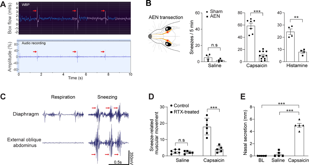
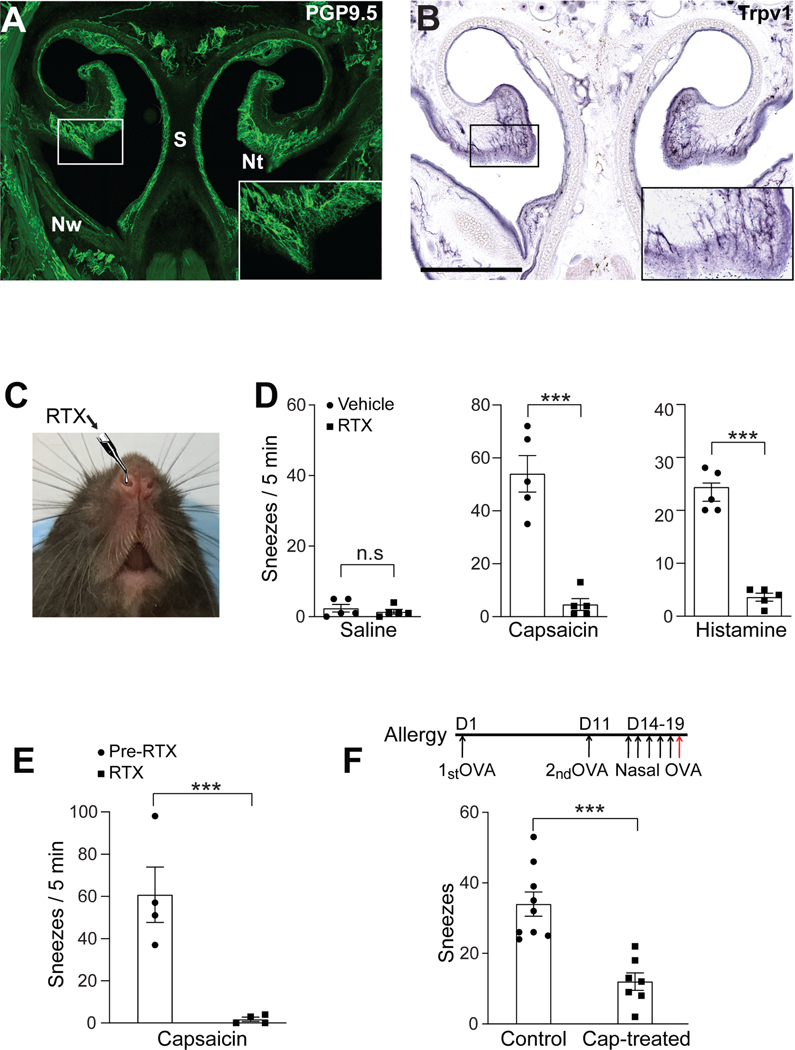
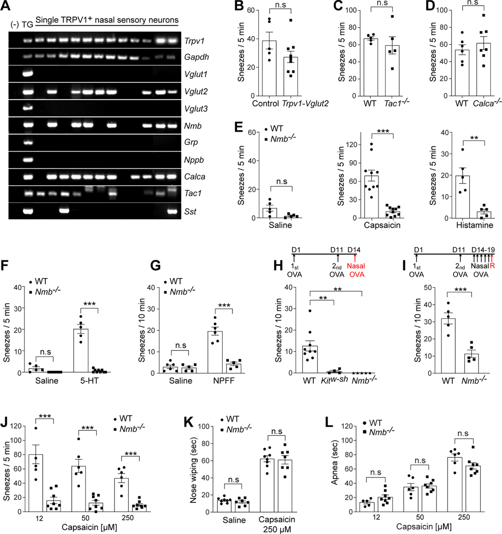
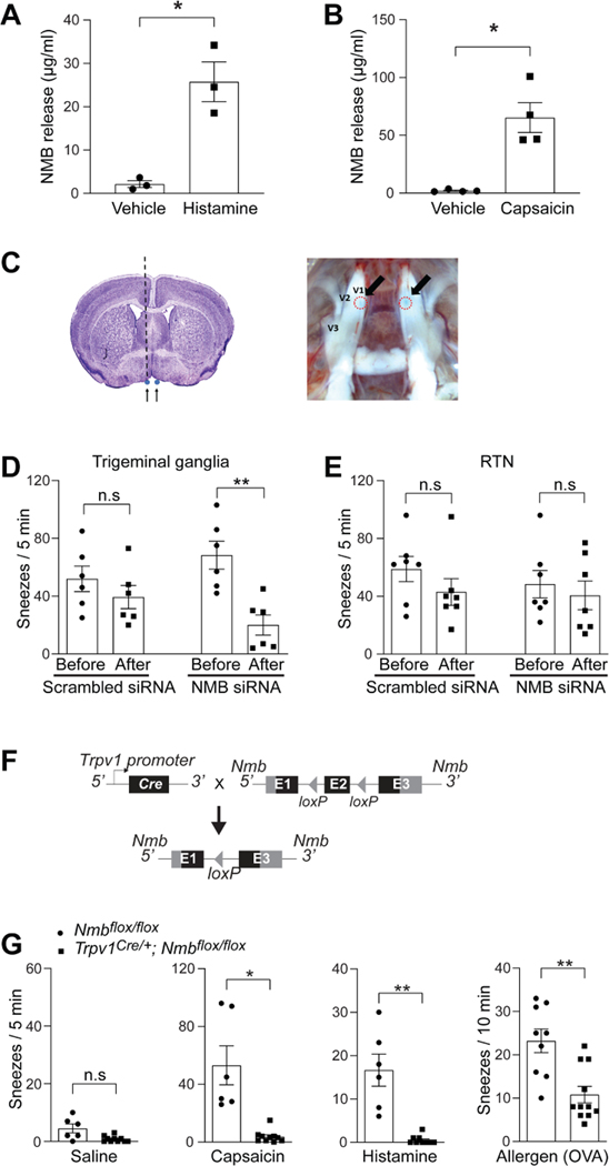
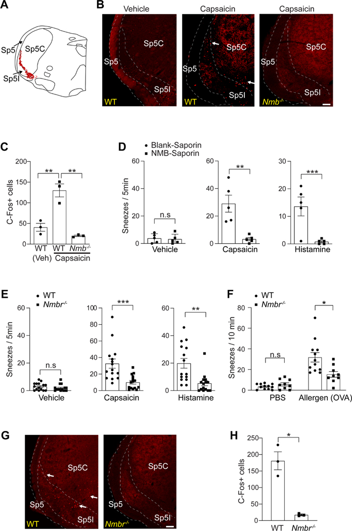
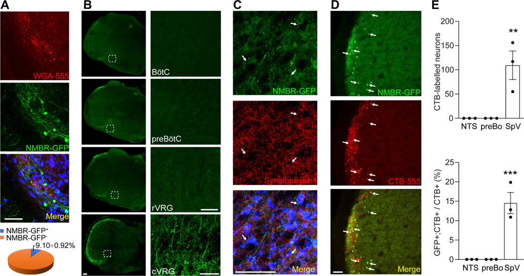
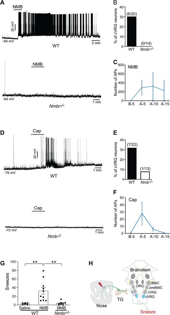
Sneezing is a vital respiratory reflex frequently associated with allergic rhinitis and viral respiratory infections. However, its neural circuit remains largely unknown. A sneeze-evoking region was discovered in both cat and human brainstems, corresponding anatomically to the central recipient zone of nasal sensory neurons. Therefore, we hypothesized that a neuronal population postsynaptic to nasal sensory neurons mediates sneezing in this region. By screening major presynaptic neurotransmitters/neuropeptides released by nasal sensory neurons, we found that neuromedin B (NMB) peptide is essential for signaling sneezing. Ablation of NMB-sensitive postsynaptic neurons in the sneeze-evoking region or deficiency in NMB receptor abolished the sneezing reflex. Remarkably, NMB-sensitive neurons further project to the caudal ventral respiratory group (cVRG). Chemical activation of NMB-sensitive neurons elicits action potentials in cVRG neurons and leads to sneezing behavior. Our study delineates a peptidergic pathway mediating sneezing, providing molecular insights into the sneezing reflex arc.

Keywords: caudal ventral respiratory group; nasal sensory neurons; neuropeptide; sneeze; sneeze-evoking region.


Conflict of interest statement

Declaration of interests The authors declare no competing interests. B.S.K. has noncompeting financial interests unrelated to this work and has served as a consultant for AbbVie, ABRAX Japan, Almirall, Cara Therapeutics, Maruho, Menlo Therapeutics, Pfizer, and Third Rock Ventures. He has participated on the advisory board for Almirall, Boehringer Ingelheim, Cara Therapeutics, Kiniksa Pharmaceuticals, Menlo Therapeutics, Regeneron Pharmaceuticals, Sanofi Genzyme, and Trevi Therapeutics. He is also founder, chief scientific officer, and stockholder of Nuogen Pharma and stockholder of Locus Biosciences.

Figures

Figure 1:
Figure 1:
Establish the mouse sneeze model. (A) Representative traces showing the simultaneous Whole Body Plethysmography (WBP) and audio recordings of a freely-moving WT mouse upon exposure to aerosolized capsaicin solution (12 μM). The WBP and audio peaks (indicated by the arrows) correlate well and indicate sneezing-like responses. (B) Transection of the anterior ethmoid nerve (AEN), which provides sensory innervation to the nose, significantly reduced sneezing-like responses to aerosolized capsaicin (12 μM) and histamine (100 mM) solution. (C) Electromyography (EMG) recording showing the representative muscle contractions of diaphragm and external oblique abdominus of mice in eupnea and during sneezing responses to nasal challenge of capsaicin (12 μM). Muscular contractions that significantly increase over regular respiratory movement and interrupt the respiratory rhythm were counted as sneezing-related responses (indicated by red arrows). (D) Nasal instillation of resiniferatoxin (RTX, 50 ng in 2 μl/ nostril) ablated nasal Trpv1+ sensory fibers and eliminated sneezing-related muscular responses to capsaicin, as revealed by EMG recording. (E) Nasal secretion was significantly increased after capsaicin-induced sneezes, compared with baseline (BL) or saline control. Each dot represents an individual mouse (n=4–11 mice/group). Data are represented as mean ± SEM. **P≤0.01, ***P≤0.001, n.s. not significant. See also Figure S1, Videos S1 and S2, and Table S1.
Figure 2:
Figure 2:
Trpv1-expressing nasal sensory fibers mediate both chemical-induced and allergy-associated sneezing. (A) The mouse nose is densely innervated by primary sensory fibers (green), as revealed by immunostaining for PGP9.5, a pan-neuronal marker. The inset shows a higher-magnification view of the boxed area. Nt, nasal turbinate; S, nasal septum; Nw, nasal wall. (B) Histochemistry for placental alkaline phosphatase (PLAP) revealed that Trpv1-expressing sensory fibers densely innervate the nasal mucosal membrane lining the nasal turbinate and septum in Trpv1PLAP/+ transgenic mice. The inset shows a higher-magnification view of the boxed area. (C) Nasal instillation of resiniferatoxin (RTX, 50 ng in 2 μl/ nostril) to desensitize/degenerate Trpv1-expressing sensory fibers in the nose. (D-E) Nasal instillation of RTX eliminated sneezing responses to aerosolized capsaicin (12 μM) and histamine (100 mM) solution, as revealed by (D) audio- and (E) WBP-recording. (F) Generation of allergic rhinitis mouse model. The red arrow indicates the intranasal instillation of allergen (ovalbumin, OVA, 0.2 μg in 2 μl PBS / nostril) for sneezing test after two systemic immunizations and five days of nasal sensitization. Allergen-induced sneezing response was significantly reduced in capsaicin-pretreated mice, compared with vehicle-treated control mice. All images shown are representatives of three biologically independent mice. Scale bars: 500 μm. Each dot represents an individual mouse (n=4–9 mice/group). Data are represented as mean ± SEM. ***P≤0.001, n.s. not significant.
Figure 3:
Figure 3:
Neuromedin B (NMB) is required for signaling sneezing. (A) Single-cell RT-PCR was performed on individual Trpv1+ nasal sensory neurons that were labeled by intranasal application of fluorescent retrograde axonal tracer. Vesicular glutamate transporter 2 (Vglut2) and neuropeptides NMB (Nmb), CGRP (Calca), and substance P (Tac1) are selectively expressed by a majority of Trpv1+ nasal neurons. In contrast, Vesicular glutamate transporter 1 and 3 (Vglut1 and Vglut3), gastrin-releasing peptide (Grp), natriuretic peptide B (Nppb), and somatostatin (Sst) are expressed by few or no Trpv1+ nasal neurons. Whole trigeminal ganglia (TG) were used as a positive control. Genomic DNA served as a negative control (−) for intron-spanning primers used for RT-PCR. (B-D) Deficiency in VGLUT2 (Vglut2), substance P (Tac1), or CGRP (Calca) does not affect the sneezing response to aerosolized capsaicin solution (12 μM), compared with control mice. (E) Deficiency in the neuropeptide NMB (Nmb) abolishes the sneezing responses to aerosolized capsaicin (12 μM) and histamine (100 mM) solution. (F-G) Nmb deficiency abolishes the sneezing responses to serotonin (5-HT, aerosolized solution (1 mM), F) and neuropeptide FF solution (NPFF, 20 nmol in 2 μl, G). (H) In acute allergic model, the sneezing responses to allergen (ovalbumin, OVA, 0.2 μg in 2 μl PBS/nostril) were abolished in mast cell-deficient Kitw-sh mice, compared with WT controls. This mast cell-dependent allergic sneezing was eliminated in Nmb−/− mice. (I) Nmb−/− mice display reduced sneezing response in chronic allergic model, compared with WT controls. The red arrow indicates the intranasal instillation of allergen (ovalbumin, OVA, 0.2 μg in 2 μl PBS / nostril) for sneezing test after five days of intranasal sensitization. (J) Inverse dose-response relationship between capsaicin and sneezing responses in WT mice. Nmb−/− mice display reduced sneezing responses to capsaicin at all the doses tested. (K) Pain-related nose wiping behavior elicited by high-dose capsaicin (250 μM) remains intact in the Nmb−/− mice, as in WT mice. (L) Capsaicin induces mouse apnea responses in a dose-dependent manner. Each dot represents an individual mouse (n=5–10 mice/group). Data are represented as mean ± SEM. ** P≤0.01, ***P≤0.001, n.s. not significant. See also Figure S2.
Figure 4:
Figure 4:
NMB is released from sensory neurons for signaling sneezing. (A-B) NMB is released from cultured trigeminal ganglia neurons of WT mice after vehicle or sneeze-inducing compounds treatment (histamine: 200 μM; capsaicin: 10 μM). Each dot represents one replicate. (C) Diagrams illustrating microinjection of siRNA into the V1 division of the trigeminal ganglia. The black dotted line represents the path of the microinjection needle. Arrows in the right panel point toward the injection sites, outlined by red dashed circles, in the trigeminal ganglia. (D) NMB-siRNA silencing in the trigeminal ganglia efficiently decreases the sneezing response to aerosolized capsaicin solution (12 μM), compared with the sneezing responses before microinjection or the non-targeting scrambled siRNA controls. (E) NMB-siRNA silencing in the retrotrapezoid nucleus (RTN) does not affect the sneezing response to aerosolized capsaicin solution (12 μM). (F) Schematic diagram illustrating the generation of Trpv1Cre/+; Nmbfl°x/fl°x mice in which Nmb was conditionally knocked out in Trpv1-lineage sensory neurons. (G) Trpv1Cre/+; Nmbfl°x/fl°x mice display significantly reduced sneezing responses to aerosolized capsaicin (12 μM), histamine (100 mM) solution and chronic allergy induced by ovalbumin (OVA, 0.2 μg in 2 μl PBS / nostril). Each dot represents an individual mouse (n=6–11 mice/group). Data are represented as mean ± SEM. * P≤0.05, ** P≤0.01, n.s. not significant. See also Figure S3.
Figure 5:
Figure 5:
NMB-sensitive postsynaptic neurons in the sneeze-evoking region mediate sneezing. (A) Diagram showing the sneeze-evoking region (i.e., the central projection of nasal sensory neurons; indicated by red dots) in the spinal trigeminal nucleus (SpV) based on previous studies. Sp5, spinal trigeminal tract; Sp5C, spinal trigeminal nucleus caudalis; Sp5I, spinal trigeminal nucleus interpolar. (B) Capsaicin-induced sneezes lead to a significant increase in the expression of c-Fos within the sneeze-evoking region of WT (indicated by arrows), compared with the saline vehicle control. Nmb-deficient mice display a significantly reduced c-Fos signal. (C) Quantification of c-Fos+ neurons in the sneeze-evoking region after saline (Veh) or capsaicin treatments. (D) Microinjection of NMB-saporin into the sneeze-evoking region abolishes the sneezing responses to aerosolized capsaicin (12 μM) and histamine (100 mM) solution. (E-F) Nmbr−/− mice display significantly reduced sneezing responses to aerosolized capsaicin (12 μM), histamine (100 mM) solution and allergen (ovalbumin, OVA, 0.2 μg in 2 μl PBS / nostril) stimuli. (G-H) Capsaicin-induced sneezes lead to the expression of c-Fos in the sneeze-evoking region of WT (indicated by arrows) but not Nmbr−/− mice. All images shown are representatives of three biologically independent mice. Scale bars: 100 μm. The total number of c-Fos+ neurons in the sneeze-evoking region was counted from ten sections per mouse (n=3 / genotype). Each dot represents an individual mouse. n=5–15 mice/group for behavioral tests. Data are represented as mean ± SEM. * P≤0.05, ** P≤0.01, *** P≤0.001, n.s. not significant.
Figure 6:
Figure 6:
The projection of NMBR+ neurons to the caudal ventral respiratory group (cVRG). (A) Representative imaging of brainstem coronal sections showing that NMBR+ neurons (green) comprise a highly restricted population in the sneeze-evoking region (red, labeled by WGA-555 applied to mouse nasal cavity). Neurons were marked by NeuN (blue). (B) Representative imaging showing that NMBR+ neurons selectively project to cVRG but no other respiratory regions including the rostral ventral respiratory group (rVRG), pre-BÖtzinger complex (preBÖtC) and BÖtzinger complex (BÖtC) as revealed by axonal tracing in NmbreGFP reporter line. Images on the right show higher-magnification view of each boxed region. (C) Representative imaging showing that NMBR-GFP+ nerve fibers (green) express the presynaptic marker synaptophysin 1 (red) and synapse with cVRG neurons (marked by NeuN, blue). (D-E) Retrograde axonal tracing by microinjection of CTB-555 into cVRG of NmbreGFP mice shows that CTB labeled NMBR-GFP+ neurons (indicated by arrows) are localized within the sneeze-evoking region in the spinal trigeminal nucleus and absent from other brain regions including the pre-BÖtzinger complex (preBÖ) and nucleus tractus solitarius (NTS). All images shown are representatives of three biologically independent mice. Scale bars: 100 μm. Each dot represents an individual mouse (n=3 mice/group). Data are represented as mean ± SEM. ** P≤0.01, *** P≤0.001. See also Figure S4.
Figure 7:
Figure 7:
The synaptic connection between NMBR+ neurons and caudal ventral respiratory group (cVRG) neurons is essential for signaling sneezing. (A) Representative traces showing that pharmacological activation of NMBR+ neurons using NMB peptide (4 μM) induces robust action potential (AP) firing in cVRG neurons of WT but not Nmbr−/− mice. (B) NMB peptide evokes robust AP firing in 30% of cVRG neurons recorded in the brainstem slices from WT mice. As a negative control, cVRG neurons of Nmbr−/− mice display no response to the same NMB treatment. (C) The number of APs fired by WT cVRG neurons 5 min before (B-5) and 0–5 min (A-5), 5–10 min (A-10), and 10–15 min (A-15) after NMB treatment. (D) Representative traces showing that capsaicin (5 μM) induces robust AP firing in cVRG neurons of WT but not Nmb−/− mice. (E) Capsaicin treatment elicits robust AP firing in 31.8% of cVRG neuron recorded from WT mice but weak or no AP firing from Nmb−/− mice. (F) The number of APs fired by WT cVRG neurons 5 min before (B-5) and 0–5 min (A-5), 5–10 min (A-10), and 10–15 min (A-15) after capsaicin treatment. (G) Microinjection of NMB peptide (1 pmol in 100 nl) into the cVRG region induces significant sneezing responses in WT but not Nmbr−/− mice. As a control, microinjection of saline does not induce sneezing. (H) Schematic diagram illustrating the neural pathway for sneezing (nasal NMB+ sensory neurons → NMBR+ neurons in the sneeze-evoking region → cVRG neurons). TG: trigeminal ganglia; rVRG: rostral ventral respiratory group; preBÖtC: pre-BÖtzinger complex; BÖtC: BÖtzinger complex; DRG: dorsal respiratory group. Each dot represents an individual mouse (n=6–10 mice/group). Data are represented as mean ± SEM. ** P≤0.01.

Comment in

  • Sneezing pathway revealed.
    Whalley K. Whalley K. Nat Rev Neurosci. 2021 Aug;22(8):455. doi: 10.1038/s41583-021-00491-3. Nat Rev Neurosci. 2021. PMID: 34226717 No abstract available.

References

    1. Batsel HL, and Lines AJ (1978). Discharge of respiratory neurons in sneezes resulting from ethmoidal nerve stimulation. Exp Neurol 58, 410–424. - PubMed
    1. Baydin A, Nural MS, Guven H, Deniz T, Bildik F, and Karaduman A. (2005). Acute aortic dissection provoked by sneeze: a case report. Emerg Med J 22, 756–757. - PMC - PubMed
    1. Bourouiba L. (2016). IMAGES IN CLINICAL MEDICINE. A Sneeze. N Engl J Med 375, e15. - PubMed
    1. Bourouiba L. (2020). Turbulent Gas Clouds and Respiratory Pathogen Emissions: Potential Implications for Reducing Transmission of COVID-19. JAMA 323, 1837–1838. - PubMed
    1. Bourouiba L, Dehandschoewercker E, and M.B.J.W. (2014). Violent expiratory events: on coughing and sneezing. J Fluid Mech 745, 27.

Publication types