

Plankton classification with high-throughput submersible holographic microscopy and transfer learning
- PMID: 34134620
- PMCID: PMC8207568
- DOI: 10.1186/s12862-021-01839-0
Plankton classification with high-throughput submersible holographic microscopy and transfer learning
Abstract
Background: Plankton are foundational to marine food webs and an important feature for characterizing ocean health. Recent developments in quantitative imaging devices provide in-flow high-throughput sampling from bulk volumes-opening new ecological challenges exploring microbial eukaryotic variation and diversity, alongside technical hurdles to automate classification from large datasets. However, a limited number of deployable imaging instruments have been coupled with the most prominent classification algorithms-effectively limiting the extraction of curated observations from field deployments. Holography offers relatively simple coherent microscopy designs with non-intrusive 3-D image information, and rapid frame rates that support data-driven plankton imaging tasks. Classification benchmarks across different domains have been set with transfer learning approaches, focused on repurposing pre-trained, state-of-the-art deep learning models as classifiers to learn new image features without protracted model training times. Combining the data production of holography, digital image processing, and computer vision could improve in-situ monitoring of plankton communities and contribute to sampling the diversity of microbial eukaryotes.
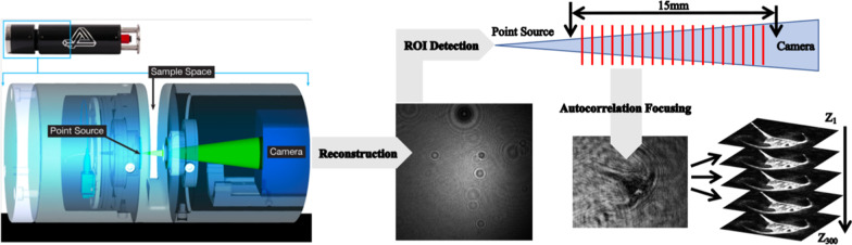
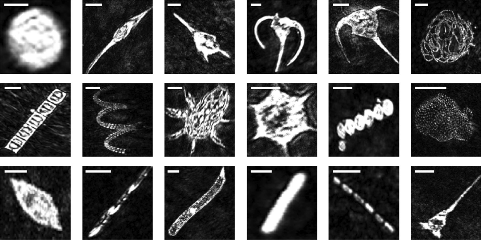
Results: Here we use a light and portable digital in-line holographic microscope (The HoloSea) with maximum optical resolution of 1.5 μm, intensity-based object detection through a volume, and four different pre-trained convolutional neural networks to classify > 3800 micro-mesoplankton (> 20 μm) images across 19 classes. The maximum classifier performance was quickly achieved for each convolutional neural network during training and reached F1-scores > 89%. Taking classification further, we show that off-the-shelf classifiers perform strongly across every decision threshold for ranking a majority of the plankton classes.
Conclusion: These results show compelling baselines for classifying holographic plankton images, both rare and plentiful, including several dinoflagellate and diatom groups. These results also support a broader potential for deployable holographic microscopes to sample diverse microbial eukaryotic communities, and its use for high-throughput plankton monitoring.
Keywords: Classification workflow; Convolutional neural networks; Deep learning; Deployable microscope; High-throughput imaging; Holographic microscopy; Plankton.
Conflict of interest statement
SM declares a competing financial interest: as the chief technical officer of 4-Deep, the creators of the Octopus and Stingray software.
Figures
References
-
- Benfield M, Grosjean P, Culverhouse P, Irigoien X, Sieracki ME, Lopez-Urrutia A, et al. RAPID: research on automated plankton identification. Oceanography. 2007;20:172–187. doi: 10.5670/oceanog.2007.63. - DOI
-
- Olson RJ, Sosik HM. A submersible imaging-in-flow instrument to analyze nano-and microplankton: imaging FlowCytobot: in situ imaging of nano- and microplankton. Limnol Oceanogr Methods. 2007;5:195–203. doi: 10.4319/lom.2007.5.195. - DOI
-
- Cowen RK, Guigand CM. In situ ichthyoplankton imaging system (ISIIS): system design and preliminary results: in situ ichthyoplankton imaging system. Limnol Oceanogr Methods. 2008;6:126–132. doi: 10.4319/lom.2008.6.126. - DOI
Publication types
MeSH terms
LinkOut - more resources
Full Text Sources
Other Literature Sources