. 2021 Jun 16;7(25):eabg7663.
doi: 10.1126/sciadv.abg7663. Print 2021 Jun.

Longitudinal high-resolution imaging through a flexible intravital imaging window

Affiliations

Longitudinal high-resolution imaging through a flexible intravital imaging window

Guillaume Jacquemin et al. Sci Adv. .

Abstract

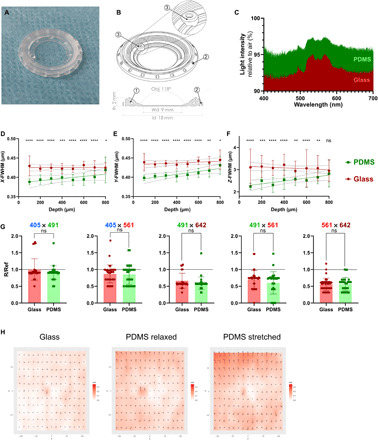
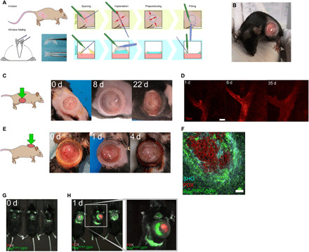
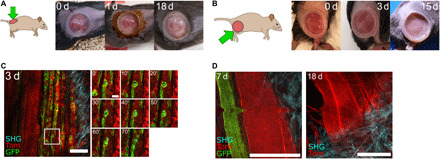
Intravital microscopy (IVM) is a powerful technique that enables imaging of internal tissues at (sub)cellular resolutions in living animals. Here, we present a silicone-based imaging window consisting of a fully flexible, sutureless design that is ideally suited for long-term, longitudinal IVM of growing tissues and tumors. Crucially, we show that this window, without any customization, is suitable for numerous anatomical locations in mice using a rapid and standardized implantation procedure. This low-cost device represents a substantial technological and performance advance that facilitates intravital imaging in diverse contexts in higher organisms, opening previously unattainable avenues for in vivo imaging of soft and fragile tissues.


Figures

Fig. 1
Fig. 1. PDMS implantable devices display microscopy-ready optical characteristics.
(A) Photograph of a PDMS intravital imaging window. (B) 3D drawing and cross-sectional view of the window; 18 mm in diameter (ld) and 2 mm in height (Ih). The device allows imaging with an angled objective (Obj) up to 118° over a working diameter (Wd) of 9 mm. At 200 mg, the PDMS window is at least 3× lighter than conventional titanium/glass windows. The angle of the skin groove (1) protects the window frame from damage and removal by the mouse. To promote healing and immobilization of the window, equally spaced holes surround the structure (2). An injection port, 0.7 mm in diameter (3), is positioned on the side of the window. (C) Light intensity measured through glass (red) or PDMS (green) windows relative to air. (D to F) PSF analysis comparing glass (red) and PDMS (green). Graphs display calculated full width at half maximum (FWHM) values in (D) X axis, (E) Y axis, or (F) Z axis over depth. Comparison between glass and PDMS FWHM at each depth was performed using a Mann-Whitney test (n > 20 measurements per section). (G) Chromatic aberration analysis of the indicated color pairs through glass (red) or PDMS (green) windows. Both glass and PDMS in every color pairs are significantly lower than ratio R/Ref < 1, indicative of colocalization (P < 0.01, one-sample Wilcoxon text). No significant differences were observed between glass and PDMS for all color pairs (P > 0.05, Mann-Whitney test). (H) Mapping of the microgrid deformations arising from glass coverslips, relaxed, or stretched (to 200%) PDMS windows compared to the microgrid alone. Vectors of deformation were calculated on each dot of the grid to generate 2D maps [md, measured deformation norms (μm); vectors represented at 50×]. Deformations induced by glass coverslips (0.037 ± 0.019 μm SD), relaxed (0.076 ± 0.031 μm SD), and stretched (0.099 ± 0.043 μm SD) PDMS windows were all below the lateral imaging resolution (0.161 μm per pixel) and the inherent aberrations of the optical system. ns, nonsignificant; *P < 0.05; **P < 0.01; ***P < 0.001; and ****P < 0.0001. All source data are provided in data files S2 and S3.
Fig. 2
Fig. 2. PDMS-based intravital windows allow high-resolution longitudinal imaging in vivo.
(A) Schematic representation of the suture-free window implantation procedure. For details, see Materials and Methods. (B) Representative photograph of a 6-week-old mouse carrying a newly implanted PDMS window over the abdominal mammary gland. (C) Photographs of a PDMS window maintained over the fourth mammary gland of a pubertal mouse for the indicated period in days (d). Representative of n > 10 mice implanted at 5 to 6 weeks of age. (D) Longitudinal tissue-scale IVM of a mammary duct expressing tdTomato (Tom) in a R26mTmG mouse for the indicated period in days (d). Scale bar, 100 μm. Maximum tissue depth achieved: 400 μm (day 35). (E) Photographs of a PDMS intravital imaging window maintained on a growing PDX tumor grafted in the interscapular region of a RagKO/KO-GFP mouse for the indicated period in days (d). Brown skin coloring is due to dermal Betadine solution. Representative of n = 7 mice. (F) IVM of tdTomato-expressing PDX tumor cells (red) engrafted in a RagKO/KO-GFP recipient mouse (host cells in green). Second harmonic generation (SHG) shows fibrillar collagen around the tumor (blue). Scale bar, 100 μm. (G and H) Near-infrared imaging (NIR-IVIS) of RagKO/KO-GFP mice bearing PDX tumors expressing tdTomato fluorescence before (G) or after (H) window implantation.
Fig. 3
Fig. 3. PDMS-based intravital windows allow high-resolution longitudinal imaging of injury induced muscle regeneration in vivo.
(A and B) Representative photographs of a PDMS intravital imaging window maintained on the back (A) or thigh (B) muscle of a mouse for the indicated period in days (d). Brown skin coloring at day 1 in (A) is due to dermal Betadine solution. Representative of n = 9 mice implanted on the back or thigh muscles. (C) IVM of thigh muscle regeneration through a PDMS imaging window 3 days (3 d) after cardiotoxin-induced muscle injury in Pax7CreERT2;R26mTmG adult mice. All cells were labeled with membrane tdTomato fluorescent protein (Tom, red). Tamoxifen-induced Cre-mediated enhanced GFP protein expression (green) in Pax7-positive muscle stem cells and their progeny. SHG imaging shows fibrillar collagen organization in blue. Time-lapse imaging shows muscle stem cell division and migration (white asterisks) over 70 min. Scale bars, 100 μm. (D) Representative IVM images of muscle fibers 7 and 18 days after implantation in a Pax7CreERT2;R26mTmG mouse, showing resolution at the cellular scale and maintenance of excellent imaging quality over time. Scale bars, 100 μm. The small bump in the image at 7 days is due to the breathing-induced movement during image acquisition.

References

    1. Pittet M. J., Weissleder R., Intravital imaging. Cell 147, 983–991 (2011). - PMC - PubMed
    1. Follain G., Mercier L., Osmani N. L., Harlepp S., Goetz J. G., Seeing is believing-multi-scale spatio-temporal imaging towards in vivo cell biology. J. Cell Sci. 130, 23–38 (2017). - PubMed
    1. Lloyd-Lewis B., Multidimensional imaging of mammary gland development: A window into breast form and function. Front. Cell Dev. Biol. 8, 203 (2020). - PMC - PubMed
    1. Nobis M., Warren S. C., Lucas M. C., Murphy K. J., Herrmann D., Timpson P., Molecular mobility and activity in an intravital imaging setting–implications for cancer progression and targeting. J. Cell Sci. 131, jcs206995 (2018). - PubMed
    1. Coste A., Oktay M. H., Condeelis J. S., Entenberg D., Intravital imaging techniques for biomedical and clinical research. Cytometry A 97, 448–457 (2020). - PMC - PubMed