. 2021 Aug;32(8):1987-2004.
doi: 10.1681/ASN.2020091373. Epub 2021 Jun 16.

Progressive Cellular Senescence Mediates Renal Dysfunction in Ischemic Nephropathy

Affiliations

Progressive Cellular Senescence Mediates Renal Dysfunction in Ischemic Nephropathy

Seo Rin Kim et al. J Am Soc Nephrol. 2021 Aug.

Abstract

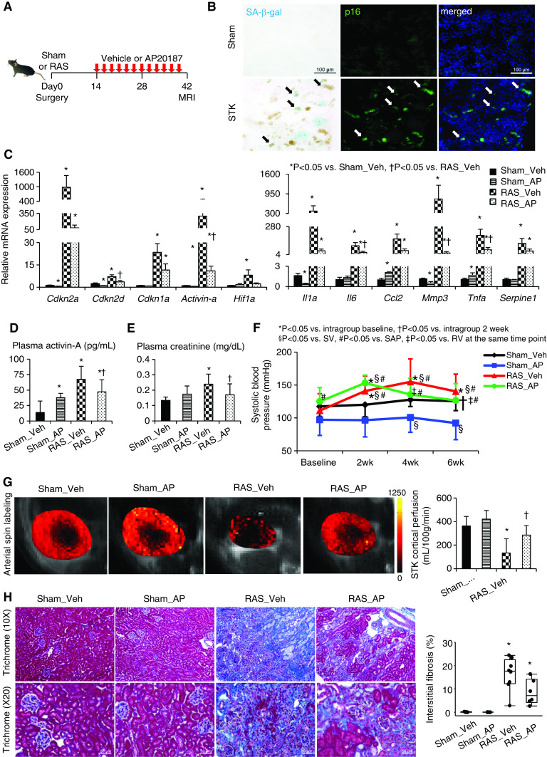
Background: Peripheral vascular diseases may induce chronic ischemia and cellular injury distal to the arterial obstruction. Cellular senescence involves proliferation arrest in response to stress, which can damage neighboring cells. Renal artery stenosis (RAS) induces stenotic-kidney dysfunction and injury, but whether these arise from cellular senescenceand their temporal pattern remain unknown.

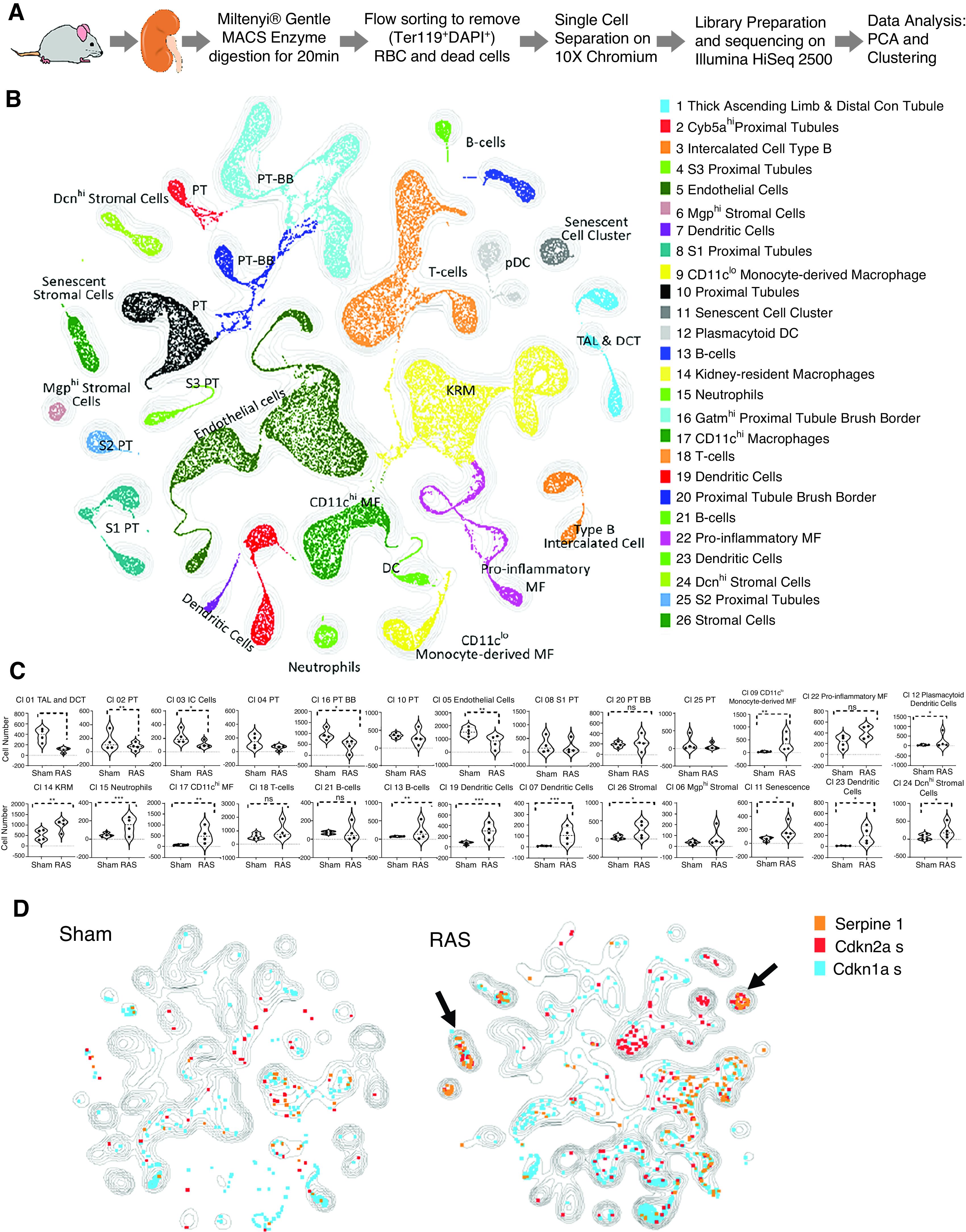
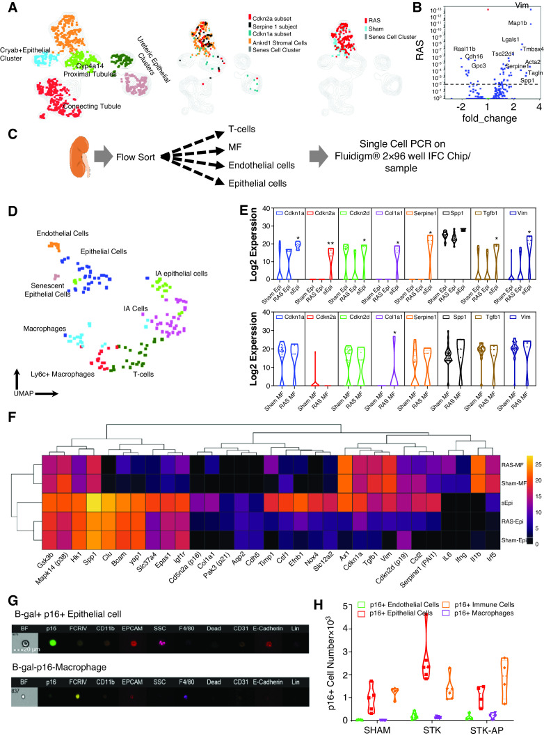
Methods: Chronic renal ischemia was induced in transgenic INK-ATTAC and wild type C57BL/6 mice by unilateral RAS, and kidney function (in vivo micro-MRI) and tissue damage were assessed. Mouse healthy and stenotic kidneys were analyzed using unbiased single-cell RNA-sequencing. To demonstrate translational relevance, cellular senescence was studied in human stenotic kidneys.

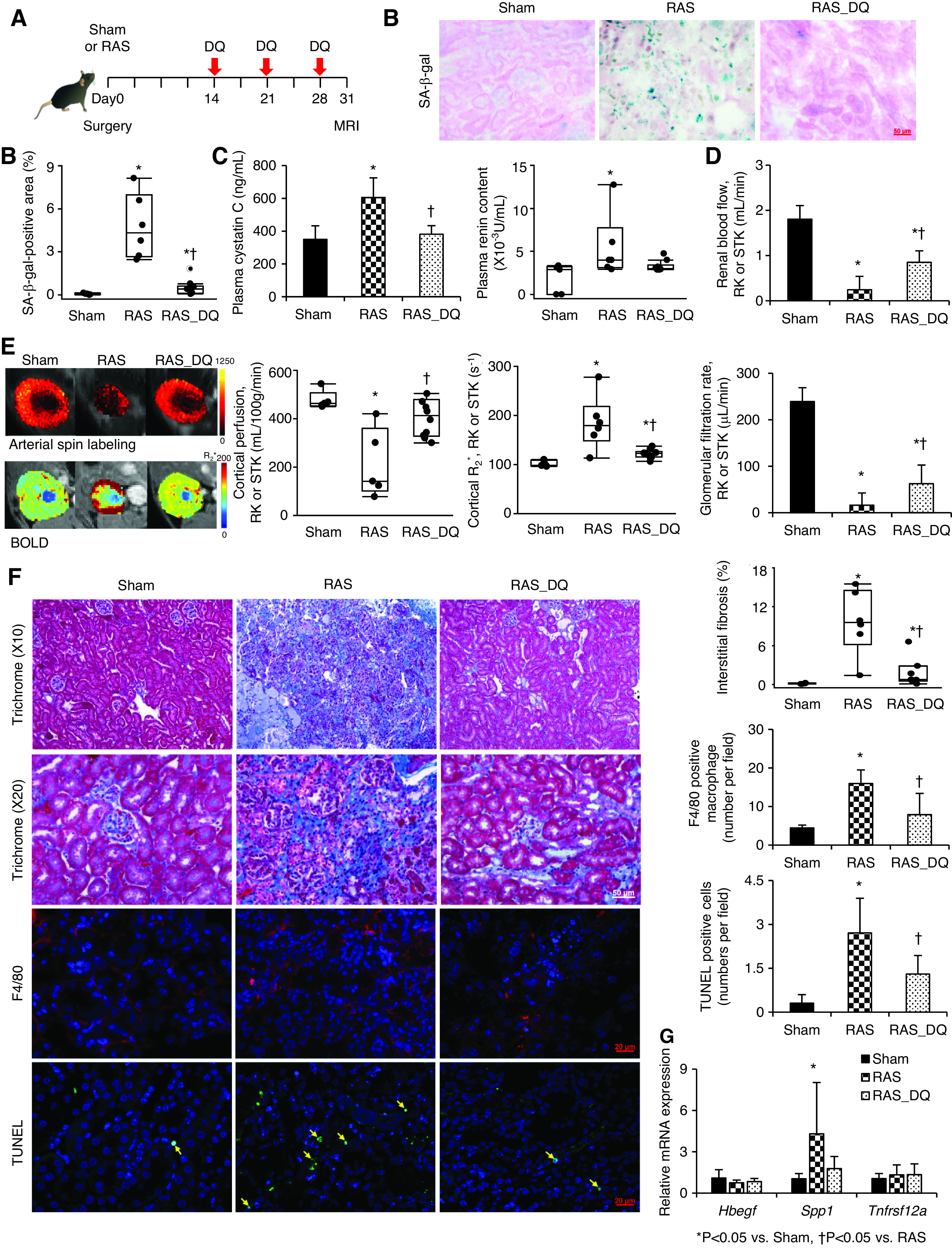
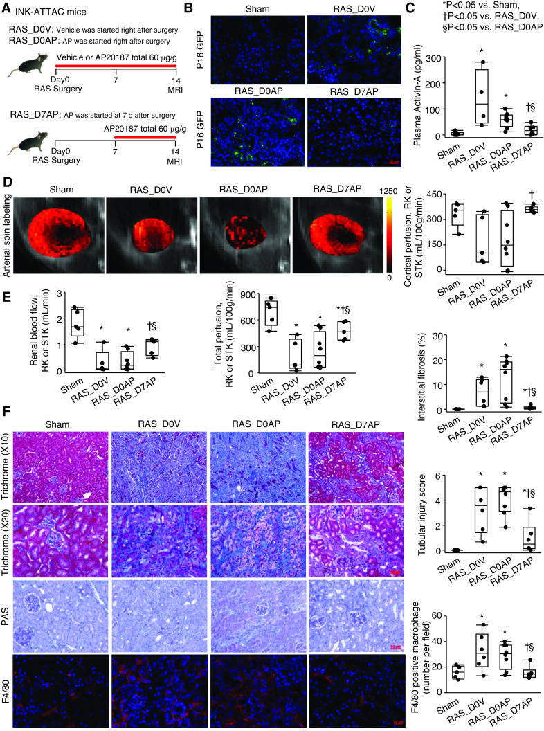
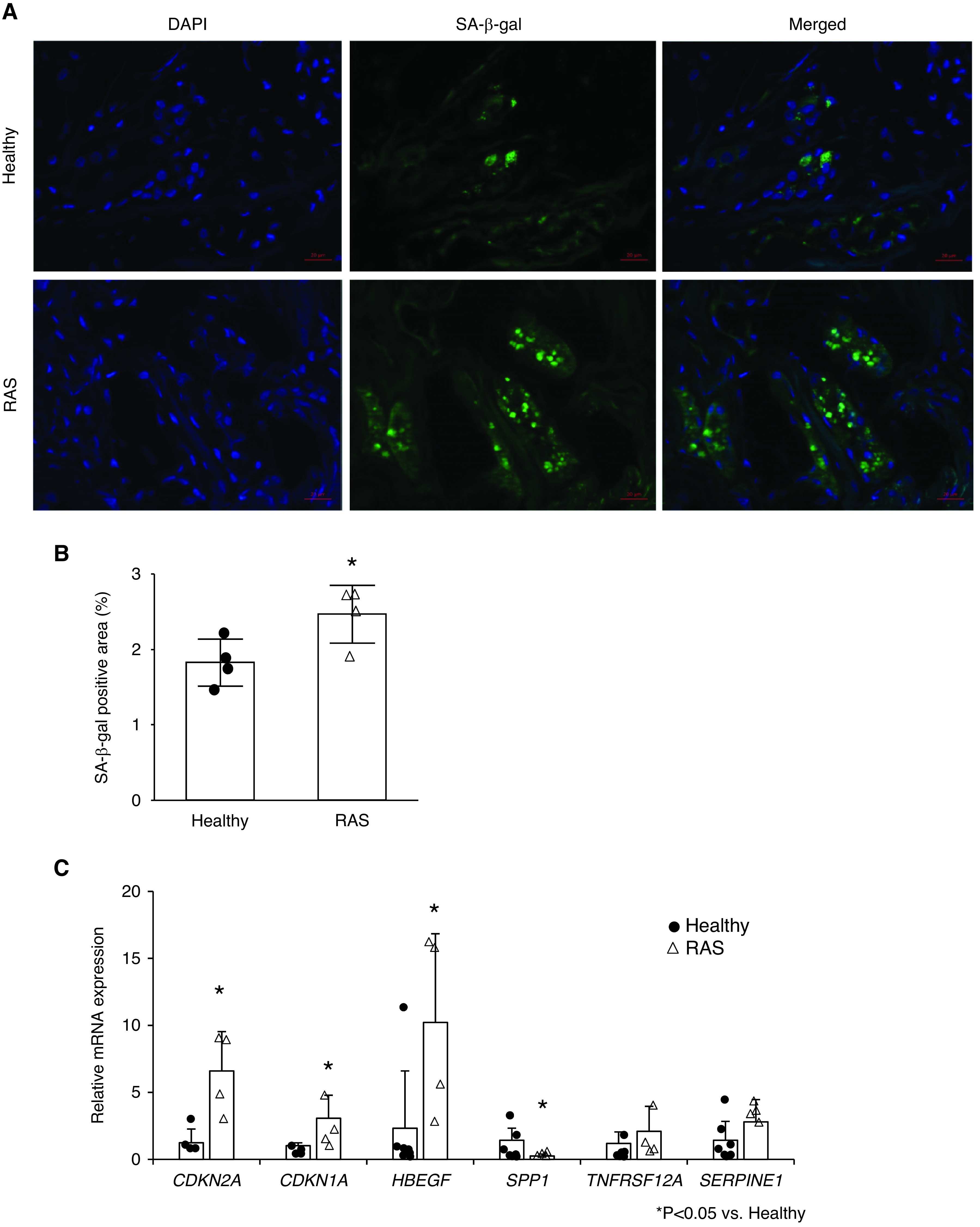
Results: Using intraperitoneal AP20187 injections starting 1, 2, or 4 weeks after RAS, selective clearance of cells highly expressing p16Ink4a attenuated cellular senescence and improved stenotic-kidney function; however, starting treatment immediately after RAS induction was unsuccessful. Broader clearance of senescent cells, using the oral senolytic combination dasatinib and quercetin, in C57BL/6 RAS mice was more effective in clearing cells positive for p21 (Cdkn1a) and alleviating renal dysfunction and damage. Unbiased, single-cell RNA sequencing in freshly dissociated cells from healthy and stenotic mouse kidneys identified stenotic-kidney epithelial cells undergoing both mesenchymal transition and senescence. As in mice, injured human stenotic kidneys exhibited cellular senescence, suggesting this process is conserved.

Conclusions: Maladaptive tubular cell senescence, involving upregulated p16 (Cdkn2a), p19 (Cdkn2d), and p21 (Cdkn1a) expression, is associated with renal dysfunction and injury in chronic ischemia. These findings support development of senolytic strategies to delay chronic ischemic renal injury.

Keywords: dasatinib; quercetin; renal artery stenosis; senescence; transcriptome.


Figures

Figure 1.
Figure 1.
Chronic ischemia induces senescence in the stenotic kidney (STK), and clearance of cells highly expressing p16Ink4a by AP improves renal function and oxygenation. (A) Experimental design in INK-ATTAC transgenic mice studied 6 weeks after sham or RAS surgeries. (B) SA-β-gal (cyan/blue) and p16 (green) colocalize (arrows) on mouse STK cryosections. Blue, 4′,6-diamidino-2-phenylindole. Renal gene expression of senescence and SASP factors using (C) real-time PCR (relative to Gapdh), (D) plasma levels of activin-A by ELISA, (E) creatinine levels using Jaffe reactions, and (F) SBP levels measured by the tail-cuff method using an automated oscillometric device in sham or RAS treated with vehicle (veh) or AP. *P<0.05 versus intragroup baseline, P<0.05 versus intragroup after 2 weeks, §P<0.05 versus sham-vehicle group, #P<0.05 versus sham-AP group, P<0.05 versus RAS-vehicle at the same time point. (G) Renal perfusion maps generated by arterial spin-labeling MRI (milliliters per 100 g per minute; brighter red, higher perfusion). (H) Masson trichrome staining and semiautomatic quantification on ten randomly chosen fields per section (using AxioVision) showed that increased STK fibrosis was slightly blunted by AP. Mean±SD or median±interquartile range (n=6–8). *P<0.05 versus sham-vehicle group, P<0.05 versus RAS-vehicle group (t tests or Wilcoxon test).
Figure 2.
Figure 2.
Clustering of sham and RAS kidneys by scRNA-seq illustrate cell types/clusters in sham and RAS kidneys. (A) Experimental design for isolating viable kidney cells. scRNA-seq data were analyzed together with all metadata. (B) Unsupervised KNetL followed by PhenoGraph mapping, showing 26 distinct cell clusters from 57,000 sham and RAS kidney cells. (C) Violin plots comparing the proportion of each cluster in sham and RAS samples. Individual populations were downsampled to approximately 7000 cells per sample, and proportions of individual populations per cluster were quantified (Mann–Whitney test). P<0.05 is considered significant. In RAS, cells were reduced in tubular and endothelial, but increased in myeloid and stromal clusters. (D) The contours illustrate and compare cell types/clusters between sham and RAS kidneys. Some genes are superimposed on these contours to highlight the cell types (listed in Figure 2B) that undergo senescence (Figure 2C) in RAS. Overlay of Cdkn2a+ senescent cells in sham and RAS show that most cells upregulating Cdkn2a are T and B lymphocytes, myeloid cells (macrophages [MF] and dendritic cells [DC]), stromal cells (arrow), and a senescent cell cluster (arrow). The senescent cell cluster (cluster 11) (arrow) expresses marker genes such as Serpine1, Cdkn2a, and Cdkn1a in RAS compared with sham. BB, brush border; Con tubule, convoluted tubule; DAPI, 4′,6-diamidino-2-phenylindole; DCT, distal convoluted tubule; KRM, kidney-resident macrophages; MACS, magnetic cell sorting; PT, proximal tubule; RBC, red blood cells; TAL, thick ascending limb of the loop of Henle.
Figure 3.
Figure 3.
Senescent cell cluster 11 is heterogenous and consists of Ankrd1+ stromal cells that express Cdkn2a, Cdkn1a, and Serpine1, suggesting senescence. (A) Reclustering of cluster 11 revealed five subclusters. Overlay of Cdkn2a+, Cdkn1a+, and Serpine1+ cells on Ankrd1+ stromal cluster (middle UMAP) shows that this cluster expresses senescent genes; overlay of RAS and sham populations shows that Ankrd1 stromal cell cluster predominantly contains RAS cells. (B) The Ankrd1+ population in RAS shows upregulation of mesenchymal genes, such as Vim, Map1b, Lgals1, Acta2, and Ankrd1, whereas epithelial cell–specific genes (e.g., Cdh16, Gpc3) are downregulated. (C) Experimental design for isolating viable epithelial cells (epi), endothelial cells, macrophages (MF), and T cells from sham and RAS kidneys. Kidneys were digested, flow sorted, and approximately 192 single cells (approximately 48 cells per population) underwent qPCR for 96 genes. (D) Data analyzed by PCA, followed by UMAP, separated cells into eight distinct clusters. Epithelial cells and macrophages from sham and RAS kidneys were compared for expression of senescence-associated genes. (E) Violin plots show that although macrophages express these genes, only epithelial cells significantly upregulate p16, p19, p21, and PAI-1 in RAS. (F) Heat map demonstrates that a portion of epithelial cells, but not macrophages, upregulate senescent genes. (G–H) Representative flow cytometry image and quantification of β-gal and p16-GFP cells expressing epithelial cell markers. IA, ischemia-associated; IFC, integrated fluidic circuits; STK, stenotic kidney.
Figure 4.
Figure 4.
DQ alleviates stenotic-kidney (STK) cellular senescence and improves function and fibrosis in mice with RAS. (A) C57/BL6 wild-type mice treated with DQ three times weekly starting 2 weeks after surgery. (B) SA-β-gal staining (blue) and quantification on renal cryosections. (C) Plasma cystatin-C (ELISA) and renin content (RIA). (D) Single-kidney RBF (MRI). (E) Renal perfusion maps by arterial spin labeling (color bar, milliliters per 100 g per minute), hypoxia by blood oxygen level–dependent (BOLD) MRI (red, increasing hypoxia; color bar, R2* per second), and GFR (MRI). (F) Masson trichrome, F4/80 (red), and TUNEL (green, arrows) staining with semiautomatic quantifications using AxioVision. Blue, 4′,6-diamidino-2-phenylindole. (G) Renal gene expression of Hbegf, Spp1, and Tnfrsf12a quantified by real-time PCR (relative to Tbp). Mean±SD or median±interquartile range (sham, n=5; RAS, n=6; RAS-DQ, n=10). *P<0.05 versus sham, P<0.05 versus RAS (two-tailed t tests or Wilcoxon test). RK, Right kidney.
Figure 5.
Figure 5.
Senescence in stenotic kidneys (STK) is initially protective, but ultimately drives persistent injury. (A) Experimental design. INK-ATTAC transgenic mice were studied 2 weeks after AP or vehicle started either immediately (RAS_D0AP and RAS_D0V, respectively) or 7 days (RAS_D7AP) after RAS surgery. (B) p16 (green) on renal cryosections. Blue, 4′,6-diamidino-2-phenylindole. (C) Levels of plasma activin-A by ELISA. (D) Renal perfusion by arterial spine labeling MRI (color bar, milliliters per 100 g per minute). (E) Single-kidney RBF and GFR by MRI. (F) Masson trichrome, Periodic acid–Schiff (PAS), F4/80 staining (red), and semiautomatic quantifications using AxioVision. Median±interquartile range (sham, n=5; RAS_D0V, n=6; RAS_D0AP, n=8; RAS_D7AP, n=6). *P<0.05 versus sham, P<0.05 versus RAS_D0V; §P<0.05 versus RAS_D0AP (Wilcoxon test). RK, right kidney.
Figure 6.
Figure 6.
Chronic ischemia induces senescence in human kidneys distal to RAS. (A) Fluorescent staining for senescence-associated β-galactosidase (SA-β-gal; green) in healthy and RAS kidneys. Blue, 4′,6-diamidino-2-phenylindole (DAPI). (B) Semiautomatic quantification of the percentage area positive for SA-β-gal. (C) Renal gene expressions of senescence-associated genes by real-time PCR (relative to TBP). Mean±SD (healthy, n=4–7; RAS, n=4). *P<0.05 versus healthy (two-tailed t tests or Wilcoxon test).

References

    1. Kim SR, Jiang K, Ferguson CM, Tang H, Chen X, Zhu X, et al.: Transplanted senescent renal scattered tubular-like cells induce injury in the mouse kidney. Am J Physiol Renal Physiol 318: F1167–F1176, 2020 - PMC - PubMed
    1. Kim SR, Jiang K, Ogrodnik M, Chen X, Zhu XY, Lohmeier H, et al.: Increased renal cellular senescence in murine high-fat diet: effect of the senolytic drug quercetin. Transl Res 213: 112–123, 2019 - PMC - PubMed
    1. Kim SR, Eirin A, Zhang X, Lerman A, Lerman LO: Mitochondrial protection partly mitigates kidney cellular senescence in swine atherosclerotic renal artery stenosis. Cell Physiol Biochem 52: 617–632, 2019 - PMC - PubMed
    1. Lerman LO, Textor SC, Grande JP: Mechanisms of tissue injury in renal artery stenosis: ischemia and beyond. Prog Cardiovasc Dis 52: 196–203, 2009 - PMC - PubMed
    1. Kwon SH, Lerman LO: Atherosclerotic renal artery stenosis: current status. Adv Chronic Kidney Dis 22: 224–231, 2015 - PMC - PubMed

Publication types

MeSH terms