. 2021 Jun 16;4(7):e202101094.
doi: 10.26508/lsa.202101094. Print 2021 Jul.

Dissection-independent production of Plasmodium sporozoites from whole mosquitoes

Affiliations

Dissection-independent production of Plasmodium sporozoites from whole mosquitoes

Joshua Blight et al. Life Sci Alliance. .

Abstract

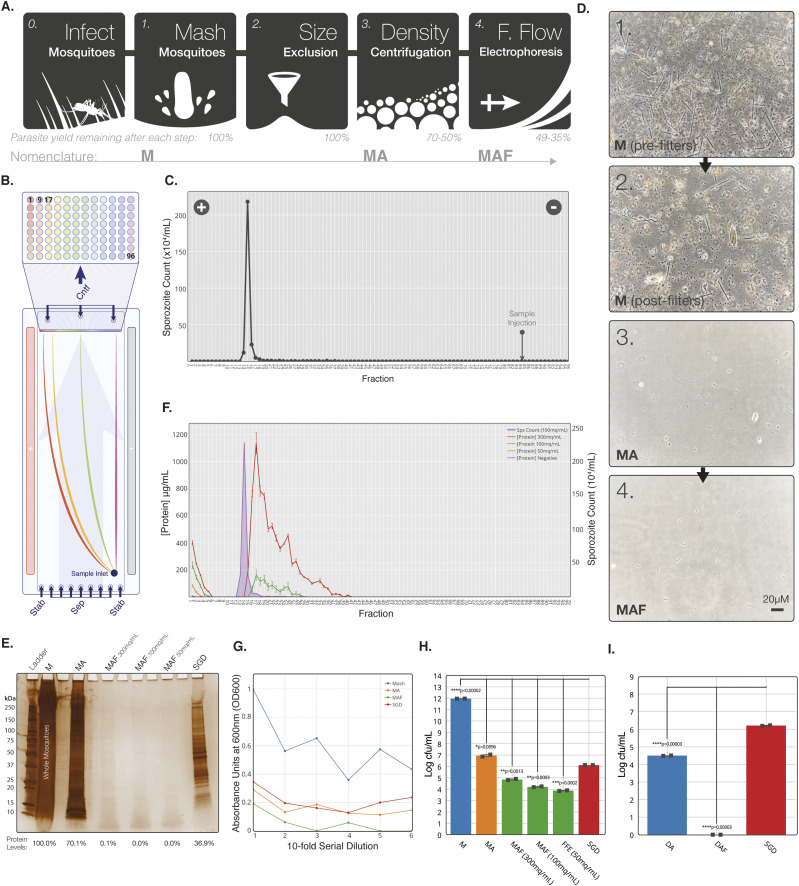
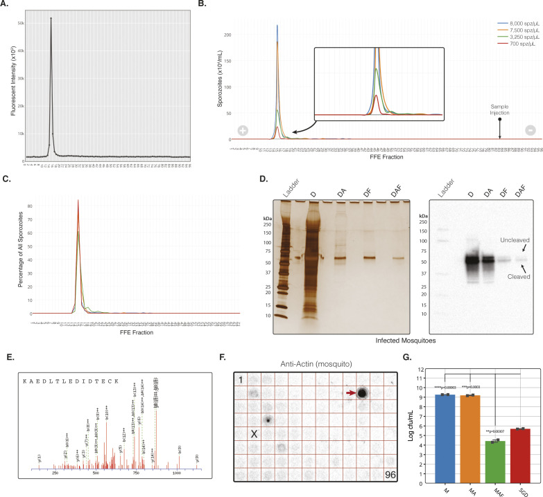
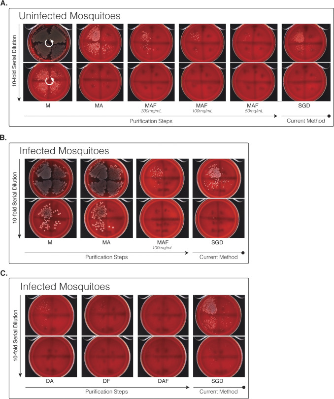
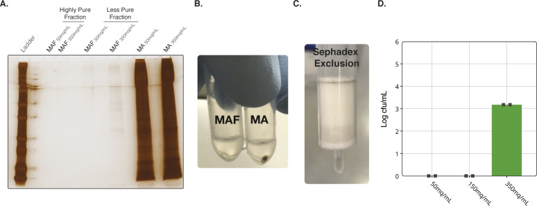
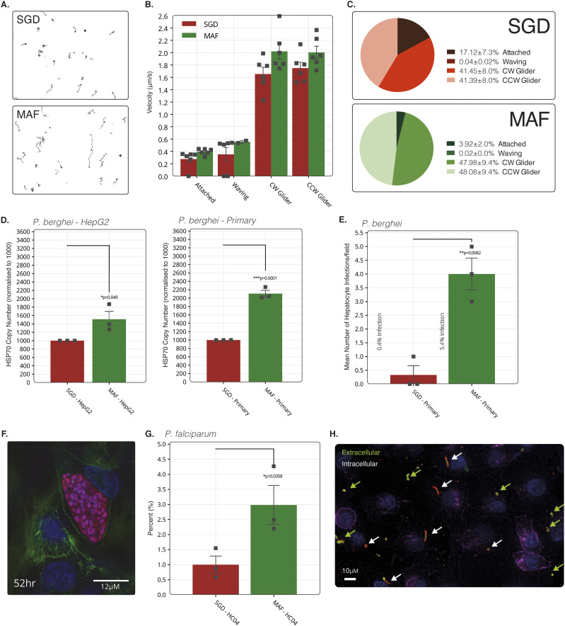
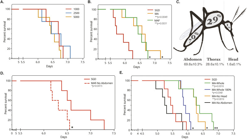
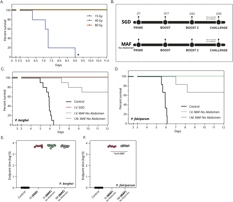
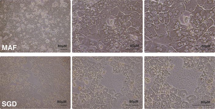
Progress towards a protective vaccine against malaria remains slow. To date, only limited protection has been routinely achieved following immunisation with either whole-parasite (sporozoite) or subunit-based vaccines. One major roadblock to vaccine progress, and to pre-erythrocytic parasite biology in general, is the continued reliance on manual salivary gland dissection for sporozoite isolation from infected mosquitoes. Here, we report development of a multi-step method, based on batch processing of homogenised whole mosquitoes, slurry, and density-gradient filtration, which combined with free-flow electrophoresis rapidly produces a pure, infective sporozoite inoculum. Human-infective Plasmodium falciparum and rodent-infective Plasmodium berghei sporozoites produced in this way are two- to threefold more infective than salivary gland dissection sporozoites in in vitro hepatocyte infection assays. In an in vivo rodent malaria model, the same P. berghei sporozoites confer sterile protection from mosquito-bite challenge when immunisation is delivered intravenously or 60-70% protection when delivered intramuscularly. By improving purity, infectivity, and immunogenicity, this method represents a key advancement in capacity to produce research-grade sporozoites, which should impact delivery of a whole-parasite based malaria vaccine at scale in the future.


Conflict of interest statement

The authors declare that they have no conflict of interest.

Figures

Figure 1.
Figure 1.. Development of a stepwise process for purification of sporozoites from whole mosquitoes.
(A) Schematic of key steps in the sporozoite purification process. (B) Schematic representation of sample separation by continuous zone electrophoresis mode. An electrophoretic buffer is run through a chamber 0.5 mm thick with a voltage applied across the flow. Sample added to the start of the chamber is carried vertically up the length of the chamber (pale blue arrow) as a voltage is applied across, separating across the horizontal length of the chamber. The outflow from the chamber is separated into 96 outlets along the horizontal length of the chamber, which drop into a 96 well plate. (C) Manual sporozoite count by haemocytometer of free-flow electrophoresis (FFE) fractions from a representative MAF sporozoite separation. Point of sample injection indicated by arrow and direction of current indicated by positive and negative symbols. (D) Bright-field images of each stage of purification from whole mosquito homogenate. All stages diluted to 7 × 10⁵ sporozoites/ml. (E) Silver stain of reducing SDS–PAGE gel with uninfected mosquitoes (four MEQs) from each step of purification. Uninfected MAF lanes are from the same fraction as the sporozoite peak fraction identified by running infected mosquitoes at the same time. (F) Protein concentration in each fraction after loading uninfected mosquito MA onto the FFE machine at three doses of mosquitoes (MAF). Sporozoite distribution (purple) from infected mosquitos loaded at 100 mq/ml is marked to allow comparison of purification. (G) End point 16-h serial dilution for each step of MAF purification. Absorbance of samples in TBS was measured at 600 nm (OD600) 16 h post-inoculation ay 37°C. All growth conducted at 37°C, 17g, using mosquitoes blood-fed on uninfected mice 21 d before MAF extraction. (H) Bacterial growth (samples normalised to MEQ of 200 mq/ml) at different stages from uninfected whole mosquito (M) origin purification. Samples were loaded onto the FFE machine at three different originating mosquito doses. (I) Bacterial growth (samples normalised to MEQ of 200 mq/ml) at different stages from infected SGD-origin purification. Experiments show the mean of two technical replicates and error bars represent SEM. All treatments compared with dissected by unpaired two-tailed t test using Bonferroni correction (H: *P < 0.01, **P < 0.002, ***P < 0.0002, ****P < 0.00002; I: *P < 0.017, **P < 0.003, ***P < 0.0003, ****P < 0.00003).
Figure S1.
Figure S1.. The MAF Purification platform additional data.
(A) Fluorescent plate read at 610 emission of free-flow electrophoresis (FFE) fractions from a representative MAF sporozoite separation. (B) Parasite distribution into FFE fractions when loaded at four different sporozoite doses (spz/ml). Quantification by haemocytometer count. (C) Sporozoite distribution based on total percent of sporozoites per fraction for each sporozoite dose. (D) Left; silver stain of infected mosquitoes from each step of purification when using dissected salivary glands for homogenisation instead of total mosquitoes. Right; Western blot against Plasmodium berghei CSP from the same samples as the silver stain (left). MAF samples in (D) where injected into the FFE machine at 100 mq/ml. All silver stains from reducing SDS–PAGE’s with samples normalised by MEQ with four MEQs loaded onto each lane. After MAF purification, the ratio of uncleaved:cleaved was 1:7.8, compared with 1:1.8 for salivary gland dissection sporozoites (assessed by densitometry). Cleaved and uncleaved bands indicated by arrows, with expected sizes of 45 and 55 kD, respectively. (E) Liquid chromatography tandem mass spectrometry (LC-MS/MS) analysis of each stage of purification. LC-MS/MS raw data searched against the Uniprot-Swissprot database using the MASCOT search algorithm identified P. berghei CSP protein exclusively with three peptides in the fraction purified by MAF only. Identification of tryptic peptide 297–312 from P. berghei CSP protein (Swissprot ID: P06915) with MASCOT score of 37 by mass spectrometry analysis. All samples normalised to 200 mq/ml. (F) Dotblot of all 96 FFE fractions in a plate layout against mosquito actin protein (A2066; Sigma-Aldrich). Actin positive fraction indicated by arrow. Peak fraction containing most sporozoites is indicated by “X,” Fraction 1 and 96 indicated. (G) Bacterial growth at different stages from infected whole mosquito (M) origin purification.
Figure S2.
Figure S2.. Blood plate agar growth of sporozoite purification steps.
(A) Bacterial growth at different steps of purification from uninfected whole mosquito homogenate. Samples were loaded onto the free-flow electrophoresis machine at three different MEQs (300, 100, and 50 mq/ml). (B) Bacterial growth at different purification steps from infected whole mosquito homogenate. (C) Bacterial growth at different stages from infected dissected salivary gland homogenate. All samples spread onto blood agar plates in eight, 10-fold serial dilutions running anti-clockwise, with plates separated into four quadrants and dilutions starting in the top right quadrant (indicated by white arrows).
Figure S3.
Figure S3.. MAF Purification using Sephadex and interval zone electrophoresis methods.
(A) Silver stain of each stage of purification illustrating the high- and low-purity fractions. All samples run at identical MEQs. (B) Images of pellets from MA and MAF purification. (C) Images of a Sephadex column after eluting sample. (D) Bacterial cfu/ml of samples injected into the free-flow electrophoresis (interval zone electrophoresis) at three concentrations of MEQs.
Figure 2.
Figure 2.. Assessment of purified sporozoite in vitro viability.
(A) Typical movement trails of MAF and salivary gland dissection (SGD) sporozoites over 600 frames at 2 Hz. (B) Sporozoite gliding motility over 600 frames at 2 Hz with a sliding nine frame average during each motility state over 600 frames at 2 Hz. (C) Comparison of the percentage of all sporozoites in each state. Sporozoite tracking represents mean of two independent replicates and six technical replicates with groups compared using an unpaired two-tailed t test. Bars represent means and error bars the SEM. A total of 10,672 and 8,370 sporozoites were counted for SGD and MAF, respectively. (D) Absolute RT-PCR quantification of parasite HSP70 housekeeping gene DNA copies normalised by host HSP60 gene in HepG2 (left) and primary rat (right) hepatocytes. Treatments for both HepG2 and primary hepatocytes were normalised to 1,000 hsp70 copies for the SGD treatment. Means of three independent replicates. (E) Mean counts of successful hepatocyte infections in primary rat hepatocytes measured by visual identification of six fields of view over 24-h time-lapse from three independent replicates. (F) Fluorescent image of late-stage schizont (52 h) captured using structured illumination microscope. Blue; nuclei, green; actin, red; mCherry parasite, pink; parasite actin (anti-5H3 [75]). (G) Means counts of initial hepatocyte invasions of Plasmodium falciparum sporozoites 4 h post infection in HC-04 at a ratio of 1:5 cells to sporozoites. SGD treatment normalised to 1. Sporozoites stained for CSP to determine intracellular or extracellular location. One independent replicate with three technical replicates. (H) Immunoflourescent staining of HC-04 cells with fixed 4 h after infection with P. falciparum sporozoites and stained with anti-CSP (extracellular = green + red, intracellular = red only), DAPI for nuclear material (blue) and phalloidin for actin (purple).
Figure S4.
Figure S4.. Ex-vivo development of MAF purified Plasmodium berghei sporozoites in rat primary hepatocytes.
(A) The ability of MAF sporozoites to infect hepatocytes in vivo but develop ex vivo was investigated. Hepatocytes were extracted by perfusion of livers that were collected from rats 14 h after i.v. injection of sporozoites into rats. These rats were infected with a total of 3 × 10⁷ GFP-expressing P. berghei sporozoites purified by MAF (whole mosquitoes) from 400 mosquitoes. Infected hepatocytes from these rats were collected by flow-sorting and subsequently plated and incubated for a period of up to 30 h. Flow sorting identified 2.83% GFP-positive cells in the extracted, perfused liver cell population. (B) Fluorescent images of GFP-positive cells collected by flow sorting 24 h after plating.
Figure S5.
Figure S5.. MAF Plasmodium berghei flow cytometry.
Flow cytometry quantification of mCherry expressing transgenic P. berghei infected primary rat hepatocytes 36 h post addition of sporozoites (total 2,808 cells in treatment, 3,906 cells in control). Data representative of three technical replicates.
Figure 3.
Figure 3.. Assessment of purified sporozoite in vivo viability.
(A) Kaplan–Meier survival curve of mice challenged i.v. with increasing doses of sporozoites from MAF. Six mice per group. End point classed as 1% parasitaemia. (B) Kaplan–Meier survival curve of mice challenged i.v. with 5,000 sporozoites from different purification steps. Six mice per group. End point classed as 1% parasitaemia, treatments compared by Mantel–Cox statistical test. (C) Sporozoite distribution of infected mosquitoes, average from 85 mosquitoes, two experimental replicates. Values show mean with SEM. Raw sporozoite numbers per mosquito are as follows: abdomen: 95,950; thorax: 42,500; head: 2,292. (D) Kaplan–Meier survival curve of mice challenged i.v. with 1,000 sporozoites from MAF-No Abdomens purified (MAF from mosquitoes with abdomens removed before homogenisation) and salivary gland dissection origin. Six mice per group. End point classed as 1% parasitaemia, treatments compared by Mantel–Cox statistical test. (E) Kaplan–Meier survival curve of mice challenged with 5,000 sporozoites obtained by MA purification from different mosquito sources or salivary gland dissection origin. Six mice per group. Death classed as 1% parasitaemia, treatments compared by Mantel–Cox statistical test.
Figure 4.
Figure 4.. Purified sporozoites as a viable vaccine.
(A) Kaplan–Meier survival curve of mice challenged i.v. with 1,000 Plasmodium berghei sporozoites from MAF-No abdomen purified and gamma irradiated. Four mice per group. End point classed as 1% parasitaemia. (B) Schematic of vaccination regime used. Sporozoites were either from salivary gland dissection or MAF-No Abdomen origin, then gamma irradiated. (C) Immunisation i.v. or i.m. of Balb/c mice with irradiated P. berghei sporozoites from either manual salivary gland (salivary gland dissection) dissection or MAF-No Abdomen. Mice given three immunisations of 40,000 sporozoites, 2 wk apart followed by challenge with five infectious mosquito bites. Ten mice per group. End point classed as 1% parasitaemia. (D) Immunisation i.v. or i.m. of Balb/c mice with irradiated Plasmodium falciparum sporozoites from MAF-No Abdomen. Mice given three immunisations of 40,000 sporozoites, 2 wk apart followed by challenge with five infectious mosquito bites. Six mice per group. End point classed as 1% parasitaemia. (E) Total titres of IgG antibodies against P. berghei sporozoite lysate in mouse serum before challenge (F) Total titres of IgG antibodies against P. falciparum sporozoite lysate in mouse serum before challenge. Squares indicate mice not protected.
Figure S6.
Figure S6.. Sporozoite-associated morphological changes in primary rat hepatocytes.
Bright-field images of primary rat hepatocytes 20 h after addition of sporozoites obtained by either MAF (top row) or salivary gland dissection (bottom row). Cultured in 1% P/S.
Figure S7.
Figure S7.. Generation and genotype analysis of the reporter line PbANKA-GFP::Luc@Pbuis4_230p.
(A) Schematic representation of the introduction of the GFP::Luciferase expression cassette into the genome of the gene insertion/marker out PbANKA parent line 1596 cl1. The gfp-luciferase fusion gene is under the control of Pbuis4 regulatory sequences (5′UTR and 3′UTR regions). DNA construct pL1962 containing the GFP::Luciferase expression cassette is integrated into the modified Plasmodium berghei p230p locus on chromosome (chr.) 3, containing the hdhfr::yfcu selectable marker (SM) cassette (black box), by double cross-over homologous recombination (DXO) at the p230p target regions (hatched boxes). Negative selection with 5-FC selects for parasites (line 2227 cl6) that have the GFP::Luciferase expression cassette introduced into the neutral p230p gene locus and the hdhfr::yfcu marker removed. Location of primers used for PCR analysis and sizes of PCR products are shown. See Tables S1 and S2 for primer sequences. (B) Conformation of correct integration of the GFP::Luciferase expression cassette into the genome by Southern analysis of pulsed-field gel–separated chromosomes and diagnostic PCR. Left panel: Hybridisation of pulsed-field gel–separated chromosomes (chr.) of the reporter line PbANKA-GFP::Luc@Pbuis4_230p shows integration of the expression cassette into the p230p gene insertion/marker out locus on chromosome (chr.) 3 by the absence of the hdhfr::yfcu SM cassette in the cloned reporter line. The Southern blot was hybridized with a mixture of two probes: one recognizing hdhfr and a control probe recognizing chr. 5. As an control parasite line, line 2117 cl1 was used with the hdhfr::yfcu SM integrated into chr. 3. Right panel: Diagnostic PCR shows the absence of the hdhfr::yfcu SM, the presence of GFP::Luciferase gene and the correct integration of the expression cassette into the genome of PbANKA-GFP::Luc@Pbuis4_230p at the 5′- and 3′-regions of p230p (5′int and 3′int). See (A) for the primers’ locations and Table S2 for primer sequences. Source data are available for this figure.

Similar articles

Cited by

References

    1. WHO (2019) World Malaria Report. Geneva: Switzerland: WHO.
    1. Duffy PE, Patrick Gorres J (2020) Malaria vaccines since 2000: Progress, priorities, products. NPJ Vaccin 5: 48. 10.1038/s41541-020-0196-3 - DOI - PMC - PubMed
    1. Chatterjee D, Cockburn IA (2021) The challenges of a circumsporozoite protein-based malaria vaccine. Expert Rev Vaccin 20: 113–125. 10.1080/14760584.2021.1874924 - DOI - PubMed
    1. RTS,S Clinical Trials Partnership (2015) Efficacy and safety of RTS,S/AS01 malaria vaccine with or without a booster dose in infants and children in Africa: Final results of a phase 3, individually randomised, controlled trial. Lancet 386: 31–45. 10.1016/S0140-6736(15)60721-8 - DOI - PMC - PubMed
    1. Roestenberg M, McCall M, Hopman J, Wiersma J, Luty AJ, van Gemert GJ, van de Vegte-Bolmer M, van Schaijk B, Teelen K, Arens T, et al. (2009) Protection against a malaria challenge by sporozoite inoculation. N Engl J Med 361: 468–477. 10.1056/NEJMoa0805832 - DOI - PubMed

Publication types

Associated data