Structural basis for late maturation steps of the human mitoribosomal large subunit
- PMID: 34135318
- PMCID: PMC8209036
- DOI: 10.1038/s41467-021-23617-8
Structural basis for late maturation steps of the human mitoribosomal large subunit
Abstract
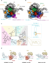
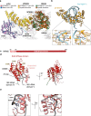
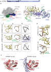
Mitochondrial ribosomes (mitoribosomes) synthesize a critical set of proteins essential for oxidative phosphorylation. Therefore, mitoribosomal function is vital to the cellular energy supply. Mitoribosome biogenesis follows distinct molecular pathways that remain poorly understood. Here, we determine the cryo-EM structures of mitoribosomes isolated from human cell lines with either depleted or overexpressed mitoribosome assembly factor GTPBP5, allowing us to capture consecutive steps during mitoribosomal large subunit (mt-LSU) biogenesis. Our structures provide essential insights into the last steps of 16S rRNA folding, methylation and peptidyl transferase centre (PTC) completion, which require the coordinated action of nine assembly factors. We show that mammalian-specific MTERF4 contributes to the folding of 16S rRNA, allowing 16 S rRNA methylation by MRM2, while GTPBP5 and NSUN4 promote fine-tuning rRNA rearrangements leading to PTC formation. Moreover, our data reveal an unexpected involvement of the elongation factor mtEF-Tu in mt-LSU assembly, where mtEF-Tu interacts with GTPBP5, similar to its interaction with tRNA during translational elongation.
Conflict of interest statement
The authors declare no competing interests.
Figures


Similar articles
-
Stepwise maturation of the peptidyl transferase region of human mitoribosomes.Nat Commun. 2021 Jun 16;12(1):3671. doi: 10.1038/s41467-021-23811-8. Nat Commun. 2021. PMID: 34135320 Free PMC article.
-
Structural basis of GTPase-mediated mitochondrial ribosome biogenesis and recycling.Nat Commun. 2021 Jun 16;12(1):3672. doi: 10.1038/s41467-021-23702-y. Nat Commun. 2021. PMID: 34135319 Free PMC article.
-
Human GTPBP5 (MTG2) fuels mitoribosome large subunit maturation by facilitating 16S rRNA methylation.Nucleic Acids Res. 2020 Aug 20;48(14):7924-7943. doi: 10.1093/nar/gkaa592. Nucleic Acids Res. 2020. PMID: 32652011 Free PMC article.
-
Insights into mitoribosomal biogenesis from recent structural studies.Trends Biochem Sci. 2023 Jul;48(7):629-641. doi: 10.1016/j.tibs.2023.04.002. Epub 2023 May 10. Trends Biochem Sci. 2023. PMID: 37169615 Review.
-
Mitochondrial ribosome assembly in health and disease.Cell Cycle. 2015;14(14):2226-50. doi: 10.1080/15384101.2015.1053672. Epub 2015 Jun 1. Cell Cycle. 2015. PMID: 26030272 Free PMC article. Review.
Cited by
-
Human mtDNA-Encoded Long ncRNAs: Knotty Molecules and Complex Functions.Int J Mol Sci. 2024 Jan 25;25(3):1502. doi: 10.3390/ijms25031502. Int J Mol Sci. 2024. PMID: 38338781 Free PMC article. Review.
-
Mitoribosome structure with cofactors and modifications reveals mechanism of ligand binding and interactions with L1 stalk.Nat Commun. 2024 May 20;15(1):4272. doi: 10.1038/s41467-024-48163-x. Nat Commun. 2024. PMID: 38769321 Free PMC article.
-
Coupling of ribosome biogenesis and translation initiation in human mitochondria.Nat Commun. 2025 Apr 17;16(1):3641. doi: 10.1038/s41467-025-58827-x. Nat Commun. 2025. PMID: 40240327 Free PMC article.
-
Stepwise maturation of the peptidyl transferase region of human mitoribosomes.Nat Commun. 2021 Jun 16;12(1):3671. doi: 10.1038/s41467-021-23811-8. Nat Commun. 2021. PMID: 34135320 Free PMC article.
-
Types and Functions of Mitoribosome-Specific Ribosomal Proteins across Eukaryotes.Int J Mol Sci. 2022 Mar 23;23(7):3474. doi: 10.3390/ijms23073474. Int J Mol Sci. 2022. PMID: 35408834 Free PMC article. Review.
References
Publication types
MeSH terms
Substances
LinkOut - more resources
Full Text Sources
Molecular Biology Databases

