. 2021 Jun 16;11(1):12680.
doi: 10.1038/s41598-021-92084-4.

Assessment with clinical data of a coupled bio-hemodynamics numerical model to predict leukocyte adhesion in coronary arteries

Affiliations

Assessment with clinical data of a coupled bio-hemodynamics numerical model to predict leukocyte adhesion in coronary arteries

Umberto Ciri et al. Sci Rep. .

Abstract

Numerical simulations of coupled hemodynamics and leukocyte transport and adhesion inside coronary arteries have been performed. Realistic artery geometries have been obtained for a set of four patients from intravascular ultrasound and angiography images. The numerical model computes unsteady three-dimensional blood hemodynamics and leukocyte concentration in the blood. Wall-shear stress dependent leukocyte adhesion is also computed through agent-based modeling rules, fully coupled to the hemodynamics and leukocyte transport. Numerical results have a good correlation with clinical data. Regions where high adhesion is predicted by the simulations coincide to a good approximation with artery segments presenting plaque increase, as documented by clinical data from baseline and six-month follow-up exam of the same artery. In addition, it is observed that the artery geometry and, in particular, the tortuosity of the centerline are a primary factor in determining the spatial distribution of wall-shear stress, and of the resulting leukocyte adhesion patterns. Although further work is required to overcome the limitations of the present model and ultimately quantify plaque growth in the simulations, these results are encouraging towards establishing a predictive methodology for atherosclerosis progress.


Conflict of interest statement

The authors declare no competing interests.

Figures

Figure 1
Figure 1
Schematic of the blood hemodynamics and leukocyte adhesion process. The wall shear stress (WSS) is the (tangential) force which blood flow applies on the artery wall and depends on the fluid viscosity μ and the shear rate (U/n where n is the normal to the arterial wall). If the WSS is large, blood will wash away leukocytes from the endothelium and impede adhesion, whereas in presence of low WSS chance of adhesion is greater. After adhesion, leukocytes transmigrate inside the arterial wall increasing the plaque area (red, green and white regions in the intravascular ultrasound image). Umean=0.384m/s is the mean bulk velocity throughout the cardiac cycle.
Figure 2
Figure 2
Leukocyte transport and adhesion inside an ideal stenotic artery. (a) Blood flow velocity Ubulk and visualizations of neutrophil concentration ρn (normalized by the reference value, 4.34×10⁹/l) throughout the cardiac cycle (T is the cardiac period). The inset in the cardiac cycle panel shows the three-dimensional geometry of the ideal artery. (b,c) adhesion of leukocytes (b, neutrophils; c lymphocytes) as a function of the distance along the artery (x=0 is the axial location of the minimum lumen area, D the diameter of the healthy tract): without tracking the particle concentration; - - - - - with concentration tracking (Eq. 1, present simulations).
Figure 3
Figure 3
Comparison between clinical data for patient 1 and simulation predictions. (a) Lumen area from VH-IVUS scans as a function of the distance along the centerline s. (b) Reduction in lumen area from baseline to follow-up, ΔAlumen=Abaseline-Afollow-up. A positive value indicates reduction of lumen area (hence plaque growth and disease progression). (c) Predicted mean rate of adhesion for three different species of leukocytes. T indicates the cardiac cycle (T=0.8s in this study), R¯ is the adhesion rate. The vertical dashed red lines indicates the location of maximum lumen area change from the baseline to the follow-up exam.
Figure 4
Figure 4
Comparison between simulation results using realistic centerline data and ideal straight centerline: (a) WSS normalized by the ideal reference value WSS04Pa; (b) rate of adhesion for neutrophils. Solid lines, realistic centerline; dashed lines, straight centerline.
Figure 5
Figure 5
Flow and WSS variability over the cross-sectional lumen area (at s/D=12.5). (a) Time-averaged WSS as a function of the azimuthal direction θ: realistic centerline; —– straight centerline. The horizontal line (–.–) denotes the adhesion threshold for neutrophils (1.2Pa). (b) Mean streamwise velocity Uξ over the lumen area for the realistic centerline case.
Figure 6
Figure 6
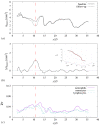
Comparison between simulation results using the arterial geometry from the baseline exam and the six-month follow-up exam: (a) WSS¯ distribution, normalized by the reference value WSS0; (b) rate of adhesion for neutrophils. Solid lines, baseline geometry; dashed lines, follow-up geometry. The inset in (b) shows particle lines for the blood flow in the follow-up geometry.

References

    1. Mozaffarian D, et al. Executive summary: Heart disease and stroke statistics-2016 update: A report from the American Heart Association. Circulation. 2016;133(4):447–454. doi: 10.1161/CIR.0000000000000366. - DOI - PubMed
    1. Libby P. Changing concepts of atherogenesis. J. Intern. Med. 2000;247:349–358. doi: 10.1046/j.1365-2796.2000.00654.x. - DOI - PubMed
    1. Malek AM, Alper SL, Izumo S. Hemodynamic shear stress and its role in atherosclerosis. JAMA. 1999;282:2035–2042. doi: 10.1001/jama.282.21.2035. - DOI - PubMed
    1. Slager CJ, et al. The role of shear stress in the generation of rupture-prone vulnerable plaques. Nat. Clin. Pract. Card. 2005;2(8):401–407. doi: 10.1038/ncpcardio0274. - DOI - PubMed
    1. Kwak BR, et al. Biomechanical factors in atherosclerosis: Mechanisms and clinical implications. Eur. Heart J. 2014;35:3013–3020. doi: 10.1093/eurheartj/ehu353. - DOI - PMC - PubMed

Publication types