

Protein residue network analysis reveals fundamental properties of the human coagulation factor VIII
- PMID: 34135429
- PMCID: PMC8209229
- DOI: 10.1038/s41598-021-92201-3
Protein residue network analysis reveals fundamental properties of the human coagulation factor VIII
Abstract
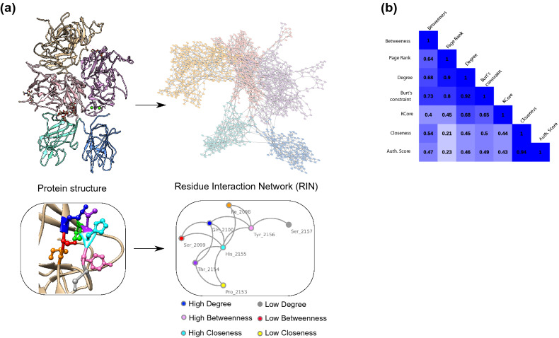
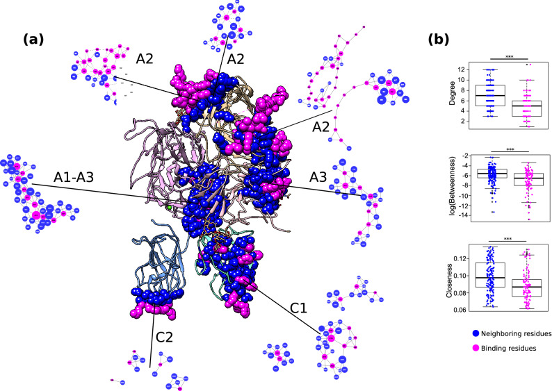
Hemophilia A is an X-linked inherited blood coagulation disorder caused by the production and circulation of defective coagulation factor VIII protein. People living with this condition receive either prophylaxis or on-demand treatment, and approximately 30% of patients develop inhibitor antibodies, a serious complication that limits treatment options. Although previous studies performed targeted mutations to identify important residues of FVIII, a detailed understanding of the role of each amino acid and their neighboring residues is still lacking. Here, we addressed this issue by creating a residue interaction network (RIN) where the nodes are the FVIII residues, and two nodes are connected if their corresponding residues are in close proximity in the FVIII protein structure. We studied the characteristics of all residues in this network and found important properties related to disease severity, interaction to other proteins and structural stability. Importantly, we found that the RIN-derived properties were in close agreement with in vitro and clinical reports, corroborating the observation that the patterns derived from this detailed map of the FVIII protein architecture accurately capture the biological properties of FVIII.
Conflict of interest statement
The authors declare no competing interests.
Figures
Similar articles
-
Structural basis for hemophilia A caused by mutations in the C domains of blood coagulation factor VIII.Thromb Haemost. 2000 Jan;83(1):78-85. Thromb Haemost. 2000. PMID: 10669159
-
The evolving understanding of factor VIII binding sites and implications for the treatment of hemophilia A.Blood Rev. 2019 Jan;33:1-5. doi: 10.1016/j.blre.2018.05.001. Epub 2018 May 24. Blood Rev. 2019. PMID: 29866493 Review.
-
Missense mutations near the N-glycosylation site of the A2 domain lead to various intracellular trafficking defects in coagulation factor VIII.Sci Rep. 2017 Mar 22;7:45033. doi: 10.1038/srep45033. Sci Rep. 2017. PMID: 28327546 Free PMC article.
-
Thirty-four novel mutations detected in factor VIII gene by multiplex CSGE: modeling of 13 novel amino acid substitutions.J Thromb Haemost. 2003 Apr;1(4):773-81. doi: 10.1046/j.1538-7836.2003.00149.x. J Thromb Haemost. 2003. PMID: 12871415
-
[Molecular genetics of hemophilia A].Medicina (B Aires). 1996;56(5 Pt 1):509-17. Medicina (B Aires). 1996. PMID: 9239887 Review. Spanish.
Cited by
-
Artificial Intelligence in the Management of Hereditary and Acquired Hemophilia: From Genomics to Treatment Optimization.Int J Mol Sci. 2025 Jun 25;26(13):6100. doi: 10.3390/ijms26136100. Int J Mol Sci. 2025. PMID: 40649878 Free PMC article. Review.
-
Computational analyses reveal fundamental properties of the AT structure related to thrombosis.Bioinform Adv. 2022 Dec 23;3(1):vbac098. doi: 10.1093/bioadv/vbac098. eCollection 2023. Bioinform Adv. 2022. PMID: 36698764 Free PMC article.
-
A graph-based machine learning framework identifies critical properties of FVIII that lead to hemophilia A.Front Bioinform. 2023 May 10;3:1152039. doi: 10.3389/fbinf.2023.1152039. eCollection 2023. Front Bioinform. 2023. PMID: 37235045 Free PMC article.
-
A Machine Learning Framework Predicts the Clinical Severity of Hemophilia B Caused by Point-Mutations.Front Bioinform. 2022 Jun 23;2:912112. doi: 10.3389/fbinf.2022.912112. eCollection 2022. Front Bioinform. 2022. PMID: 36304295 Free PMC article.
-
Using residue interaction networks to understand protein function and evolution and to engineer new proteins.Curr Opin Struct Biol. 2024 Dec;89:102922. doi: 10.1016/j.sbi.2024.102922. Epub 2024 Sep 26. Curr Opin Struct Biol. 2024. PMID: 39332048 Free PMC article. Review.
References
-
- Lee CA, Berntorp E, Hoots K. Textbook of Hemophilia. 3. Wiley; 2014.
Publication types
MeSH terms
Substances
LinkOut - more resources
Full Text Sources
Medical
Miscellaneous
