Review
. 2021 Aug;17(8):497-510.
doi: 10.1038/s41574-021-00499-w. Epub 2021 Jun 16.

The role of regulated necrosis in endocrine diseases

Affiliations
Review

The role of regulated necrosis in endocrine diseases

Wulf Tonnus et al. Nat Rev Endocrinol. 2021 Aug.

Abstract

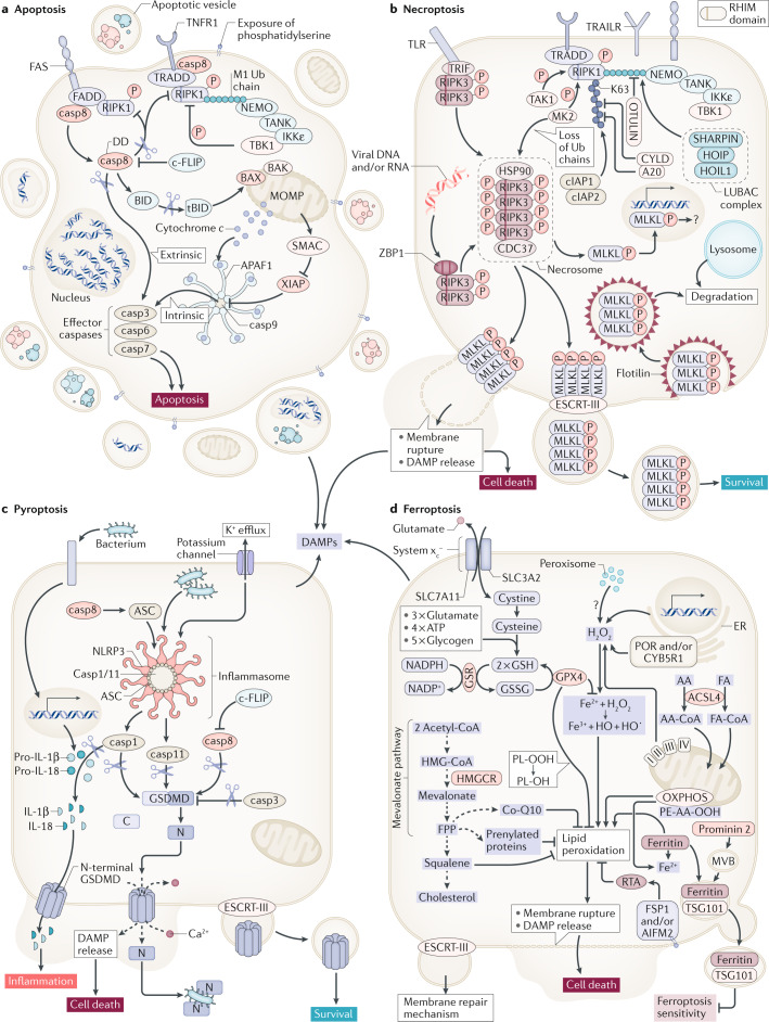
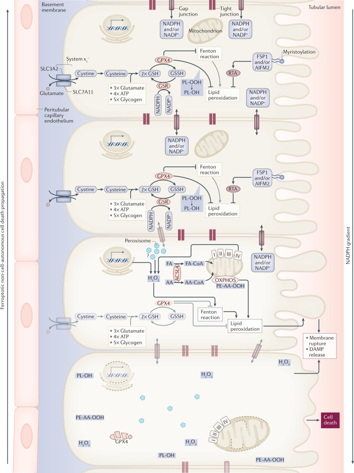
The death of endocrine cells is involved in type 1 diabetes mellitus, autoimmunity, adrenopause and hypogonadotropism. Insights from research on basic cell death have revealed that most pathophysiologically important cell death is necrotic in nature, whereas regular metabolism is maintained by apoptosis programmes. Necrosis is defined as cell death by plasma membrane rupture, which allows the release of damage-associated molecular patterns that trigger an immune response referred to as necroinflammation. Regulated necrosis comes in different forms, such as necroptosis, pyroptosis and ferroptosis. In this Perspective, with a focus on the endocrine environment, we introduce these cell death pathways and discuss the specific consequences of regulated necrosis. Given that clinical trials of necrostatins for the treatment of autoimmune conditions have already been initiated, we highlight the therapeutic potential of such novel therapeutic approaches that, in our opinion, should be tested in endocrine disorders in the future.


Conflict of interest statement

The authors declare no competing interests.

Figures

Fig. 1
Fig. 1. Regulated cell death pathways at a glance.
Apoptosis represents a non-inflammatory pathway that is mediated by caspases (part a). Two distinct signalling pathways of apoptosis, extrinsic and intrinsic apoptosis, have been characterized. While apoptosis depends on the activation of caspases, necroptosis is mediated by kinases (part b). Dependent on its RHIM domain, RIPK3 forms an amyloid-like structure referred to as the necrosome, the central signalling platform of necroptosis. Therein, RIPK3 phosphorylates the pseudokinase MLKL. By unknown mechanisms, the phosphorylated form of MLKL (pMLKL) triggers plasma membrane rupture, a process that was demonstrated to be counteracted by the membrane repair ESCRT-III complex. In contrast to apoptosis and necroptosis, inflammasomes are involved in the initiation of cell death by pyroptosis (part c). The activation of caspases (1, 4, 11) in inflammasomes results in the cleavage of pro-IL-1β, pro-IL-18 and gasdermin D (GSDMD). Finally, and without any clear connection to apoptosis, necroptosis and pyroptosis, ferroptosis is a failsafe rather than a typical cell death pathway (part d). In cellular homeostasis, H2O2 concentrations of iron-catalysed and Fenton reactions are limited by diverse cellular anti-redox systems. Glutathione peroxidase 4 (GPX4) prevents lipid peroxidation that otherwise leads to plasma membrane rupture by unknown mechanisms. By contrast, the oxidoreductase FSP1 (also known as AIFM2) prevents lipid peroxidation upon myristoylation-dependent recruitment to the plasma membrane in a GSH-independent manner. casp, caspase; DAMP, damage-associated molecular pattern; DD, death domain; DED, death effector domain; ER, endoplasmic reticulum; FA, fatty acid; PL-OH, phospholipid alcohol; PL-OOH, phospholipid hydroperoxide; RTA, radical-trapping antioxidant.
Fig. 2
Fig. 2. Synchronized regulated necrosis.
In most cases, it is currently unclear how a necrotic zone of dead cells propagates during an event of sepsis or ischaemia. Based on time-lapse videos obtained from isolated perfused renal tubules that underwent ferroptosis, the concept of synchronized regulated necrosis emerged. Although this phenomenon has not been observed in humans, necrotic tubules in the urine of patients and experimental intravital microscopy videos suggest that cell death occurs in a wave of death,. Given the connections between cells in a functional syncytium, such as a renal tubule, the adrenal gland or the myocardium, it is tempting to speculate that the intracellular redox capacity (NADPH concentration) diffuses through intercellular junctions. If the redox capacity decreases in a dying cell, an NADPH gradient forms, which leaves the closest neighbour at high risk of undergoing ferroptosis. At least from heart attacks and insults of acute kidney injury, it is now clear that the bulk of the necrotic area that occurs during myocardial infarction or tubular necrosis originates from cells that underwent ferroptosis. We speculate that similar mechanisms might occur in the adrenal gland during sepsis or during resuscitation in intensive care units and potentially in other endocrine organs. AA, arachidonic acid; DAMP, damage-associated molecular pattern; FA, fatty acid; GPX4; glutathione peroxidase 4; GSH, glutathione; GSR, glutathione-disulfide reductase; GSSG, glutathione disulfide; HMGCR, 3-hydroxy-3-methylglutaryl-CoA reductase; PL-OH, phospholipid alcohol; PL-OOH, phospholipid hydroperoxide; RTA, radical-trapping antioxidant.
Fig. 3
Fig. 3. DAMPs and DIRE and the consequences of cell death.
Regulated necrosis (necroptosis, ferroptosis and pyroptosis) and non-regulated necrosis, such as mechanical manipulation of tissue, results in the release of damage-associated molecular patterns (DAMPs) in non-endocrine cells (part a). By contrast, endocrine cells that die by necrosis are associated with the release of DAMPs and damage-induced release of endocrine factors (DIRE) (part b). Such factors include common hormones, such as cortisol, adrenaline, noradrenaline, thyroxin, tri-iodothyronine and aldosterone.

Similar articles

Cited by

References

    1. Galluzzi L, et al. Molecular mechanisms of cell death: recommendations of the nomenclature committee on cell death 2018. Cell Death Differ. 2018;25:486–541. doi: 10.1038/s41418-017-0012-4. - DOI - PMC - PubMed
    1. Sarhan M, Land WG, Tonnus W, Hugo CP, Linkermann A. Origin and Consequences of Necroinflammation. Physiol. Rev. 2018;98:727–780. doi: 10.1152/physrev.00041.2016. - DOI - PubMed
    1. Yatim N, et al. RIPK1 and NF-kappaB signaling in dying cells determines cross-priming of CD8+ T cells. Science. 2015;350:328–334. doi: 10.1126/science.aad0395. - DOI - PMC - PubMed
    1. Linkermann A, et al. Two independent pathways of regulated necrosis mediate ischemia-reperfusion injury. Proc. Natl Acad. Sci. USA. 2013;110:12024–12029. doi: 10.1073/pnas.1305538110. - DOI - PMC - PubMed
    1. Sun L, et al. Mixed lineage kinase domain-like protein mediates necrosis signaling downstream of RIP3 kinase. Cell. 2012;148:213–227. doi: 10.1016/j.cell.2011.11.031. - DOI - PubMed

Publication types

MeSH terms