. 2021 May;29(5):446-455.
doi: 10.1016/j.jsps.2021.04.007. Epub 2021 Apr 23.

Improved delivery of miR-1296 loaded cationic nanoliposomes for effective suppression of triple negative breast cancer

Affiliations

Improved delivery of miR-1296 loaded cationic nanoliposomes for effective suppression of triple negative breast cancer

Lamyaa Albakr et al. Saudi Pharm J. 2021 May.

Abstract

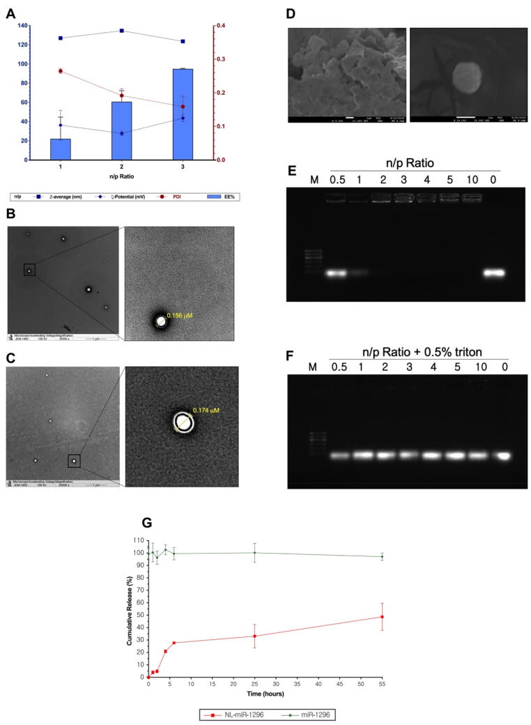
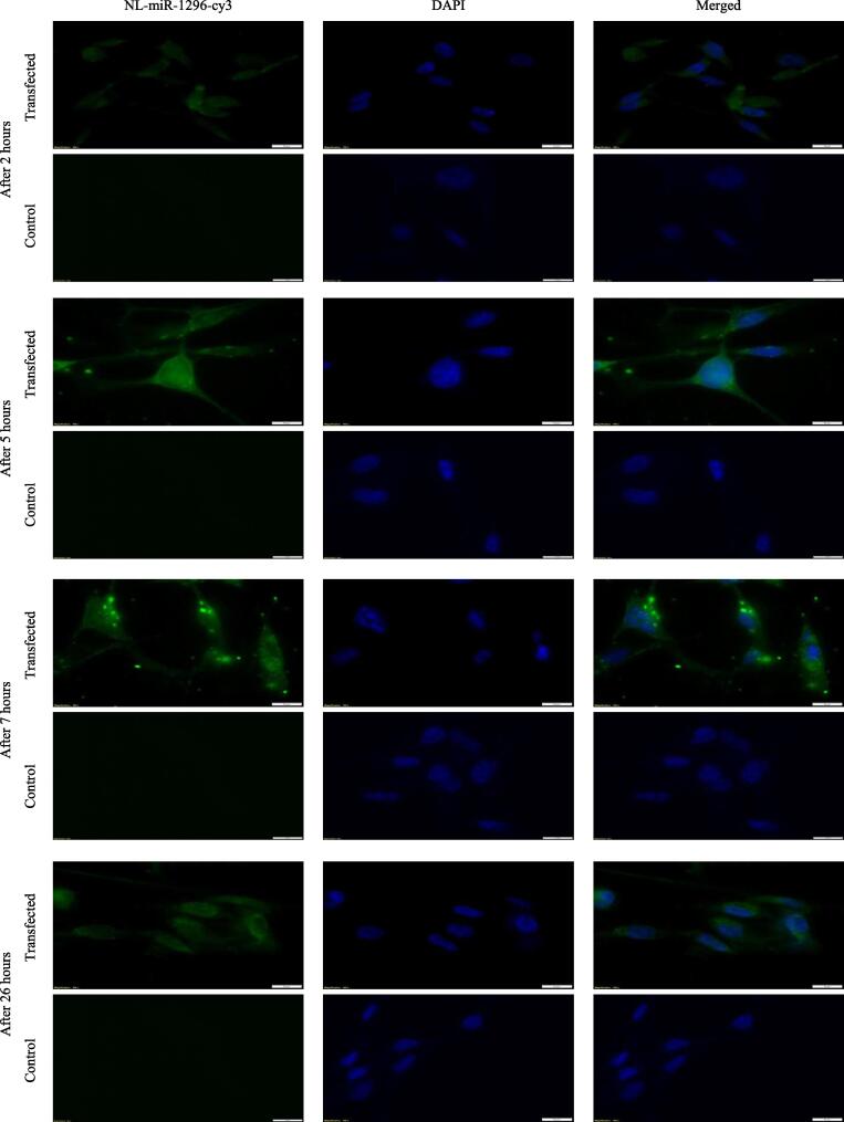
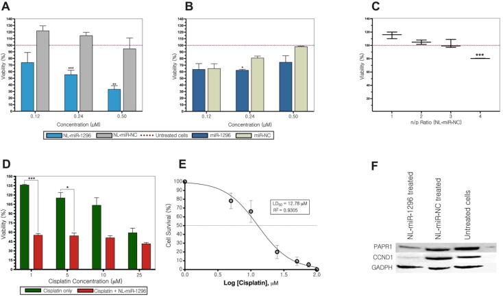
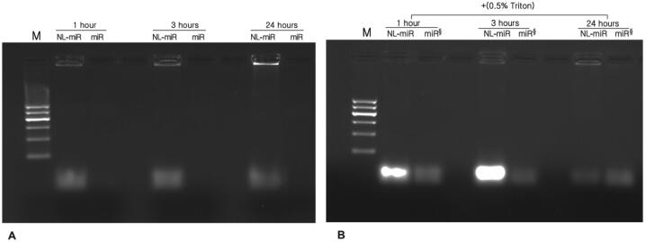
Nowadays, microRNA is considered an attractive strategy for the effective treatment of cancer. A significant delivery of microRNA for cancer therapy remains a significant obstacle to target cancer cells. The restoring microRNA-1296 (miR-1296) has immense therapeutic efficacy in triple-negative breast cancer (TNBC). TNBC is an aggressive subtype of breast tumors with the progression of malignant transformation. This study aimed to develop a cationic nanoliposome that can serve as a miR-1296 carrier and studied its efficiency in TNBC. The efficacy of miR-1296 liposomes was evaluated on its apoptotic effect, cellular uptake, and potential chemotherapy sensitization in the TNBC cell line (MDA-MB-231). For in vitro viability study, the apoptotic effect was performed to validate protein expression using Alamar blue kit and western blot. The transfection of miR-1296 into TNBC cells was also investigated using cisplatin as a TNBC resistance drug. The fluorescent miR-1296-cy3 liposome was used for cellular uptake study. The miR-liposome was successfully prepared with a particle size of 123.6 ± 1.3 nm and encapsulation efficiency of 94.33%. A dose of 0.5 uM has significantly reduced the viability of MDA-MB-231 to be 33.45%±5.29 (P < 0.001). This result was validated by down-expression of CCND1, and PARP1, the miR-1296 receptor, and apoptosis marker. The image of the miR-1296-cy3 liposome showed cytoplasmic intracellular localization. It was found high sensitization of TNBC cell line for miR-1296 liposome compared to cisplatin (P < 0.001). Future in vivo research may answer questions concerning safety and stability. This study demonstrates that miR-191 liposomes may have promising clinical applications for TNBC therapy.

Keywords: Cellular uptake; Liposomes; Triple-negative breast cancer; miR-1296.


Conflict of interest statement

The authors declare that they have no known competing financial interests or personal relationships that could have appeared to influence the work reported in this paper.

Figures

Fig. 1
Fig. 1
Characterization of NL-miR-1296. (A) Particle size, PDI, zeta potential and EE% characterization of NL-miR-1296. (B) Transmission electron microscopy of plain NL, and NL-miR-1296 (C) TEM images. (D) present scanning electron microscope overview image of prepared NL-miR-1296; (E) 1% agarose gel used to test the complexation of miR-1296 with NL at different n/p ratios. Different n/p ratios used 0.5–10, where 0 represent free miR-1296 only. All wells contain the same amount of miR-1296 (0.65 μg) but various NL part. The same n/p ratios were used but with NL were permeabilized with 0.5% triton. NL-miR-1296 were allowed 30 min to complex before running. Run was for 40 min at 80 V using TBE buffer. Ethidium bromide (0.5 μg/mL) was used to stain the gel; (F) Characterization of fresh NL-miR-1296 with different n/p ratios. Data represent mean ± SEM; (G) In vitro release of miR-1296 from NL-miR-1296. The release was evaluated in pH 7.4 using TE buffer, at 37 °C. Free miR-1296 was tested for integrity purposes. Points represent averages ± SEM.
Fig. 2
Fig. 2
Intracellular uptake of NL-miR-1296-cy3 in MDA-MB-231. About 100 thousand cells were seeded in coverslips in a 6-well cell culture plate. Next day, cells were treated with NL-miR-1296-cy3 in serum-free media (green). After 2,5,7, and 26 h, coverslips were fixed using 3.7% formaldehyde. Nuclei stained with DAPI (blue). Scale bars = 10 µm.
Fig. 3
Fig. 3
. Viability, chemotherapy-sensitization, and stability analysis of NL-miR-1296. (A) MDA-MB-231 viability after treatment with different concentrations (0.12–3 μM) of NL-miR-1296, or NL-miR-NC (B) Cells transfected using Lipofectamine. (C) Safety of nanoliposomes was analyzed using different n/p ratios (D) NL-miR-1296 sensitizes MDA-MB-231 to cisplatin treatment at a dose of as low as 1 μM. (E) A dose–response curve of cisplatin treatment in MDA-MB-231 cells (D) Down-expression of CCND1, and PARP1 in NL-miR-1296 treated MDA-MB-231 cells.
Fig. 4
Fig. 4
Effect of serum, temperature and time on NL-miR-231 stability. Serum stability was tested using 1% agarose gel. (A) simply samples of NL-miR-1296 (n/p ratio of 3) or free miR-1296 was incubated in 1:1 v/v of FBS (final concentration 50%) at 37 °C for 1, 3, and 24 h. (B) samples of the same NL-miR-1296 incubated in FBS for different periods were treated with 0.5%TritonX-100 just before the run and presented next to free miR incubated at the same time and temperature without FBS (denoted by §).
Fig. 5
Fig. 5
Effect of storage time and temperature on physical and biological activity of NL-miR-1296. (A) Interplay between stability and biological activity of NL-miR-1296 on TNBC at different time and storage temperatures. MDA-MB-231 cells were seeded in 24-well plate for 24 h. After 40 or 80 days, cells were transfected with NL-miR-1296 stored at different temperatures. Media was changed after 5 h, and viability was tested using Alamar Blue after 48 h and compared to untreated cells. Freshly prepared NL-miR-1296 was used transfect the cells as described above to represent 0-time point. Physical characterization of NL-miR-1296 stored in different temperatures at different time points was presented as (B) particle size, (C) PDI, and (D) zeta potential. Data represent the mean ± SEM of two independent studies (n = 6). (Multiple student t-test).

References

    1. Arora S., Swaminathan S.K., Kirtane A., Srivastava S.K., Bhardwaj A., Singh S., Singh A.P. Synthesis, characterization, and evaluation of poly (D, L-lactide-co-glycolide)-based nanoformulation of miRNA-150: potential implications for pancreatic cancer therapy. Int. J. Nanomed. 2014;9:2933. - PMC - PubMed
    1. Ballinger, T., Kremer, J., & Miller, K., 2016. Triple-negative breast cancer-review of current and emerging therapeutic strategies.
    1. Cappione, A., Smith, J., Mabuchi, M., Nadler, T., 2012. Rapid screening of the epidermal growth factor receptor phosphosignaling pathway via microplate-based dot blot assays. Int. J. Proteomics. - PMC - PubMed
    1. Chen G., He M., Yin Y., Yan T., Cheng W., Huang Z., Zhu W. miR-1296-5p decreases ERBB2 expression to inhibit the cell proliferation in ERBB2-positive breast cancer. Cancer Cell Int. 2017;17(1):95. - PMC - PubMed
    1. Chen Y., Gao D.-Y., Huang L. In vivo delivery of miRNAs for cancer therapy: challenges and strategies. Adv. Drug Deliv. Rev. 2015;81:128–141. - PMC - PubMed