. 2021 May 31:12:698275.
doi: 10.3389/fphar.2021.698275. eCollection 2021.

Matrix Stiffness Induces Pericyte-Fibroblast Transition Through YAP Activation

Affiliations

Matrix Stiffness Induces Pericyte-Fibroblast Transition Through YAP Activation

Feng Feng et al. Front Pharmacol. .

Abstract

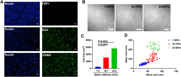
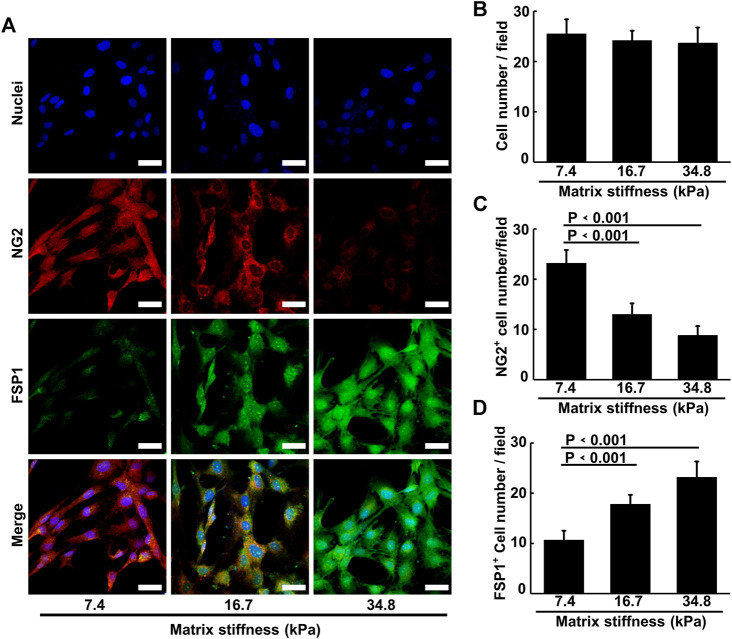
Vascular pericytes, important mural cells that retain progenitor cell properties and protect vascular integrity in healthy tissues, are often associated with tumor development, but their functions in cancer invasion remain elusive. One prominent outcome of tumor occurrence is that the microenvironment of the lesion often stiffens, which could change resident cell behavior. Here, we found pericytes are matrix stiffness-responsive and mechanical stimuli induce pericyte-fibroblast transition (PFT). Soft PA gels that mimic the stiffness of healthy tissues retain the identity and behavior of pericytes, whereas stiff PA gels that reflect the stiffness of tumorous tissues promote PFT and the mobility and invasiveness of the cells. Matrix stiffness-induced PFT depends on the activation of YAP (Yes-associated protein), a transcription factor, which, upon receiving mechanical signals, transfers from cytoplasm to nucleus to mediate cell transcriptional activities. Our result reveals a mechanism through which vascular pericytes convert to fibroblasts and migrate away from vasculatures to help tumor development, and thus targeting matrix stiffness-induced PFT may offer a new perspective to the treatment of cancer metastasis.

Keywords: blood vessel; fibroblast; hydrogel; matrix stiffness; pericyte; tumor.


Conflict of interest statement

The authors declare that the research was conducted in the absence of any commercial or financial relationships that could be construed as a potential conflict of interest.

Figures

FIGURE 1
FIGURE 1
Matrix stiffness changes the morphology of pericytes isolated from mouse lung vasculatures. (A) Representative immunofluorescence images of pericytes that are NG2 and αSMA positive but FSP1 negative. Cell nuclei were stained with DAPI. Scale bar is 200 um. (B) Morphology changes of pericytes cultured on different matrices that vary in stiffness. Scale bar is 20 um. (C) Statistical analysis of the spreading areas of pericytes on different matrices. (D) The distribution of the major axis of pericytes vs. their minor axis on different matrices.
FIGURE 2
FIGURE 2
FIRMs measurement of pericyte adhesion force on different matrix. (A) Left: TEM images of Fe4O3 nanoparticles. Scale bar is 50 nm. Right: amplification image of the encircled nanoparticle in the left image. Scale bar is 5 nm (B) Size distribution of the nanoparticles post-hydrophilization (C) The lineal relationship between the remanence signal and their mass (D) Relative remanence signal of Fe4O3 nanoparticles vs. time. The concentration of the nanoparticle used here is 30 ug/ml. (E) Adhesion force of pericytes on different matrices measured by FIRMs.
FIGURE 3
FIGURE 3
Matrix stiffness induces pericytes to migrate away from pericyte spheroid and invade their surroundings. (A) Pericyte spheroids cultured on collagen-modified PA gels. (B) Statistical analysis of invasive cell numbers of pericytes cultured on different matrices. (C) Migration velocity of pericytes cultured on different matrices as measured by wound healing assay. (D) Representative images of pericyte spheroids encircled in A. Cell nuclei were stained with DAPI. Scale bar is 90 um.
FIGURE 4
FIGURE 4
Representative images of the cytoskeletal elements of pericytes cultured on different matrices. Cell nuclei were stained with DAPI. (A) Representative images of F-actin assembling in pericytes on different matrices. Scale bar is 30 um. (B) Representative images of myosin II in pericytes on different matrices. Scale bar is 30 um.
FIGURE 5
FIGURE 5
Matrix stiffness induces pericyte-fibroblast transition. (A) Representative immunofluorescence images of pericytes on different matrices stained with NG2 and FSP1 antibody. Cell nuclei were stained with DAPI. Scale bar is 20 um. (B) Statistical analysis of pericyte cell numbers in each image field. (C) Statistical analysis of NG2 positive pericyte cell numbers in each field. (D) Statistical analysis of FSP1 positive pericyte cell numbers in each field.
FIGURE 6
FIGURE 6
Matrix stiffness induces YAP activity. (A) Representative images of YAP activation along different matrix stiffness. Nuclei were stained with DAPI. Scale bar is 20 um. (B) Statistical analysis of YAP nuclei/cytoplasm ratio along different matrix stiffness.
FIGURE 7
FIGURE 7
YAP activation inhibition prevents pericyte-fibroblast transition and abrogates adhesion enhancement of pericytes on stiff matrix. (A) Representative immunofluorescence images of pericytes stained with FSP1 and DAPI after YAP inhibition on different matrices. Scale bar is 30 um. (B) Statistical analysis of FSP1 fluorescence intensity in A. (C) FIRMs measurement of pericyte adhesion force after YAP inhibition on different matrices. (D) Representative Transwell images of mouse tumor cells after pericytes cultured on different matrices were seeded in Transwell chamber upon Matrigel but beneath mouse tumor cells. (E) In vitro angiogenesis of pericytes on different matrices.

References

    1. Bielenberg D. R., Zetter B. R. (2015). The Contribution of Angiogenesis to the Process of Metastasis. Cancer J. 21 (4), 267–273. 10.1097/ppo.0000000000000138 - DOI - PMC - PubMed
    1. Bonnans C., Chou J., Werb Z. (2014). Remodelling the Extracellular Matrix in Development and Disease. Nat. Rev. Mol. Cel Biol 15 (12), 786–801. 10.1038/nrm3904 - DOI - PMC - PubMed
    1. Calvo F., Ege N., Grande-Garcia A., Hooper S., Jenkins R. P., Chaudhry S. I., et al. (2013). Mechanotransduction and YAP-dependent Matrix Remodelling Is Required for the Generation and Maintenance of Cancer-Associated Fibroblasts. Nat. Cel Biol 15 (6), 637–646. 10.1038/ncb2756 - DOI - PMC - PubMed
    1. Chakroborty D., Sarkar C., Yu H., Wang J., Liu Z., Dasgupta P. S., et al. (2011). Dopamine Stabilizes Tumor Blood Vessels by Up-Regulating Angiopoietin 1 Expression in Pericytes and Kruppel-like Factor-2 Expression in Tumor Endothelial Cells. Proc. Natl. Acad. Sci. 108 (51), 20730–20735. 10.1073/pnas.1108696108 - DOI - PMC - PubMed
    1. Chaudhuri O., Gu L., Klumpers D., Darnell M., Bencherif S. A., Weaver J. C., et al. (2016). Hydrogels with Tunable Stress Relaxation Regulate Stem Cell Fate and Activity. Nat. Mater 15 (3), 326–334. 10.1038/nmat4489 - DOI - PMC - PubMed

LinkOut - more resources