Case Reports
. 2021 Jun 9;9(6):e04145.
doi: 10.1002/ccr3.4145. eCollection 2021 Jun.

A patient with paraganglioma undergoing laparoscopic resection: A case report

Affiliations
Case Reports

A patient with paraganglioma undergoing laparoscopic resection: A case report

Hisamichi Yoshii et al. Clin Case Rep. .

Abstract

Paraganglioma is a very rare extraadrenal nonepithelial tumor. The number of cases of laparoscopic surgery in Paraganglioma is small and controversial. This study encountered a case of successful transperitoneal laparoscopic surgery for a 56-mm paraganglioma in a 53-year-old female. Moreover, previous reports on laparoscopic surgery for paraganglioma are reviewed.

Keywords: general surgery; neurosurgery; oncology.


Conflict of interest statement

None declared.

Figures

FIGURE 1
FIGURE 1
Abdominal ultrasonography image. A 56 × 43‐mm hypoechoic tumor with a smooth margin, internal heterogeneity, and no blood flow signal at the posterior wall of the gastric corpus and tail of the pancreas
FIGURE 2
FIGURE 2
Abdominal contrast‐enhanced computed tomography images. A 56.7 × 37.9 × 54.7 mm between the posterior wall of the gastric corpus and the right margin of the abdominal aorta (red arrows)
FIGURE 3
FIGURE 3
Abdominal contrast‐enhanced magnetic resonance images. Low signal on T1‐weighted imaging, faint and heterogenous high signal on T2‐weighted imaging, and faint high signal on diffusion‐weighted images. T1‐weighted imaging, Fat‐suppressed T2‐weighted imaging, Diffusion‐weighted images
FIGURE 4
FIGURE 4
Endoscopic ultrasound image. A 47.3 × 31.3‐mm hypoechoic tumor at the posterior wall of the middle part of the gastric corpus. It is suspected to be derived from the muscular layer of the fourth layer of the gastric wall (red arrow)
FIGURE 5
FIGURE 5
Surgical findings. The pancreas was tunneled and lifted; the tumor and the retroperitoneum were then exfoliated
FIGURE 6
FIGURE 6
Surgical findings. Feeding vessels flow from the left gastric artery toward the tumor (red arrow)
FIGURE 7
FIGURE 7
Macroscopic findings. A round tumor with capsule with a clear boundary was observed, and no necrosis existed on the cut surface. Moreover, yellow consolidation was observed
FIGURE 8
FIGURE 8
Histopathological findings. Supporting tissues and capillaries were observed around the pleomorphic tumor cells and alveolar aggregates, exhibiting a Zellballen pattern
FIGURE 9
FIGURE 9
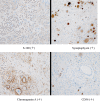
Immunohistochemistry staining. S‐100 (+), Synaptophysin (+), Chromogranin A (+), CD56 (+)

References

    1. Lam AKY. Update on adrenal tumours in 2017 World Health Organization (WHO) of endocrine tumours. Endocr Pathol. 2017;28(3):213‐227. - PubMed
    1. Yang Y, Wang G, Lu H, Liu Y, Ning S, Luo F. Haemorrhagic retroperitoneal paraganglioma initially manifesting as acute abdomen: a rare case report and literature review. BMC Surg. 2020;20(1):1‐11. - PMC - PubMed
    1. Lam K, Lo C, Wat N, Luk J, Lam K. The clinicopathological features and importance of p53, Rb, and mdm2 expression in phaeochromocytomas and paragangliomas. J Clin Pathol. 2001;54(6):443‐448. - PMC - PubMed
    1. Lam KY, Chan ACL. Paraganglioma of the urinary bladder: an immunohistochemical study and report of an unusual association with intestinal carclnold. ANZ J Surg. 1993;63(9):740‐745. - PubMed
    1. Garg A, Mishra D, Bansal M, Maharia HR, Goyal V. Right atrial paraganglioma: an extremely rare primary cardiac neoplasm mimicking myxoma. J Cardiovasc Ultrasound. 2016;24(4):334‐336. - PMC - PubMed

Publication types

LinkOut - more resources