Case Reports
. 2021 Jun 9;9(6):e04352.
doi: 10.1002/ccr3.4352. eCollection 2021 Jun.

Multifocal chondrosarcoma of the hand: Case report and review of the literature

Affiliations
Case Reports

Multifocal chondrosarcoma of the hand: Case report and review of the literature

Hunter B Jones et al. Clin Case Rep. .

Abstract

Few multifocal hand chondrosarcomas have been reported. To our knowledge, this report is the first to describe multifocal hand chondrosarcoma in a patient with no evidence of prior enchondroma, Ollier's disease, or Maffucci syndrome.

Keywords: chondrosarcoma; hand malignancy; multifocal tumor; reconstructive hand surgery.


Conflict of interest statement

Though they are not directly funding this case report, the authors would like to disclose the following support for BM: Paid teaching for TriMed. Paid teaching and consulting, as well as research support from AxoGen. Paid consulting for Baxter/Synovis and GLG. The remaining authors have nothing to disclose.

Figures

FIGURE 1
FIGURE 1
Original MRI showing an enhancing lesion with distinct areas of T2 hyperintensity and T1 hypointensity (B, C: arrows) separated by regions of T1 hyperintensity. MRI showing a T2 hyperintense and T1 hypointense (G‐I: arrows) lesion measuring 0.5 x 0.6 x 0.9 cm on the ulnar base of the 5th proximal phalanx
FIGURE 2
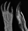
FIGURE 2
Preoperative radiographs showing abnormality in the small finger
FIGURE 3
FIGURE 3
Bone scan showing focal areas (arrows) in the in the 5th digit and in the wrist
FIGURE 4
FIGURE 4
MRI showing T1 hypointense and T2 hyperintense (arrows) 6‐mm lesion in the hook of the hamate, as well as a 3‐mm body in the ulnar body of the hamate and a 4‐mm lesion in the ulnar base of the proximal metacarpal (arrows)
FIGURE 5
FIGURE 5
Intraoperative radiographs following removal of percutaneous pins and manipulation for improved range of motion
FIGURE 6
FIGURE 6
14‐month postoperative MRI (without contrast) showing (A) T1 and (B) T2 images of the left hand
FIGURE 7
FIGURE 7
Intraoperative radiographs taken 20 months after the initial resection procedure
FIGURE 8
FIGURE 8
Fluoroscopic images taken 21 months after the initial resection procedure
FIGURE 9
FIGURE 9
Radiographs taken 28 months after initial resection procedure

References

    1. Athanasian E. Bone and Soft Tissue Tumors, Green’s Operative Hand Surgery. Philadelphia, Pennsylvania: Elsevier Churchill Livingstone; 2005.
    1. O'Connor MI, Bancroft LW. Benign and malignant cartilage tumors of the hand. Hand Clin. 2004;20(3):317‐323, vi. - PubMed
    1. Sridhar H, Vijaya M, Clement W, Srinivas C. Chondrosarcoma arising in an enchondroma of the metacarpal bone ‐ a case report. J Clin Diagn Res. 2014;8(3):142‐143. - PMC - PubMed
    1. Ollivier L, Vanel D, Leclere J. Imaging of chondrosarcomas. Cancer Imaging. 2003;4(1):36‐38. - PMC - PubMed
    1. Devaney K. Dahlin's Bone Tumors. General Aspects and Data on 11,087 Cases. Am J Surg Pathol. 1996;20(10):1298‐.

Publication types

LinkOut - more resources