. 2021 May 31:8:662530.
doi: 10.3389/fsurg.2021.662530. eCollection 2021.

A Synchrotron and Micro-CT Study of the Human Endolymphatic Duct System: Is Meniere's Disease Caused by an Acute Endolymph Backflow?

Affiliations

A Synchrotron and Micro-CT Study of the Human Endolymphatic Duct System: Is Meniere's Disease Caused by an Acute Endolymph Backflow?

Hao Li et al. Front Surg. .

Abstract

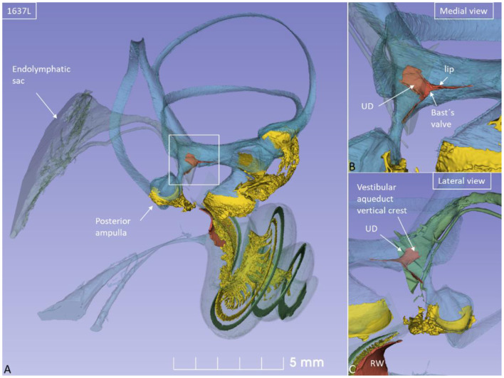
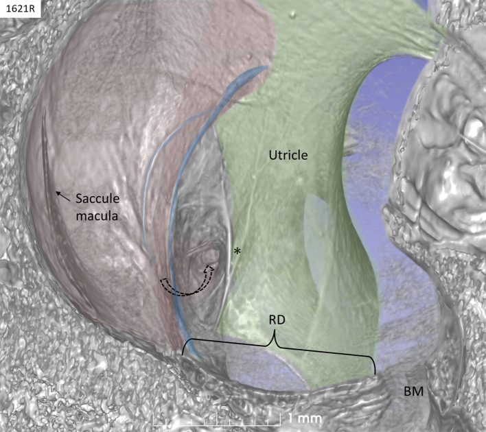
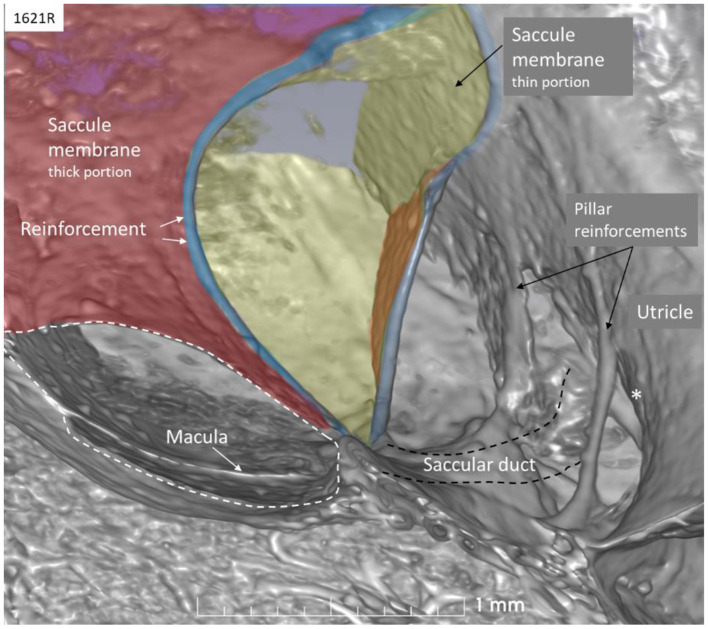
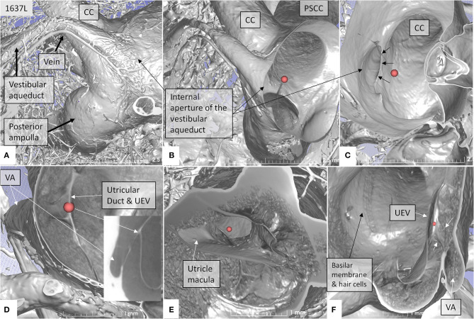
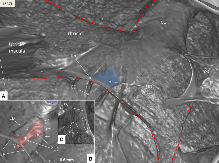
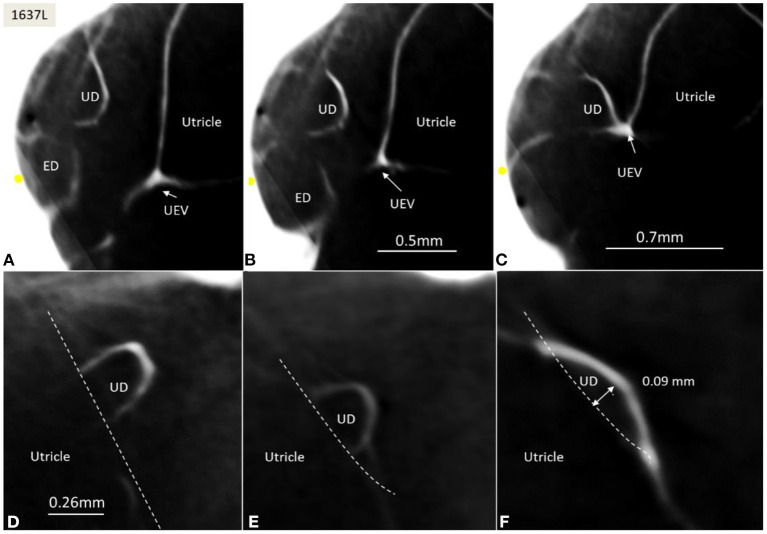
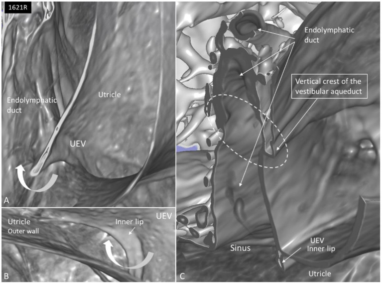
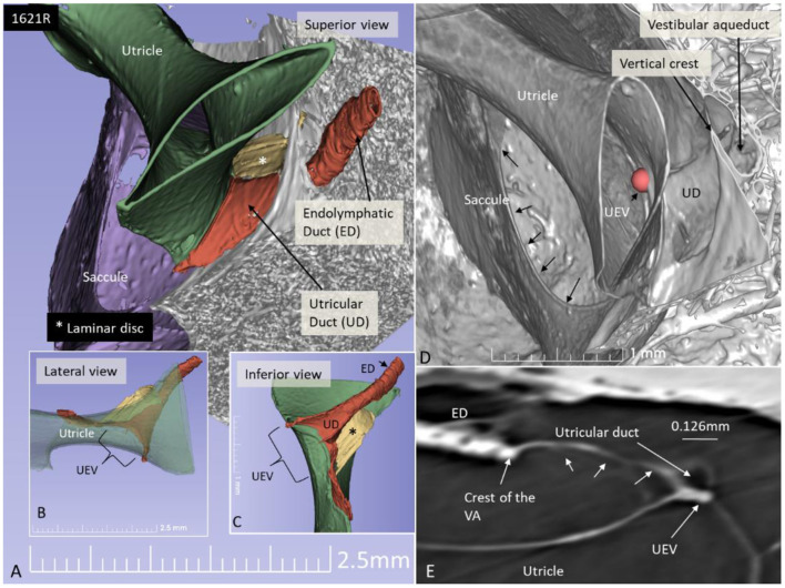
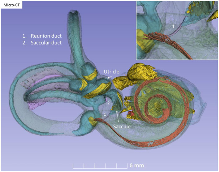
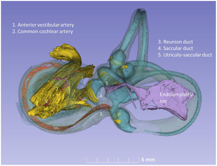
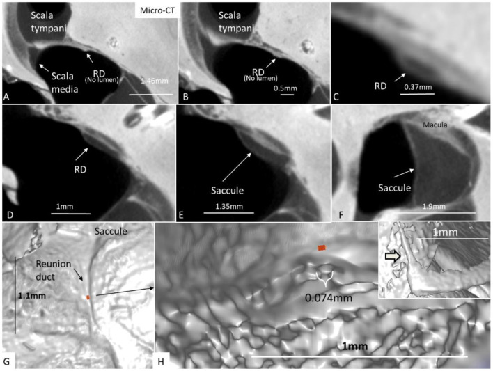
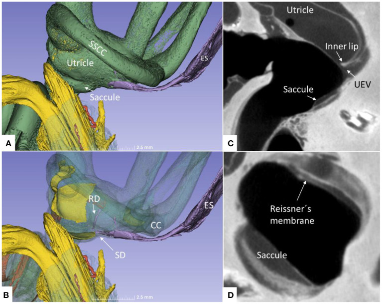
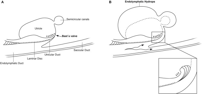
Background: The etiology of Meniere's disease (MD) and endolymphatic hydrops believed to underlie its symptoms remain unknown. One reason may be the exceptional complexity of the human inner ear, its vulnerability, and surrounding hard bone. The vestibular organ contains an endolymphatic duct system (EDS) bridging the different fluid reservoirs. It may be essential for monitoring hydraulic equilibrium, and a dysregulation may result in distension of the fluid spaces or endolymphatic hydrops. Material and Methods: We studied the EDS using high-resolution synchrotron phase contrast non-invasive imaging (SR-PCI), and micro-computed tomography (micro-CT). Ten fresh human temporal bones underwent SR-PCI. One bone underwent micro-CT after fixation and staining with Lugol's iodine solution (I2KI) to increase tissue resolution. Data were processed using volume-rendering software to create 3D reconstructions allowing orthogonal sectioning, cropping, and tissue segmentation. Results: Combined imaging techniques with segmentation and tissue modeling demonstrated the 3D anatomy of the human saccule, utricle, endolymphatic duct, and sac together with connecting pathways. The utricular duct (UD) and utriculo-endolymphatic valve (UEV or Bast's valve) were demonstrated three-dimensionally for the first time. The reunion duct was displayed with micro-CT. It may serve as a safety valve to maintain cochlear endolymph homeostasis under certain conditions. Discussion: The thin reunion duct seems to play a minor role in the exchange of endolymph between the cochlea and vestibule under normal conditions. The saccule wall appears highly flexible, which may explain occult hydrops occasionally preceding symptoms in MD on magnetic resonance imaging (MRI). The design of the UEV and connecting ducts suggests that there is a reciprocal exchange of fluid among the utricle, semicircular canals, and the EDS. Based on the anatomic framework and previous experimental data, we speculate that precipitous vestibular symptoms in MD arise from a sudden increase in endolymph pressure caused by an uncontrolled endolymphatic sac secretion. A rapid rise in UD pressure, mediated along the fairly wide UEV, may underlie the acute vertigo attack, refuting the rupture/K+-intoxication theory.

Keywords: Bast's valve; Meniere's disease; human; reunion duct; synchrotron radiation phase-contrast imaging.


Conflict of interest statement

The authors declare that the research was conducted in the absence of any commercial or financial relationships that could be construed as a potential conflict of interest.

Figures

Figure 1
Figure 1
(A) SR-PCI of a left human ear with modeling of anatomical details. Maculae and nerve structures are stained yellow. The position of the saccule and utricle and their relationship to the vestibular aqueduct (blue) are shown. (B) Medial view shows the position of the UEV relative to the internal aperture of the vestibular aqueduct. (C) Posterolateral view.
Figure 2
Figure 2
SR-PCI of a left human ear with 3D reconstructions of saccule (red) and utricle (green) (lateral view). The entrance gate to the internal opening of the VA (broken arrow) and the UEV (*) can be seen. The saccule wall contains a reinforced semilunar portion that additionally thickens (blue) against the thinner part. The thinner part and the saccular duct are difficult to discern. BM, basilar membrane. RD, reunion duct.
Figure 3
Figure 3
SR-PCI of the saccule (left ear, lateral view). The wall consists of a thick (red) and thin (yellow) part separated by bands of reinforcement (blue). The thin wall appears flaccid. The SD exits near the band and runs against the ED and the UEV (*). The utricle wall is supported by several pillars attached to the internal surface of the surrounding bony wall.
Figure 4
Figure 4
SR-PCI showing the position of the UEV relative to the internal opening of the VA in a left bone. (A) Medial view shows the entrance of the VA into the vestibule. A vascular plexus surrounds the VA that drains into the vein of the VA. (B) Anterior cropping shows the VA and the position of the UEV (red fiducial). (C) Bony algorithm shows the internal opening of the VA relative to the UEV (red fiducial). (D) Adjusting opacity gradient reveals both the UEV and the UD (red fiducial). (E) Lateral view shows the position of the utricle macula and the UEV (red fiducial). (F) Superior view and modification of gradient opacity shows the UD (white arrows) and the UEV (red fiducial). The BM and the rows of hair cells are seen. CC, common crus; UEV, utriculo-endolymphatic valve; VA, vestibular aqueduct; PSSC, posterior semicircular canal; UD, utricular duct; BM, basilar membrane.
Figure 5
Figure 5
SR-PCI of the UD (blue) at the internal aperture of the VA. (A) UEV lip (Bast's valve) is located near the floor of the utricle. The broken red line delineates parts of the vestibular labyrinth. (B) Lateral view shows the UD and the ED at the opening of the VA (small arrows). Fiducials (red) mark (1) the position of the mid-portion of the UEV, (2) where the UD reaches the base of the lip, and (3) at the division of the UD and ED. (C) Image shows the lateral disc running from the vertical crest of the VA to the UEV. The broken white lines depict the lumen of the UD. LSSC; lateral semicircular canal.
Figure 6
Figure 6
(A–C) SR-PCI of the UD and ED at the internal aperture of the VA. The UEV is closed and connected with a membranous strand against the bony wall. (D–F) Lateral view shows serial sections of the UD running against the UEV. Its lumen narrows against the valve. The diameter of the UD in (D) is 0.32 mm.
Figure 7
Figure 7
(A) SR-PCI of the left cochlea and vestibule sectioned and viewed laterally. (B) The saccule wall consist of a thicker part (small black arrows) and a thinner part (*). The UEV is located in the posterior-inferior part of the utricle (arrow). (C) The semilunar-shaped opening of the UEV is shown in higher magnification and is marked with a red fiducial. VA, vestibular aqueduct.
Figure 8
Figure 8
(A) SR-PCI and higher magnification of the UEV and the slit-like opening in the utricle. (B) The outer utricle wall is thin but can be seen reaching the inner lip of the valve. (C) Coronary section shows the internal opening of the vestibular aqueduct (circle, broken lines). A membrane disc connects the vertical crest with the UEV. The epithelial wall of the UD is not visualized. The endolymphatic duct is surrounded by several blood vessels. Sinus: sinus portion of the endolymphatic duct.
Figure 9
Figure 9
SR-PCI 3D reconstruction and model of the posterior part of the vestibule in a left ear (superior view). (A) The endolymphatic duct (ED) and utricular duct (UD) can be seen. A laminar disc (*) extends from the vertical crest of the internal opening of the vestibular aqueduct (VA) to the utricle. It also covers the UD. (B) Lateral view of the modeled UEV is viewed through a partly transparent utricle. (C) Modeled UEV is viewed from inferior. (D) UEV is shown from inside the utricle (red fiducial). The UD wall is reinforced by the laminar disc. (E) Horizontal section shows the UD and UEV. The laminar disc (small arrows) runs from the vertical crest to the UEV. It keeps the UD closely associated with the UEV. Note that the UD passes on the UEV allowing increased pressure in the UD to be transmitted and may push the valve to open.
Figure 10
Figure 10
Micro-CT and 3D modeling of a right human temporal bone (Stenver's view). The membrane labyrinth is shown in different colors after the bony capsule is made semi-transparent. The vestibular neuro-epithelium and nerves are yellow. The basilar membrane is colored red. The inset shows RD (1).
Figure 11
Figure 11
Posterior view of the modeled inner ear shown in Figure 10. The position of the saccular and utricular ducts are visualized. The internal auditory canal is shown with cranial nerves and arterial blood vessels supplying the inner ear.
Figure 12
Figure 12
(A–F) Micro-CT and serial sections show the reunion duct (RD) from the saccule to the cochlear duct. (G) Surface view of the RD. Its mid-portion is cropped and shown in (H) The RD diameter is less than a 10th of a millimeter. The inset in H shows the angle formed between the RD and the vestibular end of the cochlear duct.
Figure 13
Figure 13
(A) Superior close-up view of micro-CT shown in Figure 11. The internal aperture of the vestibular aqueduct (lilac) is seen near the common crus (CC) and the SSCC. (B) The saccular and reunion ducts are visible after making the bony capsule transparent. The RD runs almost perpendicular against the saccular duct. (C) Horizontal section of the UEV. (D) The saccule and cochlear endolymphatic space at the cecum vestibulare are shown. SSCC, superior semicircular canal; SD, saccular duct; RD, reunion duct; ES, endolymphatic sac.
Figure 14
Figure 14
(A) Principal drawing of the human utriculo-endolymphatic valve (Bast's valve) based on SR-PCI. The valve is structurally associated with the opening of the vestibular aqueduct through a laminar disc (interrupted line) emerging from the vertical crest of the vestibular aqueduct to the lip of the valve. The outer wall of the utricular duct faces the disc and forms the utricular fold. Between the inner and outer wall at the base of the lip, there is loose tissue allowing the valve to open (small arrows). (B) Hypothetical representation of the role of Bast's valve and UD in generating acute endolymphatic hypertension and hydrops in patients with MD. Increased endolymph pressure is caused by reduced reabsorption or hypersecretion in the ES, leading to a “backflow” of endolymph. It compresses the wall of the utricular duct and pushes the valve to open (inset), leading to acute EH and vertigo. Experimentally, secretion in the ES may be provoked within a short timeframe (41, 42).

References

    1. Swinburne IA, Mosaliganti KR, Upadhyayula S, Liu TL, Hildebrand DGC, Tsai TYC, et al. . Lamellar junctions in the endolymphatic sac act as a relief valve to regulate inner ear pressure. Elife. (2017) 7:e37131. 10.7554/eLife.37131 - DOI - PMC - PubMed
    1. Seymour JC. Observations on the circulation in the cochlea. J Laryngol Otol. (1954) 68:689–711. 10.1017/S0022215100050131 - DOI - PubMed
    1. Retzius G. Das Gehörorgan der Wirbelthiere: Morphologisch-Histologische STUDIEN. Stockholm: Samson and Wallin; (1884).
    1. Hensen V. Zur Morphologie der Schnecke des Menschen und der Saugetiere. Z Wiessensch Zool. (1863) 13:481.
    1. Guild SR. Circulation of the endolymph. Laryngoscope. (1927) 37:649–52. 10.1288/00005537-192709000-00004 - DOI