. 2021 May 20:15:100342.
doi: 10.1016/j.ynstr.2021.100342. eCollection 2021 Nov.

Stress induces microglia-associated synaptic circuit alterations in the dorsomedial prefrontal cortex

Affiliations

Stress induces microglia-associated synaptic circuit alterations in the dorsomedial prefrontal cortex

Taohui Liu et al. Neurobiol Stress. .

Abstract

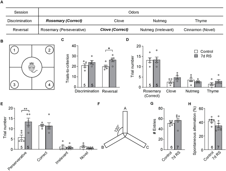
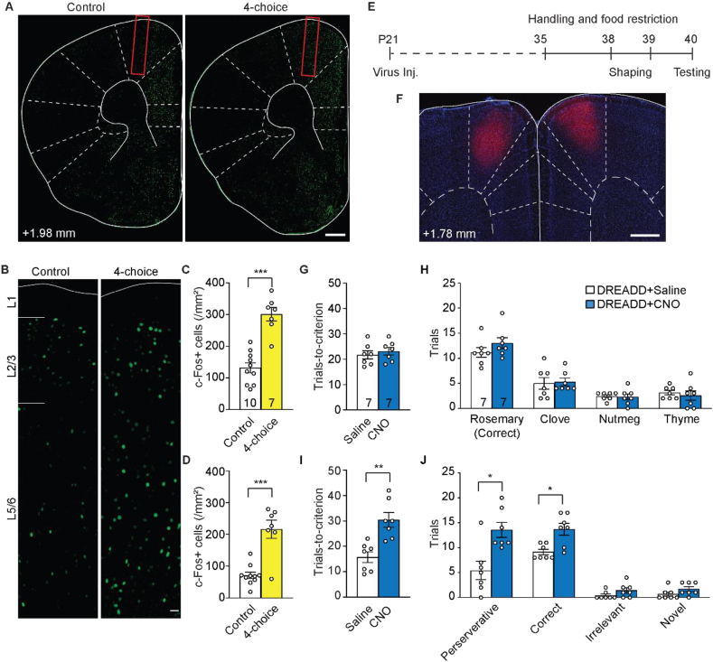
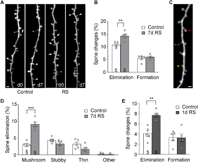
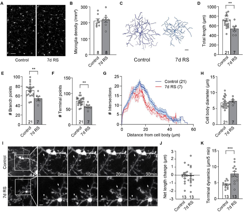
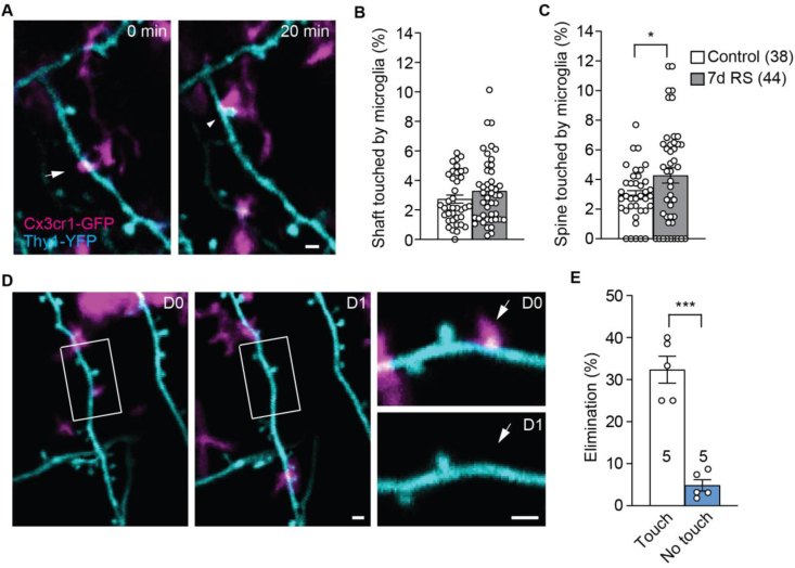
The mammalian dorsomedial prefrontal cortex (dmPFC) receives diverse inputs and plays important roles in adaptive behavior and cognitive flexibility. Stress, a major risk factor for many psychiatric disorders, compromises the structure and function of multiple brain regions and circuits. Here we show that 7-day restraint stress impairs reversal learning in the 4-choice odor discrimination test, a decision-making task requiring an intact dmPFC. In vivo two-photon imaging further reveals that stress increases dmPFC dendritic spine elimination, particularly those of the mushroom morphology, without affecting spine formation. In addition, stress alters dmPFC microglial branching complexity and elevates their terminal process dynamics. In stressed mice, dmPFC microglia contact dendrites more frequently, and dendritic spines with microglial contact are prone to elimination. In summary, our work suggests that stress-induced changes in glial-synapse interaction contributes to synaptic loss in dmPFC, resulting in neuronal circuit deficits and impaired cognitive flexibility.

Keywords: Cognitive flexibility; Dendritic spine; Microglia; Prefrontal cortex; Stress.


Conflict of interest statement

The authors declare no competing interest.

Figures

Fig. 1
Fig. 1
7d RS impairs cognitive flexibility of mice. (A) Odors used for the 4-choice odor discrimination and reversal task. (B) Schematic of the 4-choice test set-up. (C) Number of trials taken to reach the performance criterion in the discrimination and the reversal session. (D) Number of trials digging at each odor during the discrimination session. (E) Number of trials digging at each odor during the reversal session. (F) Schematic of the Y-maze arena. (G) Number of arm entries in control and RS mice. (H) Percentage of spontaneous alternations in control and RS mice. *p < 0.05, **p < 0.01. n = number of mice.
Fig. 2
Fig. 2
Silencing dmPFC increases perseverative errors in the 4-choice test. (A) Immunohistochemistry of c-Fos in the frontal cortex. (B) c-Fos labeling across layers (L) in dmPFC. Red rectangles: the dmPFC region analyzed. (C, D) Density of c-Fos + neurons in L2/3 (C) and L5/6 (D) dmPFC of mice with and without performing the 4-choice task. (E) Timeline of pharmacogenetic manipulation and behavioral task. (F) An example coronal section showing bilateral DREADD virus labeling in dmPFC. On average virus labeling spreads ±0.4 mm around the injection site along the anterior-posterior axis, and ±0.3 mm medial-laterally. (G) Number of trials taken to reach the performance criterion in the discrimination session. (H) Number of trials digging at each odor during the discrimination session. (I) Number of trials taken to reach the performance criterion in the reversal session. (J) Number of trials digging at each odor during the reversal session. *p < 0.05, **p < 0.01, ***p < 0.001. n = number of mice. Scale bars: 500 μm (A, F), 20 μm (B). (For interpretation of the references to color in this figure legend, the reader is referred to the Web version of this article.)
Fig. 3
Fig. 3
7d RS induces dendritic spine loss along apical dendrites of layer 5 pyramidal neurons in dmPFC. (A) Example of the same set of dmPFC spines imaged 7d apart in control and RS mice. Arrows: eliminated spines; arrowheads: new spines; asterisks: filopodia. Scale bar: 2 μm. (B) Percentage of spines formed and eliminated over 7d. (C) Example of different morphological categories of spines. Red: mushroom spine; green: thin spine; yellow: stubby spine. Scale bar: 2 μm. (D) Percentage of elimination of spines belonging to different morphological categories. (E) Percentage of spines formed and eliminated over 1d. *p < 0.05, **p < 0.01, ***p < 0.001. n = number of mice.
Fig. 4
Fig. 4
7d RS decreases the ramification and increases the process dynamics of microglia in dmPFC. (A) Representative images of Iba1 immunohistochemistry in dmPFC of control and RS mice. (B) Microglia density in dmPFC of control and RS mice. (C) Representative three-dimensional reconstructions of microglia from control and RS mice. (D–F) Analysis of total process length (D), the number of branching points (E), the number of terminal points (F) of dmPFC microglia in control and RS mice. (G) Sholl analysis of microglial processes in control and RS mice. (H) Microglial soma size in control and RS mice. (I) Representative time-lapse in vivo 2P images of microglia from control and RS mice. Arrows and arrowheads indicate dynamic changes. (J) Net length change of microglial terminal processes over 30 min. (K) Average terminal dynamics of microglia processes over 5 min **p < 0.01, ***p < 0.001. n = numbers of mice (B) or cells analyzed (D-H, J, K). Scale bars: 20 μm (A), 5 μm (C, I).
Fig. 5
Fig. 5
Microglia contact makes dendritic spines more prone to elimination. (A) Co-imaging dendritic spines (cyan) and microglial processes (magenta) over time shows the dynamic contact of microglia onto dendrites and spines. The arrow and the arrowhead point at dendritic shaft and spine with microglial contacts, respectively. (B) Percentage of dendritic segment length contacted by microglial processes. (C) Percentage of spines contacted by microglia processes. (D) Example of co-imaging the same dendrite and contacting microglia over 1d, showing the elimination of the spine contacted by microglia (arrow in insets). (E) Percentage of spines with and without microglial contact eliminated over 1d. *p < 0.05, ***p < 0.001. n = number of imaging regions analyzed (B, C) or mice (E). Scale bars: 1 μm. (For interpretation of the references to color in this figure legend, the reader is referred to the Web version of this article.)

References

    1. Abiega O., Beccari S., Diaz-Aparicio I., Nadjar A., Laye S., Leyrolle Q. Neuronal hyperactivity disturbs ATP microgradients, impairs microglial motility, and reduces phagocytic receptor expression triggering apoptosis/microglial phagocytosis uncoupling. PLoS Biol. 2016;14 doi: 10.1371/journal.pbio.1002466. - DOI - PMC - PubMed
    1. Arnsten A.F. Stress signalling pathways that impair prefrontal cortex structure and function. Nat. Rev. Neurosci. 2009;10:410–422. doi: 10.1038/nrn2648. - DOI - PMC - PubMed
    1. Arnsten A.F. Stress weakens prefrontal networks: molecular insults to higher cognition. Nat. Neurosci. 2015;18:1376–1385. doi: 10.1038/nn.4087. - DOI - PMC - PubMed
    1. Bachiller S., Jimenez-Ferrer I., Paulus A., Yang Y., Swanberg M., Deierborg T. Microglia in neurological diseases: a road map to brain-disease dependent-inflammatory response. Front. Cell. Neurosci. 2018;12:488. doi: 10.3389/fncel.2018.00488. - DOI - PMC - PubMed
    1. Barbas H. General cortical and special prefrontal connections: principles from structure to function. Annu. Rev. Neurosci. 2015;38:269–289. doi: 10.1146/annurev-neuro-071714-033936. - DOI - PubMed