

Injectable and thermosensitive hydrogels mediating a universal macromolecular contrast agent with radiopacity for noninvasive imaging of deep tissues
- PMID: 34136722
- PMCID: PMC8165329
- DOI: 10.1016/j.bioactmat.2021.05.013
Injectable and thermosensitive hydrogels mediating a universal macromolecular contrast agent with radiopacity for noninvasive imaging of deep tissues
Abstract
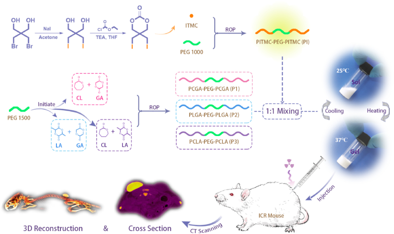
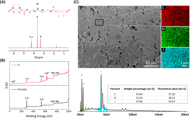
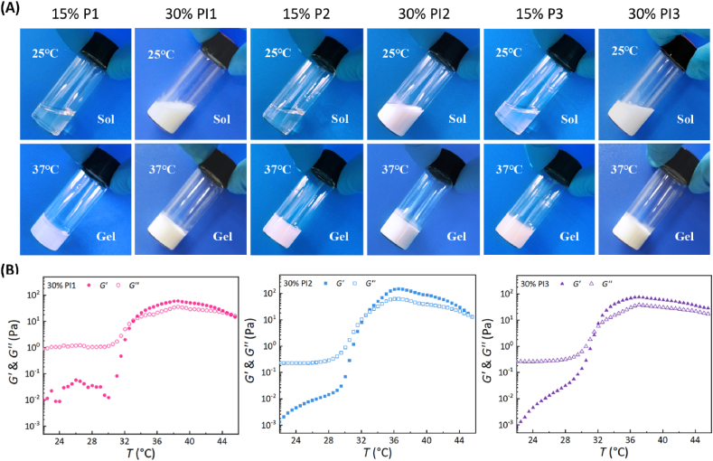
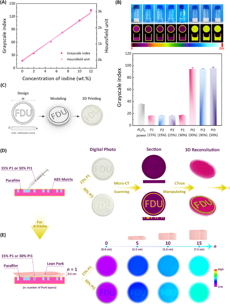
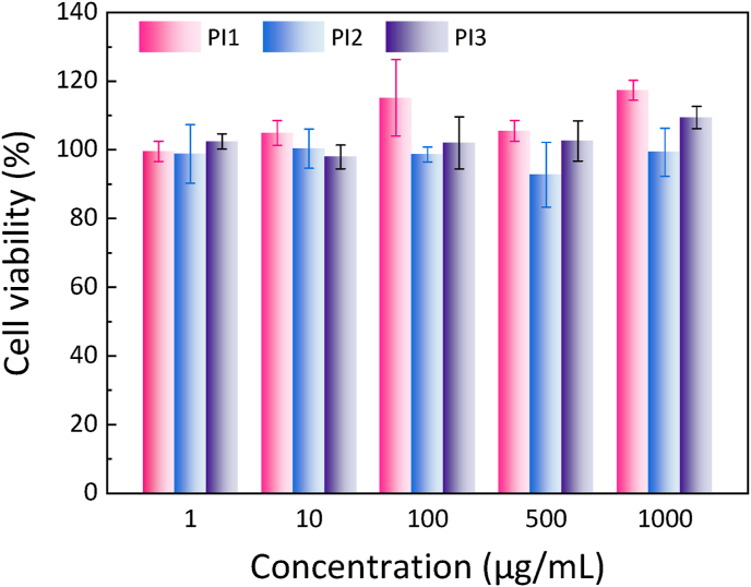
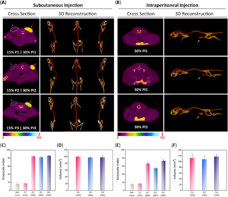
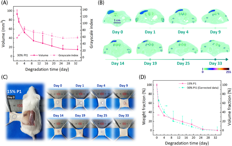
It is very challenging to visualize implantable medical devices made of biodegradable polymers in deep tissues. Herein, we designed a novel macromolecular contrast agent with ultrahigh radiopacity (iodinate content > 50%) via polymerizing an iodinated trimethylene carbonate monomer into the two ends of poly(ethylene glycol) (PEG). A set of thermosensitive and biodegradable polyester-PEG-polyester triblock copolymers with varied polyester compositions synthesized by us, which were soluble in water at room temperature and could spontaneously form hydrogels at body temperature, were selected as the demonstration materials. The addition of macromolecular contrast agent did not obviously compromise the injectability and thermogelation properties of polymeric hydrogels, but conferred them with excellent X-ray opacity, enabling visualization of the hydrogels at clinically relevant depths through X-ray fluoroscopy or Micro-CT. In a mouse model, the 3D morphology of the radiopaque hydrogels after injection into different target sites was visible using Micro-CT imaging, and their injection volume could be accurately obtained. Furthermore, the subcutaneous degradation process of a radiopaque hydrogel could be non-invasively monitored in a real-time and quantitative manner. In particular, the corrected degradation curve based on Micro-CT imaging well matched with the degradation profile of virgin polymer hydrogel determined by the gravimetric method. These findings indicate that the macromolecular contrast agent has good universality for the construction of various radiopaque polymer hydrogels, and can nondestructively trace and quantify their degradation in vivo. Meanwhile, the present methodology developed by us affords a platform technology for deep tissue imaging of polymeric materials.
Keywords: Block copolymers; In vivo degradation; Non-invasive deep tissue imaging; Radiopacity; Thermosensitive hydrogels.
© 2021 The Authors.
Conflict of interest statement
There are no conflicts of interest to declare.
Figures

Similar articles
-
Non-invasive monitoring of in vivo degradation of a radiopaque thermoreversible hydrogel and its efficacy in preventing post-operative adhesions.Acta Biomater. 2017 Jun;55:396-409. doi: 10.1016/j.actbio.2017.03.042. Epub 2017 Mar 29. Acta Biomater. 2017. PMID: 28363786
-
PEG-based thermosensitive and biodegradable hydrogels.Acta Biomater. 2021 Jul 1;128:42-59. doi: 10.1016/j.actbio.2021.04.009. Epub 2021 Apr 20. Acta Biomater. 2021. PMID: 33857694 Review.
-
Thermosensitive polymeric hydrogels as drug delivery systems.Curr Med Chem. 2013;20(1):79-94. Curr Med Chem. 2013. PMID: 23092130 Review.
-
A Custom Radiopaque Thermoresponsive Chemotherapy-Loaded Hydrogel for Intratumoural Injection: An In Vitro and Ex Vivo Assessment of Imaging Characteristics and Material Properties.Cardiovasc Intervent Radiol. 2019 Feb;42(2):289-297. doi: 10.1007/s00270-018-2103-0. Epub 2018 Nov 2. Cardiovasc Intervent Radiol. 2019. PMID: 30390105
-
Thermosensitive block copolymer hydrogels based on poly(ɛ-caprolactone) and polyethylene glycol for biomedical applications: state of the art and future perspectives.J Biomed Mater Res A. 2015 Mar;103(3):1276-90. doi: 10.1002/jbm.a.35253. Epub 2014 Jun 25. J Biomed Mater Res A. 2015. PMID: 24912941
Cited by
-
In vivo near-infrared fluorescent fibrin highlights growth of nerve during regeneration across a nerve gap.J Biomed Opt. 2022 Jul;27(7):070502. doi: 10.1117/1.JBO.27.7.070502. Epub 2022 Jul 20. J Biomed Opt. 2022. PMID: 36451699 Free PMC article.
-
Injectable PTHF-based thermogelling polyurethane implants for long-term intraocular application.Biomater Res. 2022 Dec 2;26(1):70. doi: 10.1186/s40824-022-00316-z. Biomater Res. 2022. PMID: 36461130 Free PMC article.
-
Organic Compound with Potential for X-ray Imaging Applications.ACS Omega. 2021 Sep 16;6(38):24826-24833. doi: 10.1021/acsomega.1c03671. eCollection 2021 Sep 28. ACS Omega. 2021. PMID: 34604664 Free PMC article.
-
Recent advances in regenerative biomaterials.Regen Biomater. 2022 Dec 5;9:rbac098. doi: 10.1093/rb/rbac098. eCollection 2022. Regen Biomater. 2022. PMID: 36518879 Free PMC article. Review.
-
Intracranial In Situ Thermosensitive Hydrogel Delivery of Temozolomide Accomplished by PLGA-PEG-PLGA Triblock Copolymer Blending for GBM Treatment.Polymers (Basel). 2022 Aug 18;14(16):3368. doi: 10.3390/polym14163368. Polymers (Basel). 2022. PMID: 36015626 Free PMC article.
References
-
- Lei K., Ma Q., Yu L., Ding J. Functional biomedical hydrogels for in vivo imaging. J. Mater. Chem. B. 2016;4(48):7793–7812. - PubMed
LinkOut - more resources
Full Text Sources
Other Literature Sources
