

Long non-coding ribonucleic acid urothelial carcinoma-associated 1 promotes high glucose-induced human retinal endothelial cells angiogenesis through regulating micro-ribonucleic acid-624-3p/vascular endothelial growth factor C
- PMID: 34137197
- PMCID: PMC8565426
- DOI: 10.1111/jdi.13617
Long non-coding ribonucleic acid urothelial carcinoma-associated 1 promotes high glucose-induced human retinal endothelial cells angiogenesis through regulating micro-ribonucleic acid-624-3p/vascular endothelial growth factor C
Abstract
Aims/introduction: Emerging evidence has indicated that long non-coding ribonucleic acids play important roles in the development and progression of diabetic retinopathy (DR). It is reported that urothelial carcinoma-associated 1 (UCA1) is highly expressed in diabetic lymphoendothelial cells and influences glucose metabolism in rats with DR. The aim of the present study was to explore the role of UCA1 in the mechanism of DR.
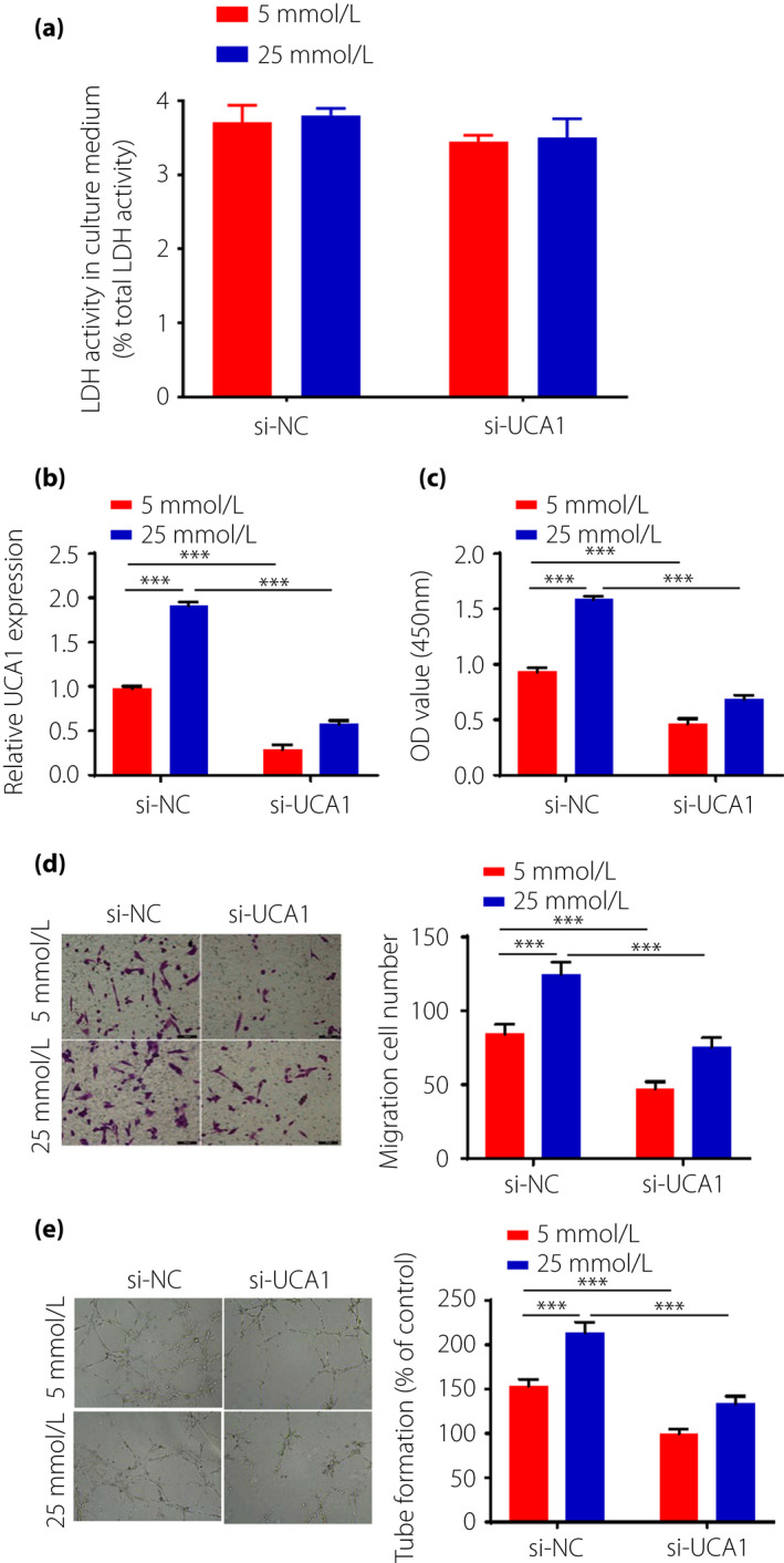
Materials and methods: Gene expression analyses in fibrovascular membranes excised from patients with DR using public microarray datasets (GSE60436). Reverse transcription polymerase chain reaction was carried out to detect UCA1, micro-ribonucleic acid (miR)-624-3p and vascular endothelial growth factor C (VEGF-C) expressions in the blood of patients and human retinal endothelial cells (HRECs). Furthermore, Cell Counting kit-8, Transwell assay, and tube formation assay were used to identify biological effects of UCA1 on HRECs proliferation, migration ability and angiogenesis in vitro.
Results: UCA1 and VEGF-C were elevated in DR patients and high glucose-induced HRECs cell lines, whereas miR-624-3p was decreased. UCA1 inhibition inhibited proliferation, angiogenesis and migration of HRECs cells under high-glucose condition. Luciferase reporter assay showed that UCA1 could sponge with miR-624-3p, which could directly target VEGF-C. Finally, we proved a pathway that UCA1 promoted cell proliferation, migration and angiogenesis through sponging with miR-624-3p, thereby upregulating VEGF-C in high-glucose-induced HRECs.
Conclusions: We identified UCA1 as an important factor associated with DR, which could regulate the expression of VEGF-C by sponging miR-624-3p in human retinal endothelial cells. Our results pave the way for further studies on diagnostic and therapeutic studies related to UCA1 in DR patients.
Keywords: Angiogenesis; Diabetic retinopathy; Urothelial carcinoma-associated 1.
© 2021 The Authors. Journal of Diabetes Investigation published by Asian Association for the Study of Diabetes (AASD) and John Wiley & Sons Australia, Ltd.
Conflict of interest statement
The authors declare no conflict of interest.
Figures


References
-
- Yu DY, Cringle SJ, Su EN, et al. Pathogenesis and intervention strategies in diabetic retinopathy. Clin Exp Ophthalmol 2001; 29: 164–166. - PubMed
-
- Wan TT, Li XF, Sun YM, et al. Recent advances in understanding the biochemical and molecular mechanism of diabetic retinopathy. Biomed Pharmacother 2015; 74: 145–147. - PubMed
-
- Hendrick AM, Gibson MV, Kulshreshtha A. Diabetic retinopathy. Prim Care 2015; 42: 451–464. - PubMed
-
- Distler JH, Hirth A, Kurowska‐Stolarska M, et al. Angiogenic and angiostatic factors in the molecular control of angiogenesis. Q J Nucl Med 2003; 47: 149–161. - PubMed
-
- Wang H, Xu X, Yin Y, et al. Catalpol protects vascular structure and promotes angiogenesis in cerebral ischemic rats by targeting HIF‐1α/VEGF. Phytomedicine 2020; 78: 153300. - PubMed
MeSH terms
Substances
LinkOut - more resources
Full Text Sources
Medical
