. 2019 Oct 31;11(1):93.
doi: 10.1007/s40820-019-0327-4.

Antiangiogenesis-Combined Photothermal Therapy in the Second Near-Infrared Window at Laser Powers Below the Skin Tolerance Threshold

Affiliations

Antiangiogenesis-Combined Photothermal Therapy in the Second Near-Infrared Window at Laser Powers Below the Skin Tolerance Threshold

Jian-Li Chen et al. Nanomicro Lett. .

Abstract

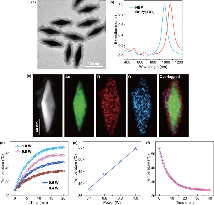
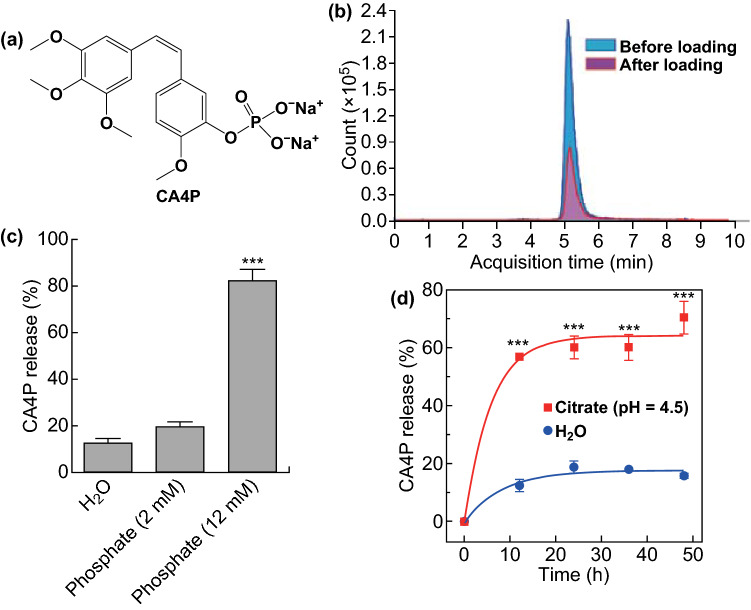
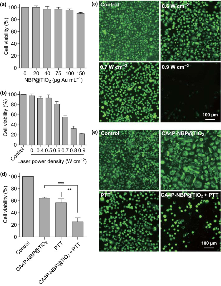
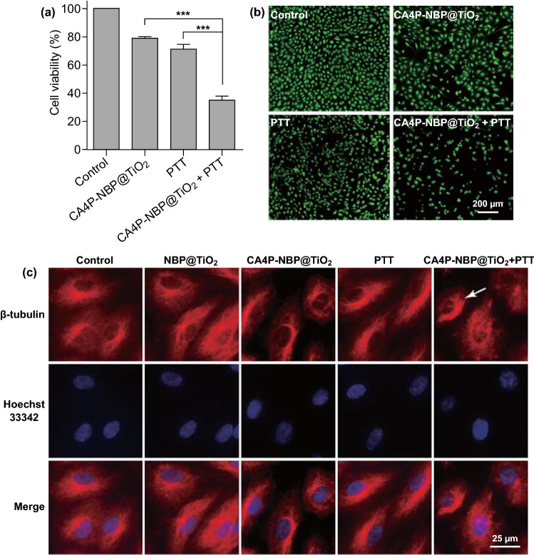
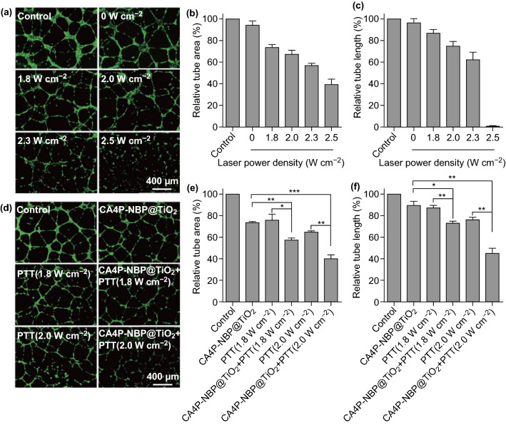
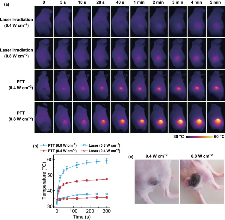
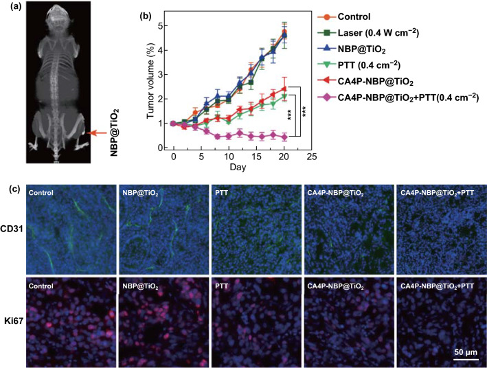
Photothermal agents with strong light absorption in the second near-infrared (NIR-II) region (1000-1350 nm) are strongly desired for successful photothermal therapy (PTT). In this work, titania-coated Au nanobipyramids (NBP@TiO2) with a strong plasmon resonance in the NIR-II window were synthesized. The NBP@TiO2 nanostructures have a high photothermal conversion efficiency of (93.3 ± 5.2)% under 1064-nm laser irradiation. They are also capable for loading an anticancer drug combretastatin A-4 phosphate (CA4P). In vitro PTT studies reveal that 1064-nm laser irradiation can efficiently ablate human lung cancer A549 cells and enhance the anticancer effect of CA4P. Moreover, the CA4P-loaded NBP@TiO2 nanostructures combined with PTT induce a synergistic antiangiogenesis effect. In vivo studies show that such CA4P-loaded NBP@TiO2 nanostructures under mild 1064-nm laser irradiation at an optical power density of 0.4 W cm-2, which is lower than the skin tolerance threshold value, exhibit a superior antitumor effect. This work presents not only the development of the NBP@TiO2 nanostructures as a novel photothermal agent responsive in the NIR-II window but also a unique combined chemo-photothermal therapy strategy for cancer therapy.

Keywords: Antiangiogenesis therapy; Core@shell nanostructures; Gold nanobipyramids; Photothermal therapy; Plasmon resonance.


Figures

Fig. 1
Fig. 1
(Au NBP)@TiO2 nanostructures. a TEM image, b extinction spectra of the uncoated NBP and NBP@TiO2 nanostructures in aqueous solutions, c HAADF-STEM and elemental mapping images of a single NBP@TiO2. The rightmost image is the overlapped image of the Au, Ti, and O elemental maps, d temperature rise curves of the NBP@TiO2 nanostructures (2 mL, 120 μg mL−1) acquired under 1064-nm laser irradiation at different optical powers for 20 min, e variation of the reached plateau temperature with the laser power, f temperature decay curve. The data points (red circles) were measured during the cooling process after the NBP/TiO2 nanostructure solution (2 mL, 120 μg mL−1) was irradiated with a 1064-nm laser at 1.0 W for 20 min. The blue line was obtained from fitting. (Color figure online)
Fig. 2
Fig. 2
NBP@TiO2 nanostructures for CA4P loading. a Molecular structure of CA4P, b LC–MS chromatograms of CA4P for the initial CA4P solution and the supernatant solution after drug loading, c desorption of CA4P in phosphate solutions and H2O after 12-h incubation, d pH- and time-dependent CA4P release profiles for the CA4P-loaded NBP@TiO2 nanostructures. The CA4P-loaded NBP/TiO2 nanostructures (120 μg Au) were dispersed in phosphate solutions (2 and 12 mM PO43−, 1 mL), citrate buffer (20 mM, pH 4.5, 1 mL), or H2O (1 mL) and incubated at 37 °C. The CA4P release percentage was calculated by measuring the drug concentration in the supernatant. The shown data represent the mean ± SEM. ***P < 0.001
Fig. 3
Fig. 3
PTT in A549 cells. a Effect of the NBP@TiO2 nanostructures at different concentrations on the viability of A549 cells, b cell viabilities of A549 cells upon PTT at different optical power densities, c calcein AM staining of the cells treated by PTT at different optical power densities as in b. A549 cells were incubated with the NBP@TiO2 nanostructures (100 μg-Au mL‒1) for 24 h, followed by 1064-nm laser irradiation at 0.4‒0.9 W cm‒2 for 5 min. The live cells were stained with green fluorescence by calcein AM, d synergistic enhancement of the cytotoxicity of the CA4P-loaded NBP@TiO2 nanostructures by combined chemotherapy and PTT, e calcein AM staining of the A549 cells after the different therapy treatments as in d. A549 cells were treated with the CA4P-loaded NBP@TiO2 nanostructures (15 nM CA4P, 100 μg-Au mL‒1) or together with 1064-nm laser irradiation at 0.7 W cm‒2 for 5 min. The data shown represent the mean ± SEM. **P < 0.01, ***P < 0.001
Fig. 4
Fig. 4
PTT-enhanced cytotoxic effect of the CA4P-loaded NBP@TiO2 nanostructures in HUVECs. a MTT assay, b calcein AM staining. The HUVECs were treated with the CA4P-loaded NBP@TiO2 nanostructures (7 nM CA4P, 12 μg-Au mL−1) for 24 h and then exposed to 1064-nm laser irradiation at 1.8 W cm−2 for 3 min. The assay and staining were performed after further incubation for 24 h. The shown data represent the mean ± SEM. ***P < 0.001, c synergistic disruption of tubulin in the HUVECs by the combined therapy. Immunofluorescent staining with β-tubulin (red) was performed on the HUVECs treated with the CA4P-loaded NBP@TiO2 nanostructures (3 nM CA4P, 12 μg-Au mL−1) in conjunction with 1064-nm laser irradiation (1.3 W cm−2, 3 min). The nuclei were stained with Hoechst 33342 (blue). In the control group, the tubulin shows a filamentous morphology. CA4P treatment induces a disordered change of tubulin and rounding of the cell morphology. The combined treatment further disrupts tubulin distribution and cell morphology (indicated by the white arrow). (Color figure online)
Fig. 5
Fig. 5
Inhibition of the endothelial tube formation by the combined chemo-photothermal therapy using the CA4P-loaded NBP@TiO2 nanostructures. a Effect of PTT at different laser power densities (1.8–2.5 W cm−2) on the HUVEC tube formation, b relative tube areas estimated from a, c relative tube lengths estimated from a, d PTT-enhanced antiangiogenesis of the CA4P-loaded NBP@TiO2 nanostructures, e relative tube areas estimated from d, f relative tube lengths estimated from d. The HUVECs were seeded in a 48-well plate that was pre-coated with Matrigel and then treated with the NBP@TiO2 nanostructures (12 μg-Au mL−1) or the CA4P-loaded NBP@TiO2 nanostructures (7 nM CA4P, 12 μg-Au mL−1), followed by 1064-nm laser irradiation for 3 min. The tubular structures were visualized with calcein AM staining, and the relative tube areas and lengths were calculated. The as-shown results are the mean ± SEM. *P < 0.05, **P < 0.01, ***P < 0.001
Fig. 6
Fig. 6
In vivo PTT. a Infrared thermal images of the A549-bearing mice, b tumor temperature change curves extracted from a. The A549-bearing mice intratumorally injected with the NBP@TiO2 nanostructures were subjected under 1064-nm laser irradiation at 0.4 or 0.8 W cm−2 for 5 min. The mice without the administration of the NBP@TiO2 nanostructures were subjected to the laser irradiation as control. The as-shown results are the mean ± SEM, c photographs of the tumor captured at 48 h after PPT at 0.4 or 0.8 W cm−2
Fig. 7
Fig. 7
In vivo antitumor effect. a Typical three-dimensional reconstructed CT image of an A549 tumor-bearing mouse after intratumoral injection with the NBP@TiO2 nanostructures. The arrow head points to the tumor site, b tumor growth in the different groups at different time points. For the laser irradiation treatment groups, the mice were intratumorally injected with the NBP@TiO2 (25 mg-Au kg−1) or the CA4P-loaded NBP@TiO2 nanostructures (25 mg-Au kg−1, 2 mg CA4P kg−1). At 24 h after the injection, the mice were subjected to 1064-nm laser irradiation (0.4 W cm−2, 5 min). The as-shown results are the mean ± SEM. ***P < 0.001, n = 6, c immunofluorescence images. The immunofluorescence staining of the tumor sections was performed to image the tumor micro-vessel density with anti-CD31 (green) and to evaluate cell proliferation with anti-Ki67 (red) at the tumor center. The nuclei were stained with Hoechst 33342 (blue). (Color figure online)

References

    1. Shanmugam V, Selvakumar S, Yeh C-S. Near-infrared light-responsive nanomaterials in cancer therapeutics. Chem. Soc. Rev. 2014;43:6254–6587. doi: 10.1039/c4cs00011k. - DOI - PubMed
    1. Weissleder R. A clearer vision for in vivo imaging. Nat. Biotechnol. 2001;19:316–317. doi: 10.1038/86684. - DOI - PubMed
    1. Smith AM, Mancini MC, Nie SM. Bioimaging: second window for in vivo imaging. Nat. Nanotechnol. 2009;4:710–711. doi: 10.1038/nnano.2009.326. - DOI - PMC - PubMed
    1. Bashkatov AN, Genina EA, Kochubey VI, Tuchin VV. Optical properties of human skin, subcutaneous and mucous tissues in the wavelength range from 400 to 2000 nm. J. Phys D-Appl. Phys. 2005;38:2543–2555. doi: 10.1088/0022-3727/38/15/004. - DOI
    1. Ding XG, Liow CH, Zhang MX, Huang RJ, Li CY, et al. Surface plasmon resonance enhanced light absorption and photothermal therapy in the second near-infrared window. J. Am. Chem. Soc. 2014;136:15684–15693. doi: 10.1021/ja508641z. - DOI - PubMed

LinkOut - more resources