Review
. 2020 May 2;12(1):103.
doi: 10.1007/s40820-020-00423-3.

Metal-Organic Framework Nanocarriers for Drug Delivery in Biomedical Applications

Affiliations
Review

Metal-Organic Framework Nanocarriers for Drug Delivery in Biomedical Applications

Yujia Sun et al. Nanomicro Lett. .

Abstract

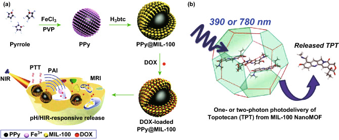
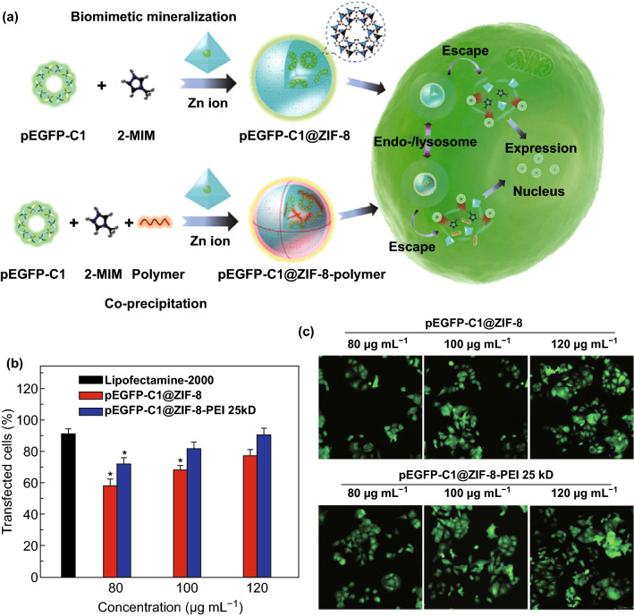
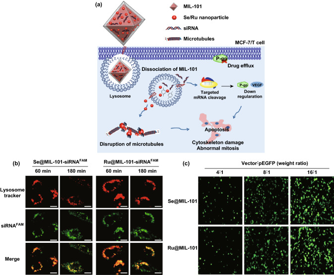
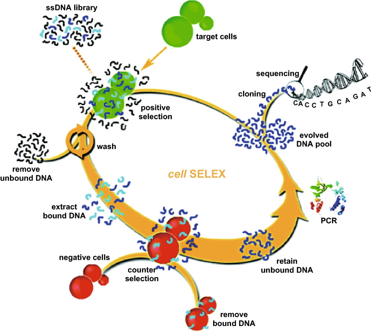
Investigation of metal-organic frameworks (MOFs) for biomedical applications has attracted much attention in recent years. MOFs are regarded as a promising class of nanocarriers for drug delivery owing to well-defined structure, ultrahigh surface area and porosity, tunable pore size, and easy chemical functionalization. In this review, the unique properties of MOFs and their advantages as nanocarriers for drug delivery in biomedical applications were discussed in the first section. Then, state-of-the-art strategies to functionalize MOFs with therapeutic agents were summarized, including surface adsorption, pore encapsulation, covalent binding, and functional molecules as building blocks. In the third section, the most recent biological applications of MOFs for intracellular delivery of drugs, proteins, and nucleic acids, especially aptamers, were presented. Finally, challenges and prospects were comprehensively discussed to provide context for future development of MOFs as efficient drug delivery systems.

Keywords: Biomedical applications; Biomolecules; Drug delivery; Drugs; Metal–organic frameworks.


Figures

Fig. 1
Fig. 1
a Schematic illustration of fabricating an integrated electrochemical biosensor for glucose using ZIF-70 as the matrix to co-immobilize MG and GDH onto the surface of electrode. Reproduced with permission from Ref. [51]. Copyright 2013, American Chemical Society. b Schematic illustration of ssDNA immobilization in Ni-IRMOF-74 series with precisely controlled channel size. Ni, C, and O atoms were labeled with green, gold, and red color, respectively. c Gradual increase in the interaction between ssDNA and MOFs with the increase in MOF channel size. Relatively weak interactions ensured the uptake, protection, and reversible release of ssDNA. Reproduced with permission from Ref. [53]. Copyright 2018, Nature Publishing Group
Fig. 2
Fig. 2
a One-step synthesis of ZIF-8 nanocrystals embedding multiple enzymes GOx and HRP. Reproduced with permission from Ref. [56]. Copyright 2015, The Royal Society of Chemistry. b Mechanism of Cyt c translocation into the MOF interior through relatively small windows. Reproduced with permission from Ref. [59]. Copyright 2012, American Chemical Society
Fig. 3
Fig. 3
a Activation of the 1D-polymer, [(Et2NH2)(In(pda)2)]n, with EDC or DCC to conjugate with EGFP. Reproduced with permission from Ref. [62]. Copyright 2011, The Royal Society of Chemistry. b DCC activation of MIL-88B-NH2(Cr) and following trypsin immobilization. Reproduced with permission from Ref. [63]. Copyright 2012, Wiley-VCH. c Schematic illustration of surface functionalization of UiO-66-N3 nanoparticles with DNA through a click reaction with DBCO-DNA. Reproduced with permission from Ref. [68]. Copyright 2014, American Chemical Society. d The crystal structure of bio-MOF-1. The MOF consists of zinc—adeninate columns linked together into a 3D framework by BPDC linkers to generate a material with 1D channels. Reproduced with permission from Ref. [77]. Copyright 2009, American Chemical Society
Fig. 4
Fig. 4
a Schematic illustration of the synthesis of PAA@ZIF-8 as the nanocarrier for DOX drug loading and pH-controlled release. b Drug release of DOX-loaded PAA@ZIF-8 at pH 5.5 and 7.4 at 37 °C. c In vitro cytotoxicity of PAA@ZIF-8, DOX-loaded PAA@ZIF-8, and free DOX against MCF-7 cells at different concentrations after 24 h. d CLSM images of MCF-7 cells incubated with DOX-loaded PAA@ZIF-8 ([DOX] = 20 μg mL−1) for 3 h (A–C), 12 h (D–F) and 24 h (G–I) at 37 °C, respectively. Columns 1–3 can be classified to cell nucleus (dyed in blue by Hoechst 33,342), DOX-loaded PAA@ZIF-8, and the merged images of both, respectively. All scale bars are 10 μm. Reproduced with permission from Ref. [34]. Copyright 2014, The Royal Society of Chemistry
Fig. 5
Fig. 5
a Encapsulation of autophagy inhibitor 3-MA into ZIF-8 nanoparticles. b Autophagic regulation proteins of xenograft tumor estimated by immunohistochemistry (scale bar: 200 μm). Reproduced with permission from Ref. [55]. Copyright 2017, American Chemical Society
Fig. 6
Fig. 6
a pH-induced one-pot synthesis of hierarchical ZIF-8 with encapsulated drug molecules. b Cell uptake studies conducted to compare the localizations of DOX@ZIF-8 and free DOX in the MDA-MB-468 cells. Reproduced with permission from Ref. [103]. Copyright 2015, American Chemical Society
Fig. 7
Fig. 7
a Schematic representation of pH-responsive ZIF-8 as a co-delivery system for overcoming MDR for efficient targeted cancer therapy: PEG-FA/(DOX + VER)@ZIF-8 synthesis; accumulation in tumors via EPR effect; internalization via FR-mediated endocytosis; pH-dependent drug release under weak acidic environments; VER-mediated MDR reversal. The biological ligand has been binded (physically touch the receptor) in order for receptor-mediated endocytosis to take place. b In vivo fluorescence imaging of B16F10 bearing mice at 1, 2, 4, 8, and 24 h after the injection of free IR820 or PEG-FA/IR820@ZIF-8. Reproduced with permission from Ref. [106]. Copyright 2017, American Chemical Society
Fig. 8
Fig. 8
a Drug loading and post-synthetic surface modification of MIL-101. b Multifunctional MIL-101 as a dual-responsive DDS for tumor-targeted drug delivery and cancer therapy. c Tumor volume change in H-22 tumor-bearing mice after treatment. d Images of the tumor after 12 days. Reproduced with permission from Ref [113]. Copyright 2015, The Royal Society of Chemistry
Fig. 9
Fig. 9
a Schematic illustration of the synthesis of PPy@MIL-100(Fe) as a pH/NIR-responsive drug carrier for dual-mode imaging and synergistic chemo-photothermal therapy. Reproduced with permission from Ref. [36]. Copyright 2017, The Royal Society of Chemistry. b MIL-100(Fe) NMOF for one- or two-photon-induced photodelivery of topotecan. Reproduced with permission from Ref. [39]. Copyright 2013, American Chemical Society
Fig. 10
Fig. 10
a Schematic illustration of the synthesis of pEGFP-C1@ZIF-8 via biomimetic mineralization and pEGFP-C1@ZIF-8-polymer via co-precipitation followed by cellular delivery and expression. b Transfection efficacy of pEGFP-C1@ZIF-8, pEGFP-C1@ZIF-8-PEI 25 kD, and lipofectamine-2000 at different concentrations. c Representative CLSM images of pEGFP-C1 expression in MCF-7 cells. Reproduced with permission from Ref. [127]. Copyright 2019, WILEY-VCH Verlag GmbH & Co. KGaA, Weinheim
Fig. 11
Fig. 11
a Mechanism of Se/Ru nanoparticles and siRNA co-delivery by MIL-101 for the reversal of drug resistance and induced apoptosis by the disruption of microtubule in MCF-7/T (Taxol-resistant) cancer cells. b Time-dependent confocal microscopy of siRNA escaped from endosomes in MCF-7/T cells. Scale bar: 5 μm. c Fluorescence microscope images of MCF-7/T cells transfected by Se@MIL-101 and Ru@MIL-101 for 24 h. Reproduced with permission from Ref. [130]. Copyright 2017, American Chemical Society
Fig. 12
Fig. 12
Schematic representation of the cell-SELEX approach for aptamer selection. Reproduced with permission from Ref. [139]. Copyright 2009, American Chemical Society
Fig. 13
Fig. 13
a Mechanism of glucose-driven release of VEGF aptamer from ZIF-8 caused by degradation of MOFs under local acidified conditions created by GOx-catalyzed aerobic oxidation of glucose to gluconic acid. b Confocal microscopy images of ZIF-8 loaded with Cy3-modified VEGF aptamer (I) and coumarin-functionalized GOx@ZIF-8 (II), and the bright field and merged image of the loaded MOF (III and IV, respectively). Reproduced with permission from Ref. [149]. Copyright 2018, American Chemical Society
Fig. 14
Fig. 14
a Schematic illustration of the synthesis of ZIF-90/protein nanoparticles and ATP-triggered protein release in the cell. b Cellular uptake efficiency of ZIF-90/GFP. c Cellular uptake efficiency of ZIF-90/GFP in the presence of different endocytosis inhibitors. d CLSM images of HeLa cells treated with ZIF-90/GFP. LysoTracker Red was used for endosome/lysosome staining. Scale bar: 10 μm. Reproduced with permission from Ref. [153]. Copyright 2019, American Chemical Society

References

    1. Meek ST, Greathouse JA, Allendorf MD. Metal–organic frameworks: a rapidly growing class of versatile nanoporous materials. Adv. Mater. 2011;23:249–267. doi: 10.1002/adma.201002854. - DOI - PubMed
    1. Zhou H-C, Long JR, Yaghi OM. Introduction to Metal–organic frameworks. Chem. Rev. 2012;112:673–674. doi: 10.1021/cr300014x. - DOI - PubMed
    1. Furukawa H, Cordova KE, O’Keeffe M, Yaghi OM. The Chemistry and applications of metal–organic frameworks. Science. 2013;341:1230444. doi: 10.1126/science.1230444. - DOI - PubMed
    1. Yaghi OM, Li G, Li H. Selective binding and removal of guests in a microporous metal–organic framework. Nature. 1995;378:703–706. doi: 10.1038/378703a0. - DOI
    1. Li H, Eddaoudi M, O'Keeffe M, Yaghi OM. Design and synthesis of an exceptionally stable and highly porous metal–organic framework. Nature. 1999;402:276–279. doi: 10.1038/46248. - DOI

LinkOut - more resources