. 2020 Feb 4;12(1):47.
doi: 10.1007/s40820-020-0384-8.

Fucoidan-Based Theranostic Nanogel for Enhancing Imaging and Photodynamic Therapy of Cancer

Affiliations

Fucoidan-Based Theranostic Nanogel for Enhancing Imaging and Photodynamic Therapy of Cancer

Mi Hyeon Cho et al. Nanomicro Lett. .

Abstract

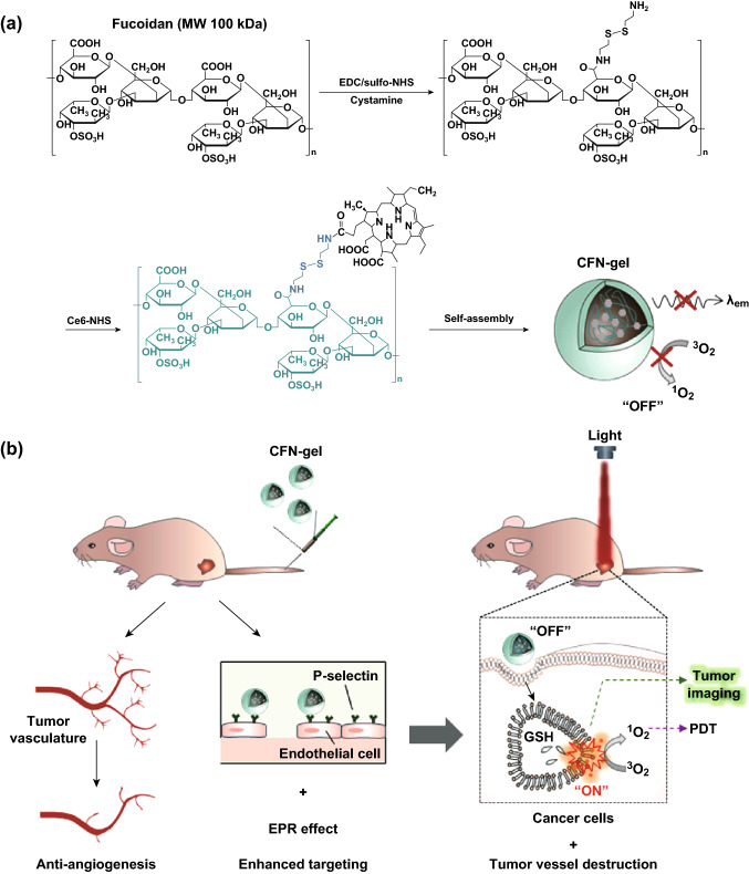
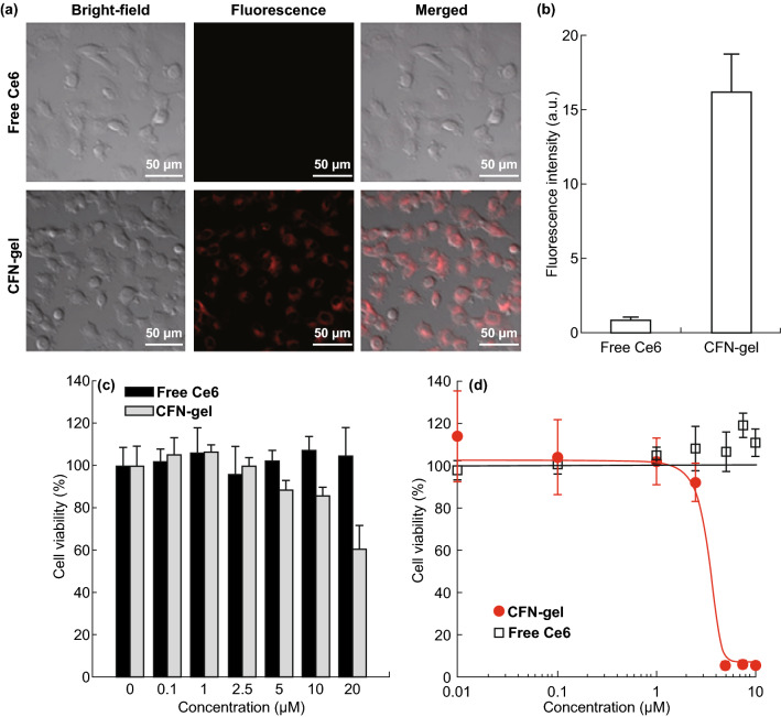
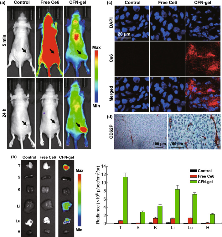
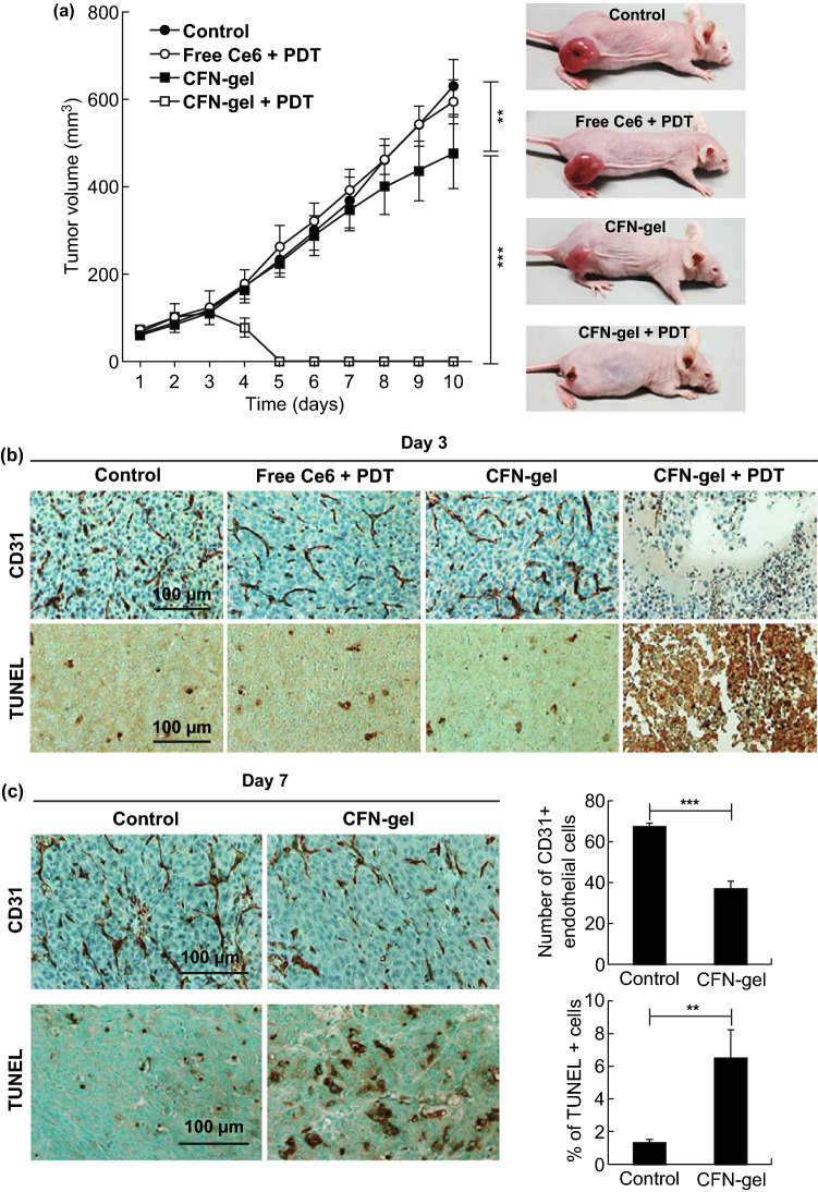
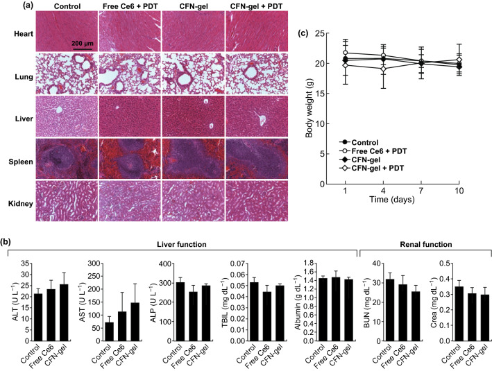
In this study, a fucoidan-based theranostic nanogel (CFN-gel) consisting of a fucoidan backbone, redox-responsive cleavable linker and photosensitizer is developed to achieve activatable near-infrared fluorescence imaging of tumor sites and an enhanced photodynamic therapy (PDT) to induce the complete death of cancer cells. A CFN-gel has nanomolar affinity for P-selectin, which is overexpressed on the surface of tumor neovascular endothelial cells as well as many other cancer cells. Therefore, a CFN-gel can enhance tumor accumulation through P-selectin targeting and the enhanced permeation and retention effect. Moreover, a CFN-gel is non-fluorescent and non-phototoxic upon its systemic administration due to the aggregation-induced self-quenching in its fluorescence and singlet oxygen generation. After internalization into cancer cells and tumor neovascular endothelial cells, its photoactivity is recovered in response to the intracellular redox potential, thereby enabling selective near-infrared fluorescence imaging and an enhanced PDT of tumors. Since a CFN-gel also shows nanomolar affinity for the vascular endothelial growth factor, it also provides a significant anti-tumor effect in the absence of light treatment in vivo. Our study indicates that a fucoidan-based theranostic nanogel is a new theranostic material for imaging and treating cancer with high efficacy and specificity.

Keywords: Activatable; Anti-angiogenic; Fucoidan; P-selectin; Theranostic nanogel.


Figures

Fig. 1
Fig. 1
a Synthetic steps of Ce6–fucoidan theranostic nanogel (CFN-gel), b schematic illustration of CFN-gel and its mode of action
Fig. 2
Fig. 2
Characterization of CFN-gel. a Hydrodynamic size distribution of CFN-gel, b TEM image of CFN-gel, c UV/Vis absorption spectra of CFN-gels in surfactant-containing (0.1 M NaOH/1% SDS) (solid line) and PBS (dotted line) solutions at a 2 µM Ce6 equivalent, d fluorescence spectra (λex 400 nm) of CFN-gel in surfactant-containing (solid line) and PBS (dotted line) solutions at 2 µM Ce6 equivalent
Fig. 3
Fig. 3
Redox-responsive turn-on of fluorescence signal and singlet oxygen generation of CFN-gel. a Fluorescence spectra of CFN-gel treated with DTT (0, 5 µM or 5 mM) for 4 h (λex = 400 nm), b time-dependent increase in SOSG fluorescence of DTT-treated CFN-gel during 670-nm laser irradiation (n = 4)
Fig. 4
Fig. 4
Surface plasmon resonance (SPR) analysis. a Sensorgram of SPR for evaluating the binding affinity between fucoidan and VEGF 165 (equilibrium dissociation rate constant (KD) = 953.0 nM) and CFN-gel and VEGF 165 (KD = 756.1 nM), b sensorgram of SPR for evaluating the binding affinity between fucoidan and P-selectin (KD = 69.71 nM), and between CFN-gel and P-selectin (KD = 718.9 nM)
Fig. 5
Fig. 5
a NIR fluorescence images of free Ce6 and CFN-gel-treated HT1080 cancer cells. The HT1080 cancer cells were treated with free Ce6 or CFN-gel at a 2 µM Ce6 equivalent for 6 h. After washing the cells, NIR fluorescence images (λex = 633 nm, λem = 650, using a long-pass filter) were obtained. The red color indicates the fluorescence signals generated from Ce6, b quantitative analysis of fluorescence intensities in the free Ce6 and CFN-gel-treated HT1080 cells (n = 4), c in vitro dark cytotoxicity of the HT1080 cells after treatment with free Ce6 and CFN-gel at various concentrations, d in vitro phototoxicity of the HT1080 cells after treatment with free Ce6 and CFN-gel at various concentrations upon 670-nm light irradiation (50 mW cm−2, 10 J cm−2). The IC50 of the CFN-gel is 2.73 µM. (Color figure online)
Fig. 6
Fig. 6
In vivo evaluation of selective tumor imaging in xenograft tumor models. a NIR fluorescence images of PBS-, free Ce6- or CFN-gel-treated mice. After 5 min and 24 h of intravenous injection of PBS (100 μL), free Ce6 (5 mg Ce6 kg−1) or a CFN-gel (5 mg Ce6 equiv. kg−1), NIR fluorescence images of the HT1080 tumor-bearing mice were obtained (λex = 660/20 nm, λem = 710/40 nm), b left: ex vivo NIR fluorescence images of the tumors and major organs (T: tumor, S: spleen, K: kidney, Li: liver, Lu: lung, H: heart) at 24 post-injection. Right: quantitative analysis comparison of fluorescent intensity in the tissues (n = 3 per group), c confocal fluorescence microscopic images of tumor sections prepared 24 h post-injection. Nuclei in the tumor sections were stained using DAPI. The fluorescence of DAPI (λex = 405 nm, λem = 420–480 nm) and Ce6 (λex = 633 nm, λem = 647–754 nm) were pseudocolored in blue and red, respectively, d immunohistochemical staining images of CD62P expression (i.e., P-selectin) in the tumor sections. (Color figure online)
Fig. 7
Fig. 7
Anti-tumor activity of CFN-gel combined with PDT in vivo. a Left: tumor growth curves of mice. Control group (n = 7), free Ce6 + PDT (n = 5), CFN-gel (n = 7) and CFN-gel + PDT (n = 5). PBS, free Ce6 or Ce6–fucoidan was injected intravenously on day 1. Light illumination (670 nm, 50 mW cm−2, 20 J cm−2) of the tumors was conducted on day 2 for PDT. Right: representative photographs of the mice obtained on day 10, b immunohistochemical staining of CD31 (for tumor blood vessels) and TUNEL (for apoptotic cell death) of tumor sections collected from different groups of mice at 24 h after PDT, c left: micrographs of immunohistochemical staining of CD31 and TUNEL-stained tumor sections collected from control and CFN-gel groups of mice at day 7. Right: quantitative analysis of CD31 and TUNEL assays in the tumor sections. **P < 0.01, ***P < 0.001
Fig. 8
Fig. 8
In vivo biocompatibility of CFN-gel. a Representative H&E-stained images of major organs collected from different treatment groups on day 10, b blood chemistry data of mice in the PBS-, free Ce6- and CFN-gel-treated groups on day 10 (n = 4), c change in average body weight of mice in different groups over time. No apparent signs of toxicity in organs, blood biomarkers and body weights were observed in any treatment group

References

    1. Dolmans DE, Fukumura D, Jain RK. Photodynamic therapy for cancer. Nat. Rev. Cancer. 2003;3:380–387. doi: 10.1038/nrc1071. - DOI - PubMed
    1. Celli JP, Spring BQ, Rizvi I, Evans CL, Samkoe KS, Verma S, Poque BW, Hasan T. Imaging and photodynamic therapy: mechanisms, monitoring, and optimization. Chem. Rev. 2010;110:2795–2838. doi: 10.1021/cr900300p. - DOI - PMC - PubMed
    1. Juzeniene A. Chlorin e6-based photosensitizers for photodynamic therapy and photodiagnosis. Photodiagn. Photodyn. Ther. 2009;6:94–96. doi: 10.1016/j.pdpdt.2009.06.001. - DOI - PubMed
    1. Fernandez JM, Bilgin MD, Grossweiner LI. Singlet oxygen generation by photodynamic agents. J. Photochem. Photobiol. B. 1997;33:131–140. doi: 10.1016/S1011-1344(96)07349-6. - DOI
    1. Bonnett R. Photosensitizers of the porphyrin and phthalocyanine series for photodynamic therapy. Chem. Soc. Rev. 1995;24:19–33. doi: 10.1039/CS9952400019. - DOI

LinkOut - more resources