

Effect of maternal oxytocin on umbilical venous and arterial blood flows during physiological-based cord clamping in preterm lambs
- PMID: 34138957
- PMCID: PMC8211207
- DOI: 10.1371/journal.pone.0253306
Effect of maternal oxytocin on umbilical venous and arterial blood flows during physiological-based cord clamping in preterm lambs
Abstract
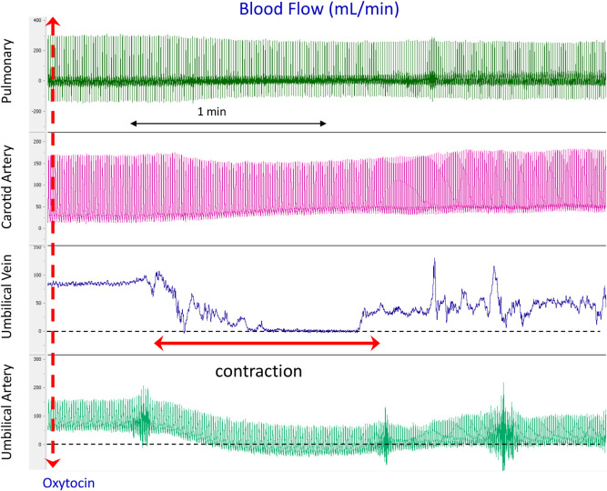
Background: Delayed umbilical cord clamping (UCC) after birth is thought to cause placental to infant blood transfusion, but the mechanisms are unknown. It has been suggested that uterine contractions force blood out of the placenta and into the infant during delayed cord clamping. We have investigated the effect of uterine contractions, induced by maternal oxytocin administration, on umbilical artery (UA) and venous (UV) blood flows before and after ventilation onset to determine whether uterine contractions cause placental transfusion in preterm lambs.
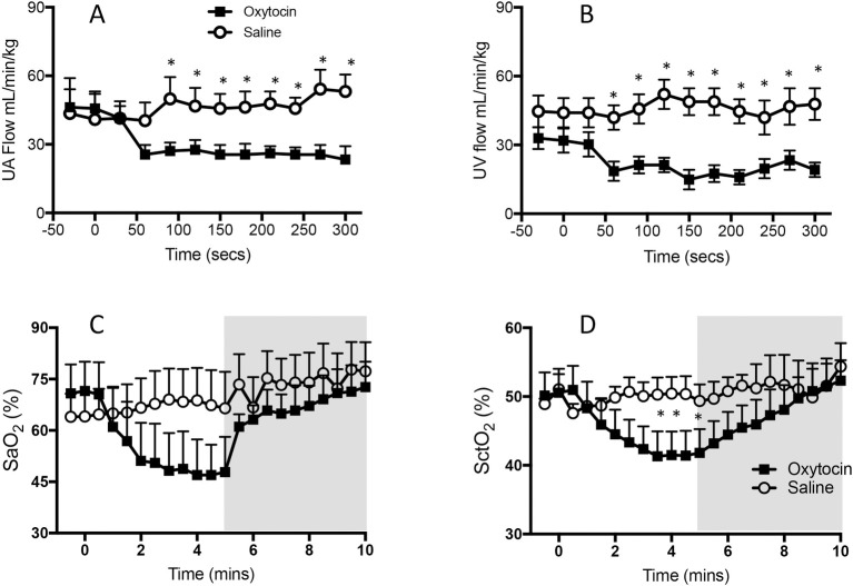
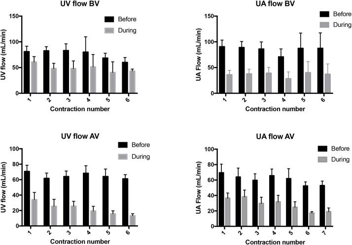
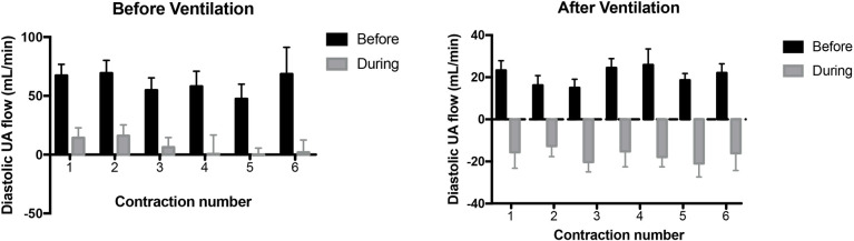
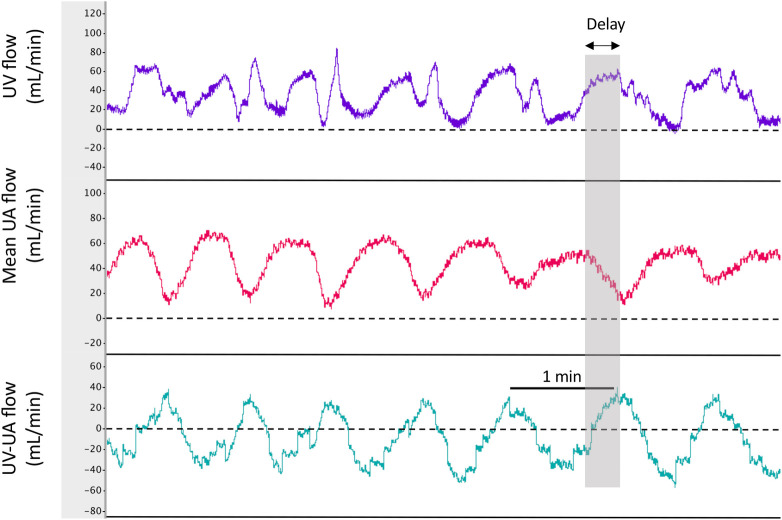
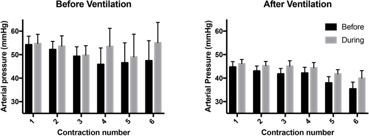
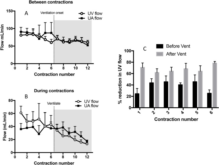
Methods and findings: At ~128 days of gestation, UA and UV blood flows, pulmonary arterial blood flow (PBF) and carotid arterial (CA) pressures and blood flows were measured in three groups of fetal sheep during delayed UCC; maternal oxytocin following mifepristone, mifepristone alone, and saline controls. Each successive uterine contraction significantly (p<0.05) decreased UV (26.2±6.0 to 14.1±4.5 mL.min-1.kg-1) and UA (41.2±6.3 to 20.7 ± 4.0 mL.min-1.kg-1) flows and increased CA pressure and flow (47.1±3.4 to 52.8±3.5 mmHg and 29.4±2.6 to 37.3±3.4 mL.min-1.kg-1). These flows and pressures were partially restored between contractions, but did not return to pre-oxytocin administration levels. Ventilation onset during DCC increased the effects of uterine contractions on UA and UV flows, with retrograde UA flow (away from the placenta) commonly occurring during diastole.
Conclusions: We found no evidence that amplification of uterine contractions with oxytocin increase placental transfusion during DCC. Instead they decreased both UA and UV flow and caused a net loss of blood from the lamb. Uterine contractions did, however, have significant cardiovascular effects and reduced systemic and cerebral oxygenation.
Conflict of interest statement
The authors have declared that no competing interests exist.
Figures
Similar articles
-
Effect of body position and ventilation on umbilical artery and venous blood flows during delayed umbilical cord clamping in preterm lambs.Arch Dis Child Fetal Neonatal Ed. 2017 Jul;102(4):F312-F319. doi: 10.1136/archdischild-2016-311159. Epub 2016 Nov 8. Arch Dis Child Fetal Neonatal Ed. 2017. PMID: 27827796 Free PMC article.
-
Umbilical cannulation optimizes circuit flows in premature lambs supported by the EXTra-uterine Environment for Neonatal Development (EXTEND).J Physiol. 2018 May 1;596(9):1575-1585. doi: 10.1113/JP275367. Epub 2018 Mar 2. J Physiol. 2018. PMID: 29392729 Free PMC article.
-
Effect of spontaneous breathing on umbilical venous blood flow and placental transfusion during delayed cord clamping in preterm lambs.Arch Dis Child Fetal Neonatal Ed. 2020 Jan;105(1):26-32. doi: 10.1136/archdischild-2018-316044. Epub 2019 May 15. Arch Dis Child Fetal Neonatal Ed. 2020. PMID: 31092674 Free PMC article.
-
The timing of umbilical cord clamping at birth: physiological considerations.Matern Health Neonatol Perinatol. 2016 Jun 13;2:4. doi: 10.1186/s40748-016-0032-y. eCollection 2016. Matern Health Neonatol Perinatol. 2016. PMID: 27298730 Free PMC article. Review.
-
Italian Recommendations for Placental Transfusion Strategies.Front Pediatr. 2018 Dec 3;6:372. doi: 10.3389/fped.2018.00372. eCollection 2018. Front Pediatr. 2018. PMID: 30560107 Free PMC article. Review.
Cited by
-
Transitional circulation and hemodynamic monitoring in newborn infants.Pediatr Res. 2024 Aug;96(3):595-603. doi: 10.1038/s41390-022-02427-8. Epub 2023 Jan 2. Pediatr Res. 2024. PMID: 36593283 Free PMC article. Review.
-
Clinical Experiences and Mechanism of Action with the Use of Oxytocin Injection at Parturition in Domestic Animals: Effect on the Myometrium and Fetuses.Animals (Basel). 2023 Feb 20;13(4):768. doi: 10.3390/ani13040768. Animals (Basel). 2023. PMID: 36830555 Free PMC article. Review.
-
Physiological-Based Cord Clamping: When the Baby Is Ready for Clamping.Neonatology. 2024;121(5):547-552. doi: 10.1159/000540667. Epub 2024 Aug 28. Neonatology. 2024. PMID: 39197438 Free PMC article. Review.
References
-
- Crossley KJ, Allison BJ, Polglase GR, Morley CJ, Davis PG, Hooper SB. Dynamic changes in the direction of blood flow through the ductus arteriosus at birth. J Physiol. 2009;587(Pt 19):4695–704. Epub 2009/08/14. doi: 10.1113/jphysiol.2009.174870 ; PubMed Central PMCID: PMC2768022. - DOI - PMC - PubMed
-
- Rabe H, Diaz-Rossello JL, Duley L, Dowswell T. Effect of timing of umbilical cord clamping and other strategies to influence placental transfusion at preterm birth on maternal and infant outcomes. The Cochrane database of systematic reviews. 2012;8:CD003248. doi: 10.1002/14651858.CD003248.pub3 . - DOI - PubMed
Publication types
MeSH terms
Substances
Grants and funding
LinkOut - more resources
Full Text Sources
Miscellaneous
