. 2021 Jul 9;49(12):7179-7188.
doi: 10.1093/nar/gkab531.

Mechanical diversity and folding intermediates of parallel-stranded G-quadruplexes with a bulge

Affiliations

Mechanical diversity and folding intermediates of parallel-stranded G-quadruplexes with a bulge

Yashuo Zhang et al. Nucleic Acids Res. .

Abstract

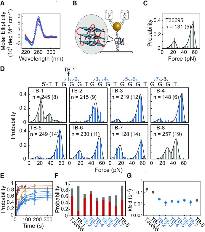
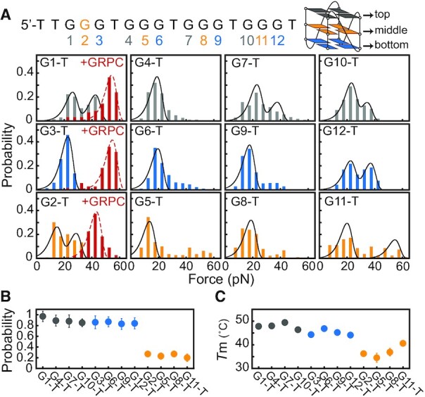
A significant number of sequences in the human genome form noncanonical G-quadruplexes (G4s) with bulges or a guanine vacancy. Here, we systematically characterized the mechanical stability of parallel-stranded G4s with a one to seven nucleotides bulge at various positions. Our results show that G4-forming sequences with a bulge form multiple conformations, including fully-folded G4 with high mechanical stability (unfolding forces > 40 pN), partially-folded intermediates (unfolding forces < 40 pN). The folding probability and folded populations strongly depend on the positions and lengths of the bulge. By combining a single-molecule unfolding assay, dimethyl sulfate (DMS) footprinting, and a guanine-peptide conjugate that selectively stabilizes guanine-vacancy-bearing G-quadruplexes (GVBQs), we identified that GVBQs are the major intermediates of G4s with a bulge near the 5' or 3' ends. The existence of multiple structures may induce different regulatory functions in many biological processes. This study also demonstrates a new strategy for selectively stabilizing the intermediates of bulged G4s to modulate their functions.


Figures

Figure 1.
Figure 1.
Mechanical stability of G4s bearing a 1 nt bulge. (A) CD spectra of T30695 (red) and TB-1 to TB-8 DNA (cyan). (B) Schematic of the magnetic tweezers experiments. The G4-forming sequence is sandwiched between two dsDNA handles. (C) Unfolding force distribution of T30695. (D) Unfolding force distributions of TB-1 to TB-8. Data were fitted by Bell's model. n represents the total number of unfolding events, where the number in the bracket represents the total number of molecules of each sequence. (E) The time evolution of the folding probability pfold(t). Data represent the mean ± SD from three different DNA tethers. TB-1 (red), TB-8 (orange), TB-2 to TB-7 (cyan), and T30695 (gray). The TB-1 shows the fastest refolding among TB-1 to TB-8. (F) The steady-state folding probability pst. The red columns represent the fraction of fully-folded G4s (unfolding forces > 40 pN) and the gray columns represent the less stable states (unfolding forces < 40 pN). (G) Apparent folding rates kfold of T30695 and TB-1 to TB-8.
Figure 2.
Figure 2.
G4-forming sequences with a bulge near the 5′ or 3′ end mainly form GVBQs. (A) Unfolding force distributions of TB-1 to T7B-1 and TB-8 to T7B-8 (gray column). Stabilization of GVBQs by 0.5 μM GRPC (red columns). The total number of unfolding events and molecules are shown in Supplementary Table S3. (B) Average unfolding step sizes. (C) The guanine-RHAU23 peptide conjugate (GRPC) binds and stabilizes GVBQs. (D) DMS footprinting of the T3B-1 sequence in the presence of 0.5 μM GRPC.
Figure 3.
Figure 3.
Mechanical stability of GVBQs. (A) Unfolding force distributions of the G1-T to G12-T. The G-to-T substitutions at the top, bottom, and middle tetrads are shown in gray, blue, and orange columns, respectively. Stabilization of GVBQs by 0.5 μM GRPC (red columns). (B) The steady-state folding probability pst. (C) Melting temperatures (Tms) of G1-T to G12-T DNA measured in 20 mM KCl.
Figure 4.
Figure 4.
Mechanical diversity of G4s with a bulge in the middle. (A, B) Unfolding step sizes and unfolding force distributions of TB-2 to T7B-2 (A) and TB-3 to T7B-3 (B). The red lines and arrows present the total number of nucleotides in fully-folded G4s. (C) Unfolding force distribution of T2B-3 measured in the presence of 0.5 μM GRPC (red columns). (D) The steady-state folding probability pst. The data of T7B-2 and T7B-3 represent the folding probability measured at 300 s. The red columns represent the fraction of fully-folded G4s (unfolding forces > 40 pN), and the gray columns represent the less stable states (unfolding forces < 40 pN).
Figure 5.
Figure 5.
G4-forming sequences with a bulge can form multiple structures, including fully-folded G4s, partially-folded G4s, and misfolded. Sequences with a bulge near the 5′ and 3′ end mainly form partially-folded intermediates and slowly convert to fully-folded G4s (upper panel). The major intermediates are GVBQs which can be selectively stabilized by GRPC. A ≥2 nt middle bulge significantly reduced the folding probability and increase the unfolded or misfolded fractions (lower panel).

References

    1. Gellert M., Lipsett M.N., Davies D.R.. Helix formation by guanylic acid. Proc. Natl Acad. Sci. U.S.A. 1962; 48:2013–2018. - PMC - PubMed
    1. Sen D., Gilbert W.. Formation of parallel four-stranded complexes by guanine-rich motifs in DNA and its implications for meiosis. Nature. 1988; 334:364–366. - PubMed
    1. Davis J.T. G-quartets 40 years later: from 5′-GMP to molecular biology and supramolecular chemistry. Angew. Chem. Int. Ed. 2004; 43:668–698. - PubMed
    1. Rhodes D., Lipps H.J.. G-quadruplexes and their regulatory roles in biology. Nucleic Acids Res. 2015; 43:8627–8637. - PMC - PubMed
    1. Hansel-Hertsch R., Di Antonio M., Balasubramanian S.. DNA G-quadruplexes in the human genome: detection, functions and therapeutic potential. Nat. Rev. Mol. Cell Biol. 2017; 18:279–284. - PubMed

Publication types