. 2021 Jul 1;35(13-14):992-1004.
doi: 10.1101/gad.348508.121. Epub 2021 Jun 17.

Epitranscriptomic addition of m6A regulates HIV-1 RNA stability and alternative splicing

Affiliations

Epitranscriptomic addition of m6A regulates HIV-1 RNA stability and alternative splicing

Kevin Tsai et al. Genes Dev. .

Abstract

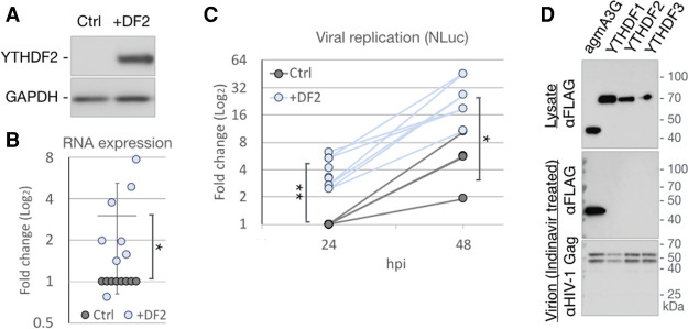
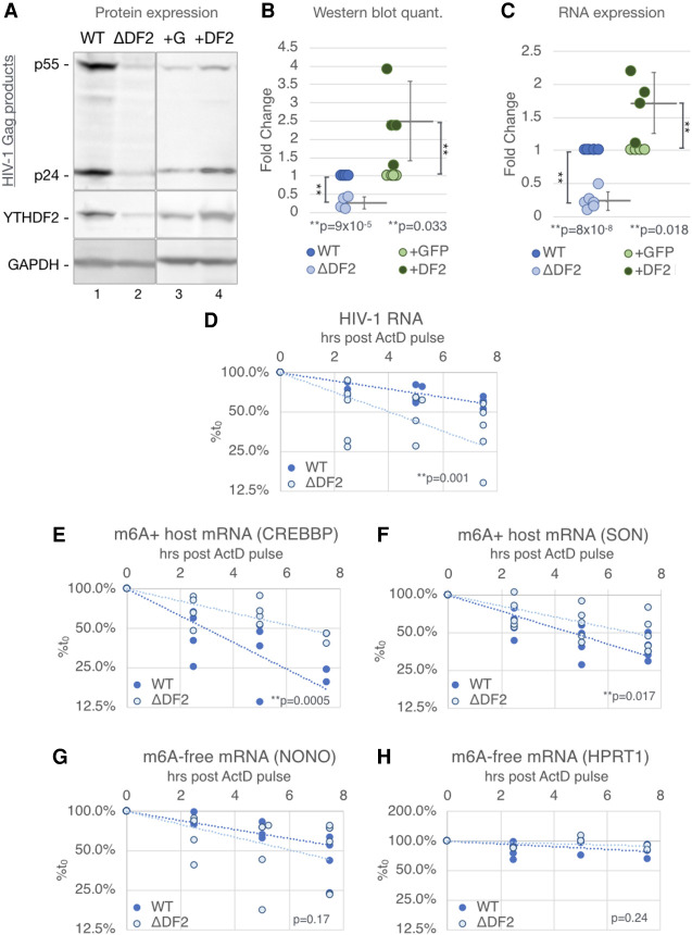
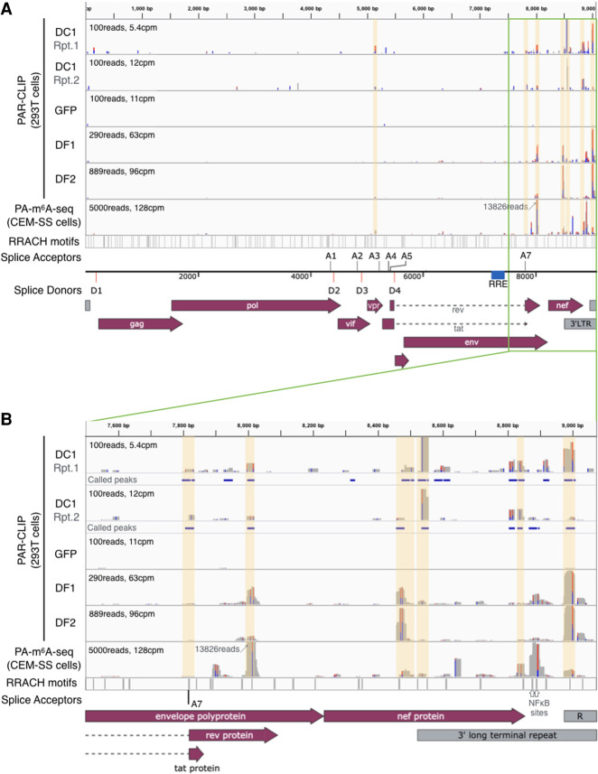
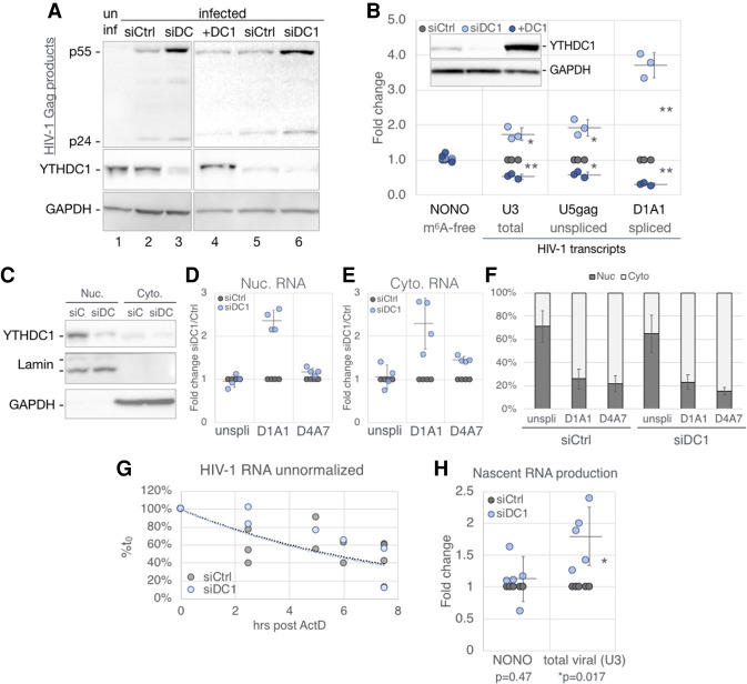
Previous work has demonstrated that the epitranscriptomic addition of m6A to viral transcripts can promote the replication and pathogenicity of a wide range of DNA and RNA viruses, including HIV-1, yet the underlying mechanisms responsible for this effect have remained unclear. It is known that m6A function is largely mediated by cellular m6A binding proteins or readers, yet how these regulate viral gene expression in general, and HIV-1 gene expression in particular, has been controversial. Here, we confirm that m6A addition indeed regulates HIV-1 RNA expression and demonstrate that this effect is largely mediated by the nuclear m6A reader YTHDC1 and the cytoplasmic m6A reader YTHDF2. Both YTHDC1 and YTHDF2 bind to multiple distinct and overlapping sites on the HIV-1 RNA genome, with YTHDC1 recruitment serving to regulate the alternative splicing of HIV-1 RNAs. Unexpectedly, while YTHDF2 binding to m6A residues present on cellular mRNAs resulted in their destabilization as previously reported, YTHDF2 binding to m6A sites on HIV-1 transcripts resulted in a marked increase in the stability of these viral RNAs. Thus, YTHDF2 binding can exert diametrically opposite effects on RNA stability, depending on RNA sequence context.

Keywords: HIV-1; RNA; RNA stability; YTHDC1; YTHDF2; epitranscriptomic regulation; splicing.


Figures

Figure 1.
Figure 1.
Global depletion of m6A in T cells suppresses HIV-1 RNA and protein expression. Three separate clones of the CD4+ CEM T-cell line overexpressing ALKBH5 (A1, A3, and A12) were compared with WT CEM cells (Ctrl). (A) HIV-1 Gag expression was analyzed by Western blot at 2 dpi after single cycle HIV-1 infection of + ALKBH5 CEM cells or control cells (one WT and the others expressing the irrelevant GFP-targeted Cas9 [ΔG]). Also analyzed were ALKBH5 (the overexpressed epitope tagged ALKBH5 runs slightly slower than endogenous ALKBH5) and GAPDH, as a loading control. (B) ALKBH5 band intensities from A were quantified and are shown relative to control cells, set at 1. (C) The m6A content of poly(A)+ mRNAs from Ctrl and +ALKBH5 cells was quantified by ELISA (n = 5). (D) Quantification of the HIV-1 Gag band intensities (p24 + p55) from A. (E) Aliquots of the samples from C were analyzed for viral RNA expression levels by qRT-PCR. The m6A-free cellular transcript (NONO) served as a negative control. Statistical analyses by two-tailed Student's t-test. Error bars indicate SD. (**) P < 0.01.
Figure 2.
Figure 2.
YTHDF2 enhances HIV-1 replication, yet is not packaged into virions. (AC) 293T cells, transfected with either empty vector (Ctrl) or a YTHDF2 expression plasmid (+DF2), were infected with NL-NLuc reporter virus and collected at 24 hpi for Western blot detection of overexpressed YTHDF2 (A) or qRT-PCR analysis of viral RNA expression (B). n = 8. (C) Quantification of virally encoded NLuc as a measure of viral replication at 24 hpi (n = 8) and 48 hpi (n = 4). (D) 293T cells were cotransfected with NL-NLuc along with expression vectors expressing FLAG-tagged YTHDF readers or agmA3G and treated with the HIV-1 protease inhibitor Indinavir. Lysates of the virus producer cells (top panel) and purified virions from the supernatant media (two bottom panels) were analyzed by Western blot for the FLAG-tagged YTHDF or agmA3G proteins and the HIV-1 Gag protein. Statistical analysis by two-tailed Student's t-test. (*) P < 0.05, (**) P < 0.01. . Error bars indicate SD.
Figure 3.
Figure 3.
The cytosolic m6A reader YTHDF2 enhances HIV-1 RNA stability while destabilizing m6A+ host mRNAs in CD4+ T cells. YTHDF2 knockout CD4+ T cells (ΔDF2), WT control cells (WT), and cells transduced with a lentiviral YTHDF2 or GFP expression vector (+DF2 or +G) were used for the following single-cycle infection assays. (A) Viral Gag protein expression levels analyzed by Western blot. (B) Quantification of protein band intensities of Western blots as shown in A (n = 4). (C) Viral RNA expression assayed by qRT-PCR (WT & ΔYTHDF2 n = 6, +GFP & +YTHDF2 n = 5). (DH) Stability of RNA transcripts assayed by treating infected WT or ΔDF2 cells at 2 dpi with actinomycin D (ActD), quantifying the transcript at the indicated time points by qRT-PCR, shown as percentage of the RNA level at time point 0. (D) Stability of HIV-1 transcripts. (E,F) Stability of host m6A+ transcripts known to be destabilized by YTHDF2, including CREBBP and SON. (G,H) Stability of m6A-free host transcripts NONO and HPRT1. Statistical analysis of data in B and C used the two-tailed Student's t-test. Error bars indicate SD. For D–H, slopes of regression lines were compared by ANCOVA, n = 4–5.
Figure 4.
Figure 4.
The nuclear m6A reader YTHDC1 binds HIV-1 RNA at previously mapped m6A sites. PAR-CLIP was performed on 293T cells transfected with FLAG-GFP or FLAG-YTHDC1, and infected with HIV-1. Sequencing reads were mapped to the HIV-1 genome, with two independent repeats of YTHDC1 PAR-CLIP (DC1 lanes) shown alongside previously published YTHDF1 and YTHDF2 PAR-CLIP (DF1 and DF2 lanes) and m6A mapping results (PA-m6A-seq lane) (Kennedy et al. 2016; Courtney et al. 2019b). A schematic of the HIV-1 genome is shown in A, with the 3′ end (3′ of 7500 bp) shown in B where most m6A sites are located. Locations of the m6A motif 5′-RRACH-3′ are shown in the bottom lane, with the location of viral splice donors and acceptors indicated. Significant YTHDC1 peaks called by PARalyzer are shown as blue bars below each DC1 lane in B. PARalyzer-called peaks that overlap with m6A sites and 5′-RRACH-3′ motifs are highlighted in yellow.
Figure 5.
Figure 5.
YTHDC1 regulates HIV-1 gene expression with no detectable effect on RNA nuclear export. 293T cells transfected with nontargeting (siCtrl, gray) or YTHDC1-targeting (siDC1, light blue) siRNAs or were transfected with a YTHDC1 expression vector (+DC1, dark blue) or empty vector. HIV-1 single-cycle infections were performed for the following analyses: (A) Viral Gag protein expression assayed by Western blot, costained with a YTHDC1 antibody. (B) Viral RNA levels assayed by qRT-PCR, n = 3, with the m6A-free host NONO mRNA as a control. A representive Western blot shown in the top left inset depicts the validation of YTHDC1 levels for samples used in this panel. (CF) Subcellular fractionation assay of infected siCtrl and siDC1 cells. (C) Western blot validation of fractionation, stained for YTHDC1, nuclear Lamin A/C, and cytosolic GAPDH. (DF) HIV-1 transcript alternatively spliced isoforms were quantified by qRT-PCR with primers targeting unspliced (unspli, U5-gag), and the D1/A1 and D4/A7 splice junctions, calculated as fold change of siDC1 over siCtrl in the nuclear (D), and cytosolic fraction (E). (F) The same RNA quantification as in D and E calculated as percent nuclear and percent cytoplasmic. (G) Stability of RNA transcripts in siCtrl and siDC1 cells assayed by treating infected cells at 2 dpi with ActD and the viral RNA levels from the indicated time points analyzed by qRT-PCR and shown as percentage of the RNA level at time point 0. (H) Production of nascent HIV-1 transcripts in infected siCtrl and siDC1 cells was measured by pulsing cells with the nucleoside analog 4SU for 1.5 h. The 4SU+ transcripts were then isolated and quantified by qRT-PCR. Statistical analysis used Student's t-test. Error bars indicate SD. (*) P < 0.05, (**) P < 0.01.
Figure 6.
Figure 6.
YTHDC1 regulates the alternative splicing of HIV-1 RNAs. Viral transcript spliced isoforms in infected siCtrl, siDC1, and +DC1 cells 48 hpi were analyzed by PrimerID RNA-seq. n = 3. (A) Schematic of HIV-1 splice donors and acceptors relative to the three major classes of spliced viral RNAs. Top arrows depict the primers used to amplify sequences for RNA-seq, including the common 5′ forward primer in blue, the reverse 4-kb primer in green, and the reverse 1.8-kb primer in red spanning the D4/A7 splice junction. A random reverse primer was also used but is not shown. (B) Spliced transcripts are given as a percentage of all transcripts (1.8-kb + 4-kb class read counts/total read counts). (C) Percent of fully spliced transcripts over all spliced transcripts (1.8-kb/[1.8-kb + 4-kb] read counts). (DF) Splice acceptor usage assayed using the common forward primer in conjunction with the random reverse primer (D), 1.8-kb reverse primer (E), and 4-kb reverse primer (F). Use of acceptor A3 results in long transcripts that are biased against when amplifying using the 1.8- or 4-kb reverse primers, A3 usage is thus only shown with the random reverse primer. (G) Percent occurrence of D1/A7 splices. (H) Percent occurrence of A2-using spliced RNAs that subsequently splice from D3. Statistical analysis used Student's t-test. Error bars indicate SD. (*) P < 0.05, (**) P < 0.01.

References

    1. Adachi A, Gendelman HE, Koenig S, Folks T, Willey R, Rabson A, Martin MA. 1986. Production of acquired immunodeficiency syndrome-associated retrovirus in human and nonhuman cells transfected with an infectious molecular clone. J Virol 59: 284–291. 10.1128/JVI.59.2.284-291.1986 - DOI - PMC - PubMed
    1. Alarcón CR, Goodarzi H, Lee H, Liu X, Tavazoie S, Tavazoie SF. 2015. HNRNPA2B1 is a mediator of m6A-dependent nuclear RNA processing events. Cell 162: 1299–1308. 10.1016/j.cell.2015.08.011 - DOI - PMC - PubMed
    1. Bartosovic M, Molares HC, Gregorova P, Hrossova D, Kudla G, Vanacova S. 2017. N6-methyladenosine demethylase FTO targets pre-mRNAs and regulates alternative splicing and 3′-end processing. Nucleic Acids Res 45: 11356–11370. 10.1093/nar/gkx778 - DOI - PMC - PubMed
    1. Bogerd HP, Doehle BP, Wiegand HL, Cullen BR. 2004. A single amino acid difference in the host APOBEC3G protein controls the primate species specificity of HIV type 1 virion infectivity factor. Proc Natl Acad Sci 101: 3770–3774. 10.1073/pnas.0307713101 - DOI - PMC - PubMed
    1. Bokar JA, Shambaugh ME, Polayes D, Matera AG, Rottman FM. 1997. Purification and cDNA cloning of the AdoMet-binding subunit of the human mRNA (N6-adenosine)-methyltransferase. RNA 3: 1233–1247. - PMC - PubMed

Publication types