. 2021 Jun 17;11(1):12818.
doi: 10.1038/s41598-021-92172-5.

A new deep learning algorithm of 12-lead electrocardiogram for identifying atrial fibrillation during sinus rhythm

Affiliations

A new deep learning algorithm of 12-lead electrocardiogram for identifying atrial fibrillation during sinus rhythm

Yong-Soo Baek et al. Sci Rep. .

Abstract

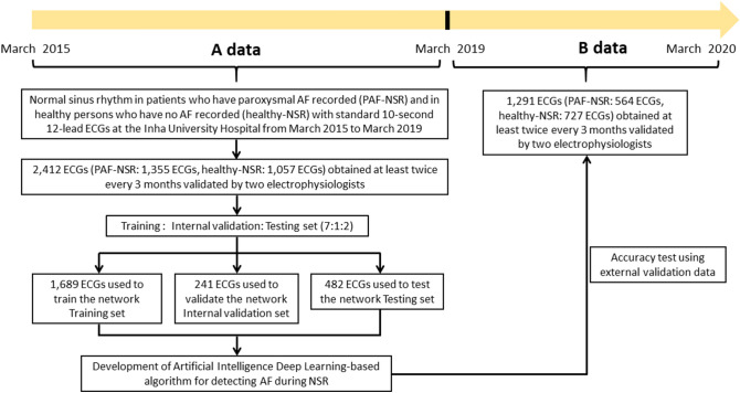
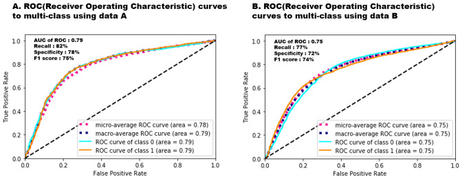
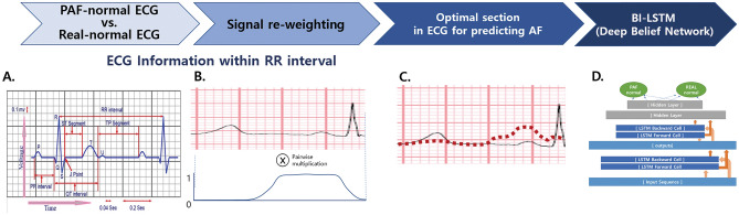
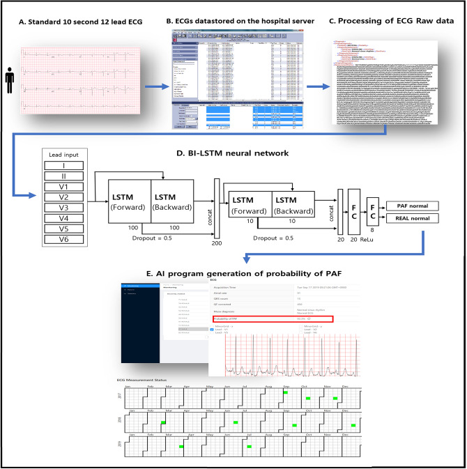
Atrial fibrillation (AF) is the most prevalent arrhythmia and is associated with increased morbidity and mortality. Its early detection is challenging because of the low detection yield of conventional methods. We aimed to develop a deep learning-based algorithm to identify AF during normal sinus rhythm (NSR) using 12-lead electrocardiogram (ECG) findings. We developed a new deep neural network to detect subtle differences in paroxysmal AF (PAF) during NSR using digital data from standard 12-lead ECGs. Raw digital data of 2,412 12-lead ECGs were analyzed. The artificial intelligence (AI) model showed that the optimal interval to detect subtle changes in PAF was within 0.24 s before the QRS complex in the 12-lead ECG. We allocated the enrolled ECGs to the training, internal validation, and testing datasets in a 7:1:2 ratio. Regarding AF identification, the AI-based algorithm showed the following values in the internal and external validation datasets: area under the receiver operating characteristic curve, 0.79 and 0.75; recall, 82% and 77%; specificity, 78% and 72%; F1 score, 75% and 74%; and overall accuracy, 72.8% and 71.2%, respectively. The deep learning-based algorithm using 12-lead ECG demonstrated high accuracy for detecting AF during NSR.


Conflict of interest statement

The authors declare no competing interests.

Figures

Figure 1
Figure 1
Patient flow diagram showing the selection of the study population and the creation of the study datasets. ECGs were allocated to the training, internal validation, and testing datasets in a 7:1:2 ratio to assure a robust and reliable dataset. PAF paroxysmal atrial fibrillation; ECG electrocardiogram.
Figure 2
Figure 2
Multiclass ROC curves with deep neural networks applied in the internal and external datasets. The micro-average and macro-average AUC derived from the ROC curve of the AI algorithm is calculated during the internal and external validations (0.78 [95% CI 0.76–0.80], 0.79 [95% CI 0.78–0.80] for internal validation dataset; 0.75 [95% CI 0.74–0.76], 0.75 [95% CI 0.74–0.76] for external validation dataset). ROC receiver operating characteristic; AUC area under the ROC curve. Class 0: healthy-NSR, Class 1: PAF-NSR.
Figure 3
Figure 3
The serial changes of the probability of PAF using AI deep learning algorithm program according to acquired ECG dates. A 72-year-old man diagnosed with PAF is calculated to have AF with probabilities of 90% and 100% by the AI program during normal sinus rhythm, observed after his AF episode had terminated (red lightning bolt). Moreover, he was calculated to have AF with probabilities of 6.3% and 10% when there was no AF symptom. AF symptom is defined as palpitations, fatigue, dizziness, dyspnea, chest pain, and anxiety during AF. AI artificial intelligence; ECG electrocardiogram; NSR normal sinus rhythm; PAF paroxysmal atrial fibrillation.
Figure 4
Figure 4
Optimal section for AF detection during NSR. (a) The experiment for the optimal sample size to identify AF was performed at a certain range of the R–R interval, where we reweighted the input EEG signal f(t) using the window function g(t) (b). The reweighted signal h(t) is computed by the equation h(t) = f(t) × g(t) and illustrated by the red dotted curve (c). This process clarifies the value ranges that particularly affect the trained model along the time-axis. The bi-directional connection is added so that the time flow can be considered in forward and backward passes, and long short-term memory is used to maintain a series of information in the short and long terms (d). NSR normal sinus rhythm, AF paroxysmal atrial fibrillation; LSTM long short-term memory.
Figure 5
Figure 5
Description of the artificial intelligence algorithm for detecting PAF. All raw ECG data were stored as XML documents using the MUSE data management system in a relational database server. PAF probability is calculated through our developed AI algorithm using an RNN with two-dimensional convolution (red box). AI artificial intelligence; ECG electrocardiogram; LSTM long short-term memory; PAF paroxysmal atrial fibrillation; RNN recurrent neural network.

References

    1. Kim D, et al. 10-year nationwide trends of the incidence, prevalence, and adverse outcomes of non-valvular atrial fibrillation nationwide health insurance data covering the entire Korean population. Am. Heart J. 2018;202:20–26. doi: 10.1016/j.ahj.2018.04.017. - DOI - PubMed
    1. Gladstone DJ, et al. Atrial fibrillation in patients with cryptogenic stroke. N. Engl. J. Med. 2014;370:2467–2477. doi: 10.1056/NEJMoa1311376. - DOI - PubMed
    1. Sanna T, et al. Cryptogenic stroke and underlying atrial fibrillation. N. Engl. J. Med. 2014;370:2478–2486. doi: 10.1056/NEJMoa1313600. - DOI - PubMed
    1. Freedman B, Schnabel R, Calkins H. Opportunistic electrocardiogram screening for atrial fibrillation to prevent stroke. JAMA Cardiol. 2019;4:91–92. doi: 10.1001/jamacardio.2018.4335. - DOI - PubMed
    1. Perez MV, et al. Large-scale assessment of a smartwatch to identify atrial fibrillation. N. Engl. J. Med. 2019;381:1909–1917. doi: 10.1056/NEJMoa1901183. - DOI - PMC - PubMed

Publication types