

Highlighting COVID-19: What the imaging exams show about the disease
- PMID: 34141092
- PMCID: PMC8188839
- DOI: 10.4329/wjr.v13.i5.122
Highlighting COVID-19: What the imaging exams show about the disease
Abstract
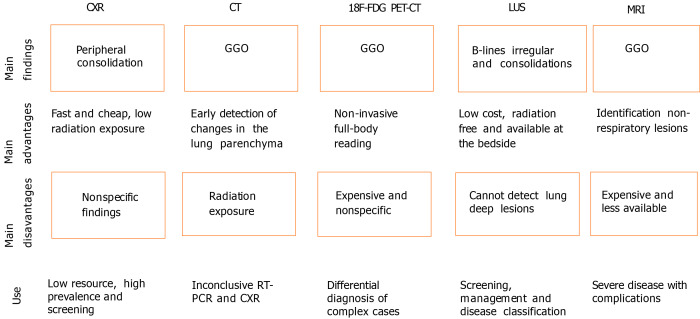
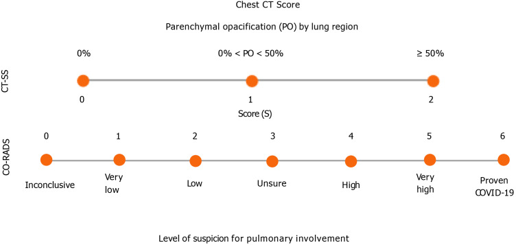
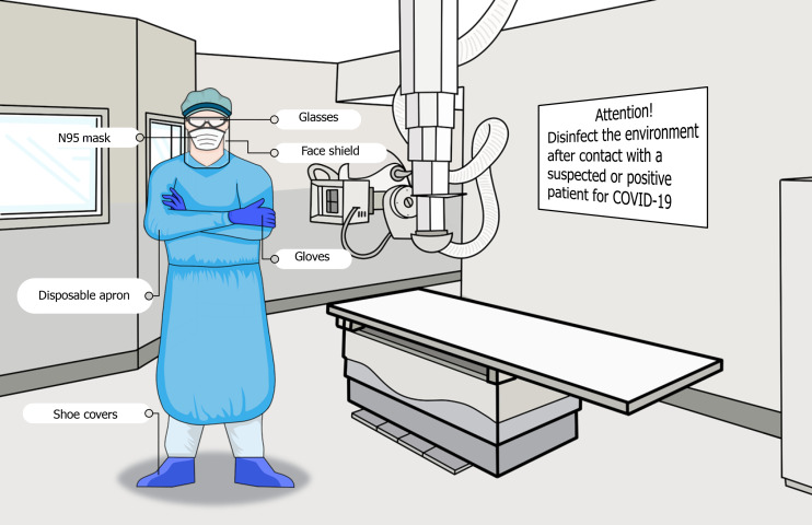
Coronavirus disease 2019 (COVID-19), a global emergency, is caused by severe acute respiratory syndrome coronavirus 2. The gold standard for its diagnosis is the reverse transcription polymerase chain reaction, but considering the high number of infected people, the low availability of this diagnostic tool in some contexts, and the limitations of the test, other tools that aid in the identification of the disease are necessary. In this scenario, imaging exams such as chest X-ray (CXR) and computed tomography (CT) have played important roles. CXR is useful for assessing disease progression because it allows the detection of extensive consolidations, besides being a fast and cheap method. On the other hand, CT is more sensitive for detecting lung changes in the early stages of the disease and is also useful for assessing disease progression. Of note, ground-glass opacities are the main COVID-19-related CT findings. Positron emission tomography combined with CT can be used to evaluate chronic and substantial damage to the lungs and other organs; however, it is an expensive test. Lung ultrasound (LUS) has been shown to be a promising technique in that context as well, being useful in the screening and monitoring of patients, disease classification, and management related to mechanical ventilation. Moreover, LUS is an inexpensive alternative available at the bedside. Finally, magnetic resonance imaging, although not usually requested, allows the detection of pulmonary, cardiovascular, and neurological abnormalities associated with COVID-19. Furthermore, it is important to consider the challenges faced in the radiology field in the adoption of control measures to prevent infection and in the follow-up of post-COVID-19 patients.
Keywords: COVID-19; Pandemic; Pneumonia; Radiology; SARS-CoV-2; Tomography.
©The Author(s) 2021. Published by Baishideng Publishing Group Inc. All rights reserved.
Conflict of interest statement
Conflict-of-interest statement: No potential conflicts of interest. No financial support.
Figures
References
-
- Huang C, Wang Y, Li X, Ren L, Zhao J, Hu Y, Zhang L, Fan G, Xu J, Gu X, Cheng Z, Yu T, Xia J, Wei Y, Wu W, Xie X, Yin W, Li H, Liu M, Xiao Y, Gao H, Guo L, Xie J, Wang G, Jiang R, Gao Z, Jin Q, Wang J, Cao B. Clinical features of patients infected with 2019 novel coronavirus in Wuhan, China. Lancet. 2020;395:497–506. - PMC - PubMed
-
- Zhou F, Yu T, Du R, Fan G, Liu Y, Liu Z, Xiang J, Wang Y, Song B, Gu X, Guan L, Wei Y, Li H, Wu X, Xu J, Tu S, Zhang Y, Chen H, Cao B. Clinical course and risk factors for mortality of adult inpatients with COVID-19 in Wuhan, China: a retrospective cohort study. Lancet. 2020;395:1054–1062. - PMC - PubMed
Publication types
LinkOut - more resources
Full Text Sources
Miscellaneous
