

doi: 10.1016/j.jds.2020.12.001.
Epub 2020 Dec 15.
Pindborg tumor: Not all Congo red-positive amyloid-like areas show apple-green birefringence under polarizing microscope
Affiliations
- PMID: 34141122
- PMCID: PMC8189889
- DOI: 10.1016/j.jds.2020.12.001
Item in Clipboard
Pindborg tumor: Not all Congo red-positive amyloid-like areas show apple-green birefringence under polarizing microscope
J Dent Sci.
2021 Jul.
No abstract available
Keywords: Amyloid; Calcification; Congo red Stain; Pericoronal radiolucency; Pindborg tumor.
Conflict of interest statement
The authors have no conflicts of interest relevant to this article.
Figures
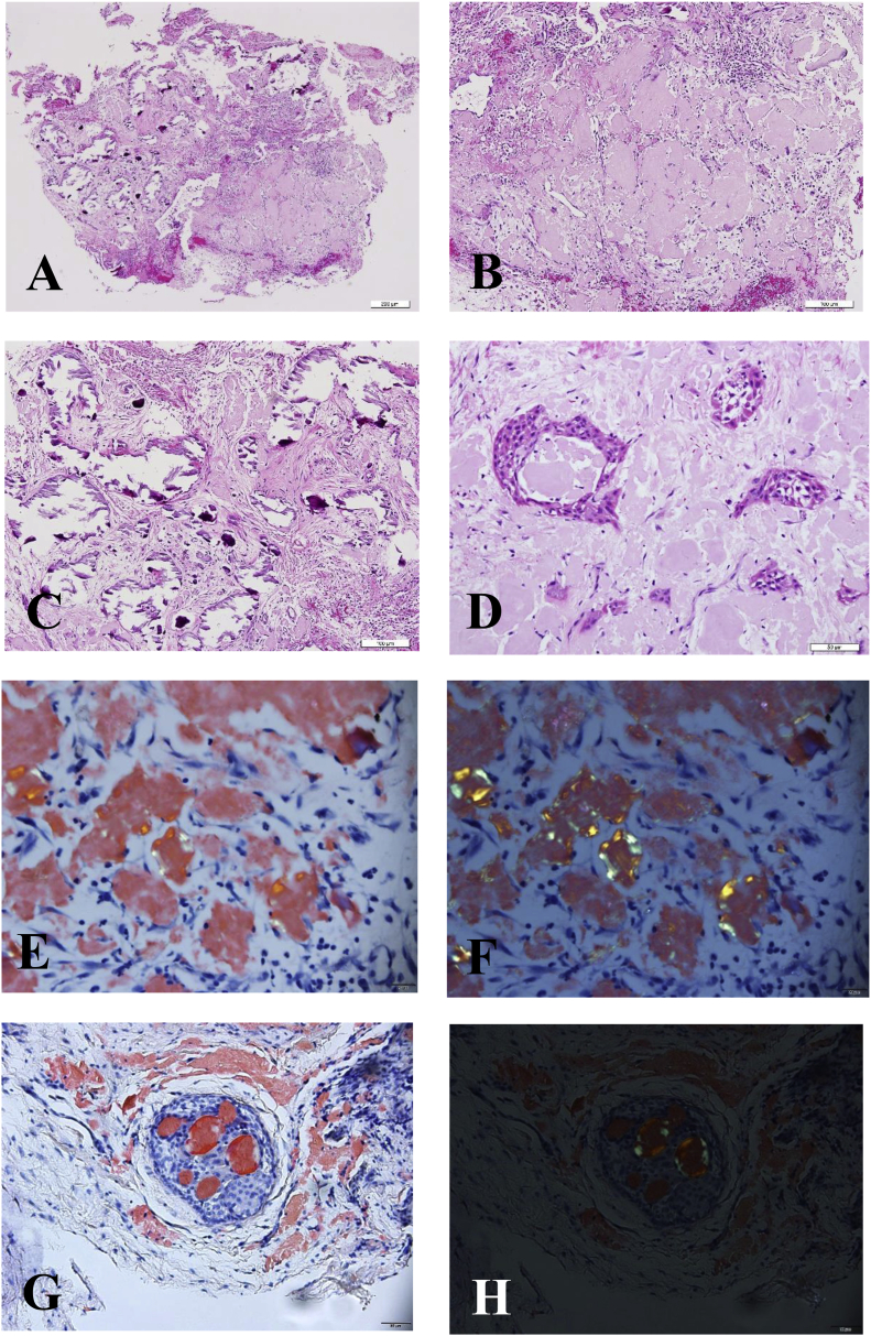
Histopathological and histochemical-stained microphotographs of the Pindborg tumor. (A) Low-power microphotograph showing that the tumor was composed of many areas or globules of amorphous, eosinophilic, and amyloid-like material (right lower part) and some foci of calcification (left half and right upper part) in a fibrous connective tissue stroma. (B) Medium-power microphotograph exhibited many areas or globules of amorphous, eosinophilic, and amyloid-like material in a fibrous connective tissue stroma. (C) Medium-power microphotograph demonstrating mainly some foci of calcification in a fibrous connective tissue stroma. (D) High-power microphotograph showing nests or islands of odontogenic epithelial cells in a background of eosinophilic and amyloid-like areas. (Hematoxylin and eosin stain; original magnification; A, 4 × ; B and C, 10 × ; D, 20 × ). (E, F, G, and H) By Congo red stain, the amyloid-like areas were brick-red and only very small focal spots of the brick-red areas exhibited apple-green birefringence under polarizing microscope (F and H). When the amyloid-like globules were surrounded by a big mass of odontogenic epithelial cells, it might mimic a cribriform appearance (G and H) (Congo red stain, original magnification; E and F, 10 × ; G and H, 20 × ). (For interpretation of the references to color in this figure legend, the reader is referred to the Web version of this article.)
References
-
- Neville B.W., Damm D.D., Allen C.M., Chi A.C. Odontogenic cyst and tumors. In: Neville B.W., Damm D.D., Allen C.M., Chi A.C., editors. Oral and maxillofacial pathology. 4th ed. Elsevier; St Louis: 2016. pp. 666–668.
-
- Mitra S., Kaur G., Nada R., Mohindra S. Pindborg tumor presenting as a nasal polyp: immunohistology and ultrastructural features of a rare case, with review of the literature. Int J Surg Pathol. 2016;24:568–572. - PubMed
LinkOut - more resources
Full Text Sources
