. 2021 Apr 3;10(5):e1203-e1209.
doi: 10.1016/j.eats.2021.01.013. eCollection 2021 May.

Arthroscopic Bursa-Augmented Rotator Cuff Repair: A Vasculature-preserving Technique for Subacromial Bursal Harvest and Tendon Augmentation

Affiliations

Arthroscopic Bursa-Augmented Rotator Cuff Repair: A Vasculature-preserving Technique for Subacromial Bursal Harvest and Tendon Augmentation

Deepak N Bhatia. Arthrosc Tech. .

Abstract

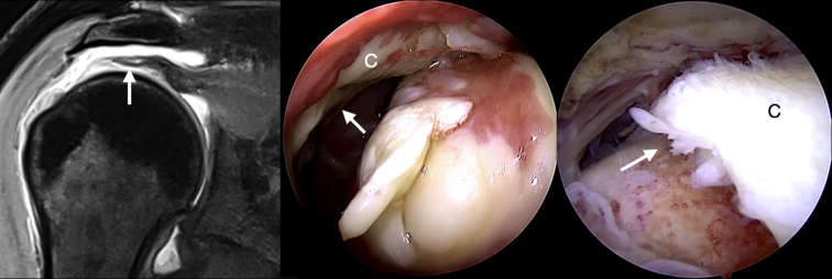
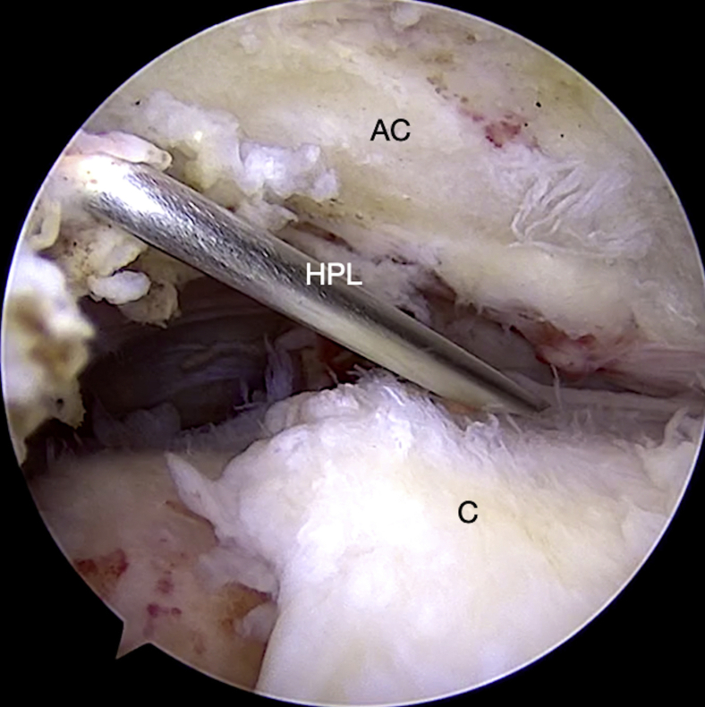
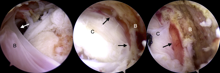
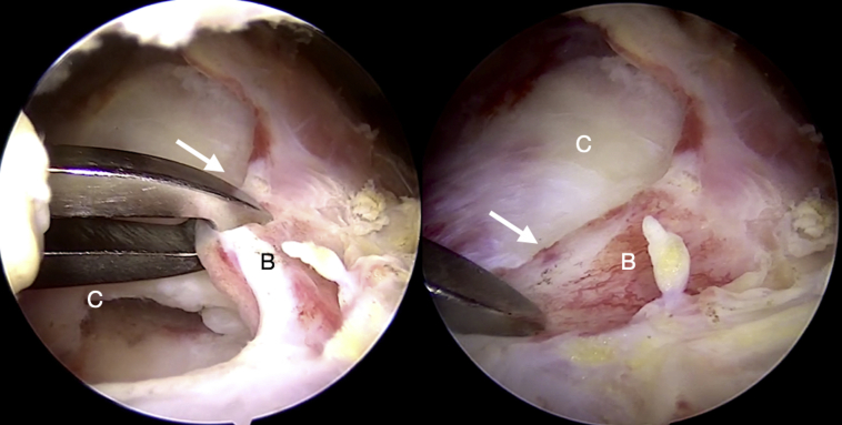
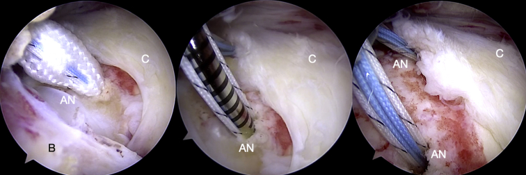
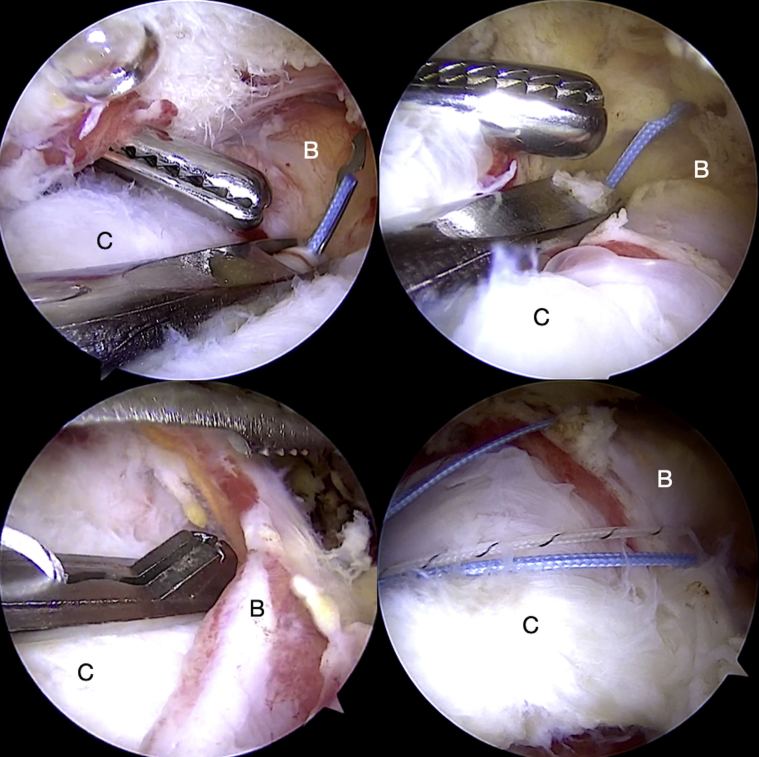
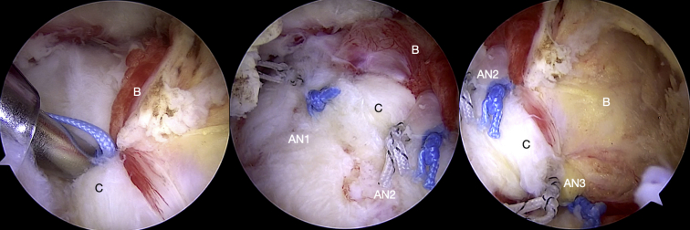
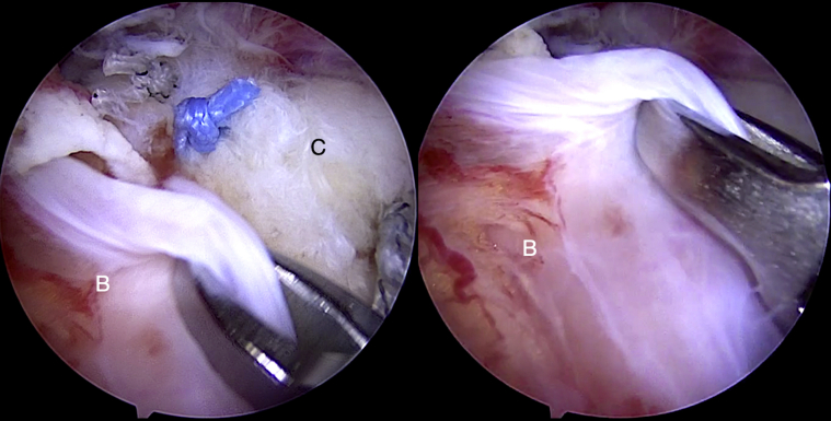
Rotator cuff repairs are associated with suboptimal outcomes and possibly greater incidence of retears if the biological healing environment is compromised. Strategies to optimize tendon-bone healing include the use of bioinductive scaffolds and regenerative stem cell therapy. The subacromial bursa has been shown to have significant pluripotent stem cell potency for tendon healing and has the advantage of easy accessibility and no added cost. However, a reproducible surgical technique for bursal mobilization, harvest, and vascularity preservation has not been described. We describe our technique for vasculature-preserving bursal mobilization and harvest of the entire posterosuperior and lateral subacromial bursa, and its use in rotator cuff repair augmentation is presented. The technique involves mobilization of the bursa as a continuous layer by maintain its medial and lateral vascularity. The bursa is advanced laterally, and the "vascular bursal duvet" and cuff tendons are repaired together as a tendon-bursa unit.


Figures

Fig 1
Fig 1
Magnetic resonance image and arthroscopic view (posterior portal) of a full-thickness rotator cuff tear in a left shoulder (arrow) is shown. The torn tendon (C) is atrophic and retracted.
Fig 2
Fig 2
An HPL is created using an outside-in technique and the needle should be seen passing obliquely in a superolateral–inferomedial direction. This angulation preserves the lateral bursal attachment and provides an unobstructed view of the rotator cuff tear (C) and bursa for further repair. (AC, acromion, left shoulder; HPL, high posterolateral portal.)
Fig 3
Fig 3
Arthroscopic view through the high posterolateral portal (left shoulder) demonstrates the entire extent of the subacromial bursal layer (B) and the rotator cuff (C). The lateral attachment of B to deltoid fascia (left image) and medial attachments (middle image) are preserved (arrows). The deep vascularity (right image, arrow) along the bursal layer can be visualized.
Fig 4
Fig 4
Mobility of the bursal layer (B) is assessed by applying traction at the superior edge (arrow) and assessing coverage obtained. This helps to determine suture passage points for optimal coverage of the repaired tendon. (C, torn cuff tendon, left shoulder, high posterolateral portal.)
Fig 5
Fig 5
All-suture anchors (AN) are passed along the medial aspect of the tuberosity in a single row configuration (left to right images). The 3-mm self-punching anchors help in preservation of the lateral bursal attachment (B, left image), and a small aperture is necessary thereby preserving more tuberosity bone. (C, torn cuff tendon, left shoulder, high posterolateral portal view.)
Fig 6
Fig 6
Sutures are seen passing through the cuff tendon (C) and bursa (B) together as a composite (top images), and separately through the bursa alone (bottom left). Final view after passing sutures is demonstrated (bottom right, left shoulder, high posterolateral portal view).
Fig 7
Fig 7
Sliding knots are used to advance the bursa (B) laterally over the repaired tendon (C) at its edge (left image). The final view shows the anterior (middle image) and posterior aspects (right image) of the repaired and augmented cuff tendons. The “vascular bursal duvet” is seen adequately covering the repaired tendons. (AN1, anterior anchor; AN2, middle anchor; AN3, posterior anchor, left shoulder, high posterolateral portal view.)
Fig 8
Fig 8
The lateral aspect of the subacromial bursal layer (B) is thin and structurally weak (left image). This aspect of the bursa may be used to cover the repaired cuff if necessary (right image), however, there is a risk of potential disruption of this fragile layer. (C, torn cuff tendon, left shoulder, high posterolateral portal view.)

References

    1. Schlegel T.F., Abrams J.S., Bushnell B.D., Brock J.L., Ho C.P. Radiologic and clinical evaluation of a bioabsorbable collagen implant to treat partial-thickness tears: A prospective multicenter study. J Shoulder Elbow Surg. 2018;27:242–251. - PubMed
    1. Morikawa D., Muench L.N., Baldino J.B., et al. Comparison of preparation techniques for isolating subacromial bursa-derived cells as a potential augment for rotator cuff repair. Arthroscopy. 2020;36:80–85. - PubMed
    1. Dyrna F., Zakko P., Pauzenberger L., McCarthy M.B., Mazzocca A.D., Dyment N.A. Human subacromial bursal cells display superior engraftment versus bone marrow stromal cells in murine tendon repair. Am J Sports Med. 2018;46:3511–3520. - PMC - PubMed
    1. Chillemi C., Petrozza V., Franceschini V., et al. The role of tendon and subacromial bursa in rotator cuff tear pain: A clinical and histopathological study. Knee Surg Sports Traumatol Arthrosc. 2016;24:3779–3786. - PubMed
    1. Uhthoff H.K., Sarkar K. Surgical repair of rotator cuff ruptures. The importance of the subacromial bursa. J Bone Joint Surg Br. 1991;73:399–401. - PubMed

LinkOut - more resources