Surgical treatment of four segment lumbar spondylolysis: A case report
- PMID: 34141808
- PMCID: PMC8173437
- DOI: 10.12998/wjcc.v9.i17.4408
Surgical treatment of four segment lumbar spondylolysis: A case report
Abstract
Background: Four-level lumbar spondylolysis is extremely rare. So far, only 1 case has been reported in the literature.
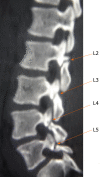
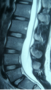
Case summary: A 19-year-old man presented with severe back pain irresponsive to conservative therapies for 2 years. Lumbar radiographs and two-dimensional computed tomography scan showed four segment lumbar spondylolysis on both sides of L2-L5. Lumbar magnetic resonance imaging showed normal signal in all lumbar discs. Because daily activities were severely limited, surgery was recommended for the case. The patient underwent four-level bilateral isthmic repair at L2-L5. During surgery, L2-L5 isthmi were curetted bilaterally, freshened, and then grafted with autologous iliac bone that was bridged and compressed with a pedicular screw connected to a sub-laminar hook by a short rod. The symptoms of back pain almost disappeared. He has been followed-up for 96 mo, and his symptoms have never recurred. Fusion was found in all repaired isthmi 14 mo after surgery according to evaluation of lumbar radiography and computed tomography scan.
Conclusion: We report here 1 case of four-level lumbar spondylolysis that was treated successfully with direct isthmic repair.
Keywords: Case report; Isthmic repair; Lumbar spondylolysis; Pedicle screw-hook system.
©The Author(s) 2021. Published by Baishideng Publishing Group Inc. All rights reserved.
Conflict of interest statement
Conflict-of-interest statement: The authors declare that they have no current financial arrangement or affiliation with any organization that may have a direct influence on their work.
Figures


Similar articles
-
Application of a new anatomic hook-rod-pedicle screw system in young patients with lumbar spondylolysis: A pilot study.World J Clin Cases. 2022 Jun 16;10(17):5680-5689. doi: 10.12998/wjcc.v10.i17.5680. World J Clin Cases. 2022. PMID: 35979102 Free PMC article.
-
Direct repair of multiple levels lumbar spondylolysis by pedicle screw laminar hook and bone grafting: clinical, CT, and MRI-assessed study.J Spinal Disord Tech. 2007 Jul;20(5):399-402. doi: 10.1097/01.bsd.0000211253.67576.90. J Spinal Disord Tech. 2007. PMID: 17607107 Clinical Trial.
-
[Direct repair of adolescent lumbar spondylolysis using a pedicle screw-laminar hook system by paramedian approach].Zhongguo Gu Shang. 2011 Aug;24(8):687-9. Zhongguo Gu Shang. 2011. PMID: 21928681 Chinese.
-
Double-level lumbar spondylolysis and spondylolisthesis: A retrospective study.J Orthop Surg Res. 2018 Mar 16;13(1):55. doi: 10.1186/s13018-018-0723-3. J Orthop Surg Res. 2018. PMID: 29548343 Free PMC article. Review.
-
Acute progression of spondylolysis to isthmic spondylolisthesis in an adult.Spine (Phila Pa 1976). 2002 Aug 15;27(16):E370-2. doi: 10.1097/00007632-200208150-00023. Spine (Phila Pa 1976). 2002. PMID: 12195078 Review.
References
-
- Fredrickson BE, Baker D, McHolick WJ, Yuan HA, Lubicky JP. The natural history of spondylolysis and spondylolisthesis. J Bone Joint Surg Am. 1984;66:699–707. - PubMed
-
- Ravichandran G. Multiple lumbar spondylolyses. Spine (Phila Pa 1976) 1980;5:552–557. - PubMed
-
- Darnis A, Launay O, Perrin G, Barrey C. Surgical management of multilevel lumbar spondylolysis: a case report and review of the literature. Orthop Traumatol Surg Res. 2014;100:347–351. - PubMed
-
- Gu SX, Ma Y, Chen X, Cai XJ, Cui X, Bao D, Huang FS, Luo ZP, Li DW, Luo XB, Li LT. Re : He B, Yan L, Guo H, et al. The difference in superior adjacent segment pathology after lumbar posterolateral fusion by using 2 different pedicle screw insertion techniques in 9-year minimum follow-up. Spine (Phila Pa 1976) 2014;39:1093-8. Spine (Phila Pa 1976) 2014;39:E1493. - PubMed
-
- Ogawa H, Nishimoto H, Hosoe H, Suzuki N, Kanamori Y, Shimizu K. Clinical outcome after segmental wire fixation and bone grafting for repair of the defects in multiple level lumbar spondylolysis. J Spinal Disord Tech. 2007;20:521–525. - PubMed
Publication types
LinkOut - more resources
Full Text Sources

