Review
. 2021 Jun 5:8:100361.
doi: 10.1016/j.ejro.2021.100361. eCollection 2021.

Dots, lines, contours, and ends: An image-based review of esophageal pathology

Affiliations
Review

Dots, lines, contours, and ends: An image-based review of esophageal pathology

Nandan Keshav et al. Eur J Radiol Open. .

Abstract

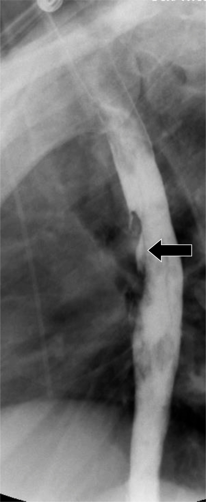
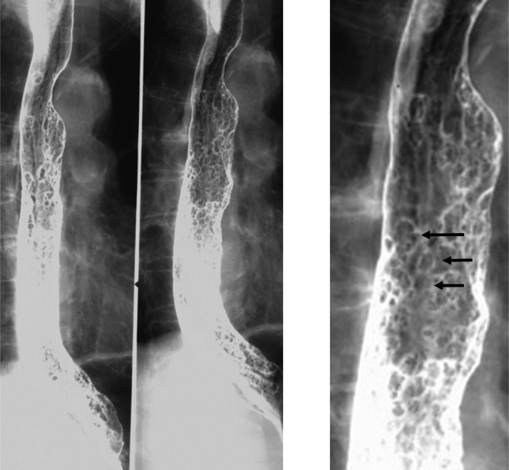
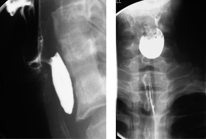
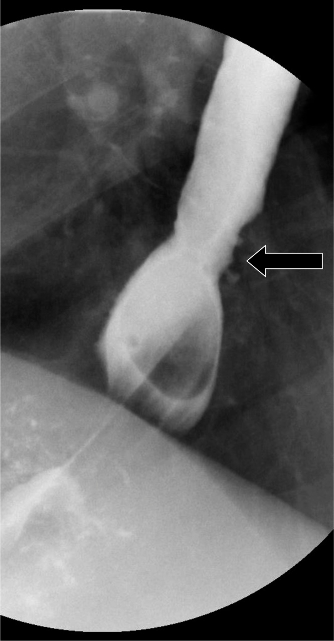
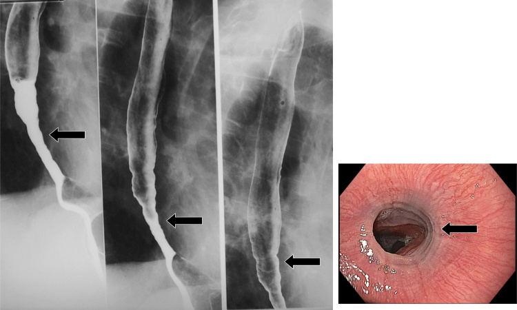
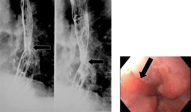
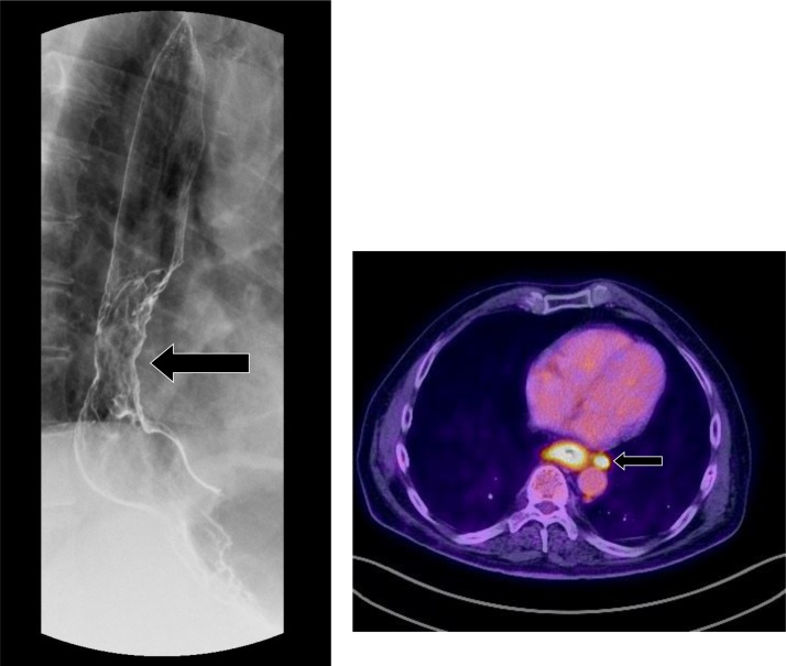
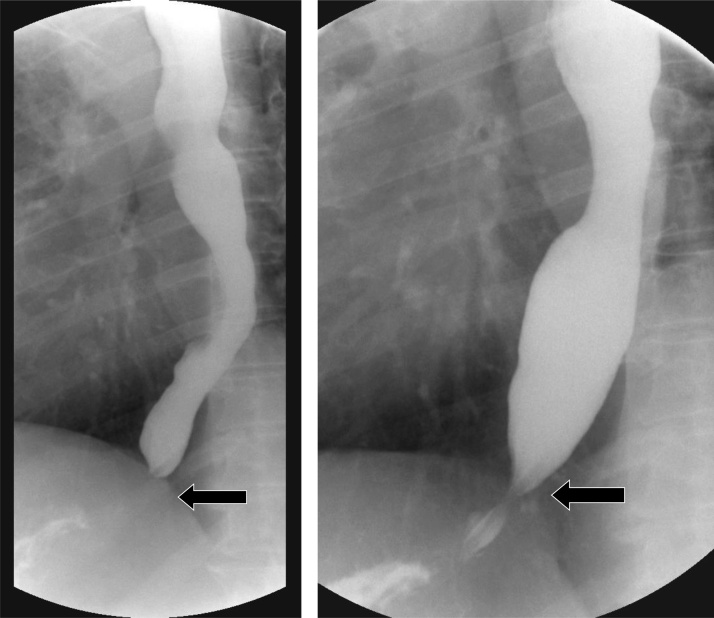
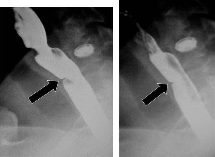
Esophageal pathologies encountered on fluoroscopic examination may pose a diagnostic challenge to the interpreting Radiologist. Understanding the varied imaging appearances of esophageal pathology requires a thorough understanding of barium esophagography. This article reviews the various fluoroscopic imaging findings of different esophageal pathologies by describing an approach to image interpretation centered on dots, lines, contours, and ends. By utilizing this approach, the Radiologist will be better positioned to reconcile seemingly disparate pathologies into a cogent and succinct differential diagnosis.

Keywords: Fluoroscopy; Oesophageal patterns; Oesophaghus; Oesophagram.


Conflict of interest statement

There are no known conflicts of interest associated with this publication and there has been no financial support for this work. All authors explicitly approve the content within this manuscript.

Figures

Fig. 1
Fig. 1
Normal double contrast esophagram. Note the featureless mucosa (inset).
Fig. 2
Fig. 2
Intraluminal esophageal dots refer to distinct intraluminal foci on double contrast exam, which may appear either as bright spots or focal lucencies/cavities.
Fig. 3
Fig. 3
Herpes esophagitis. Note the presence of small, discrete ulcers (arrows).
Fig. 4
Fig. 4
Drug-induced esophagitis. Note the characteristic location within the mid esophagus, where the left mainstem bronchus or aortic arch serves as an external buttress (arrow).
Fig. 5
Fig. 5
Crohn’s esophagitis. Diffuse irregularity and small ulcers are present within the esophagus. A small-bowel follow through exam from the same patient demonstrates a terminal ileal stricture (arrow).
Fig. 6
Fig. 6
CMV esophagitis. Large, ovoid, and flat ulcers are present (arrow). Corresponding endoscopic findings (inset) demonstrate the ulcers (arrow).
Fig. 7
Fig. 7
Candida esophagus. Note the presence of multiple plaques in a linear course (arrow).
Fig. 8
Fig. 8
Advanced candidiasis. The plaques may be become fulminant and ulcerated, with the esophagus possessing a shaggy and irregular appearance. Note endoscopic correlate (inset).
Fig. 9
Fig. 9
Glycogen acanthosis. There are numerous, tiny, raised plaques which may coalesce and form a finely nodular or cobblestone mucosal pattern.
Fig. 10
Fig. 10
Esophageal papillomatosis. Note the innumerable filling defects (arrows).
Fig. 11
Fig. 11
Esophageal varices. Note the serpiginous filling defects (arrow). This appearance on fluoroscopy may mimic varicoid esophageal carcinoma; however, varices change appearance with time. Endoscopic correlate of esophageal varices is also shown (inset, arrows).
Fig. 12
Fig. 12
Feline esophagus. Note the transient, transverse, thinly spaced striations.
Fig. 13
Fig. 13
Eosinophilic esophagitis. The esophagram demonstrates long segment symmetric narrowing. Endoscopic correlate of the ringed esophagus variant from a different patient is also shown (inset).
Fig. 14
Fig. 14
Normal contour deformities of the esophagus. The aortic arch, left mainstem bronchus, and left atrium form gentle indentations of the esophageal contour.
Fig. 15
Fig. 15
A pictorial summary of esophageal diverticula. Also see Table 4.
Fig. 16
Fig. 16
Zenker’s diverticulum. This is located posteriorly at the pharyngoesophageal junction, and inferior to the cricopharyngeus muscle.
Fig. 17
Fig. 17
Killian-Jameson diverticulum. Note the characteristic outpouching from the lateral wall of the proximal cervical esophagus.
Fig. 18
Fig. 18
Esophageal traction diverticulum. Note the ancillary finding of calcified granulomas within the spleen on upper GI from the same patient (arrow), which may support the diagnosis.
Fig. 19
Fig. 19
Epiphrenic pulsion diverticulum. Note the characteristic outpouching within the distal esophagus. There is an associated small sliding hiatal hernia (arrow).
Fig. 20
Fig. 20
Pseudodiverticulosis (arrow) with distal esophageal stricture.
Fig. 21
Fig. 21
A more pronounced case of pseudodiverticulosis.
Fig. 22
Fig. 22
Vascular ring. Note the impression on the posterior esophagus (arrow). This is secondary to a right aortic arch with aberrant right subclavian artery.
Fig. 23
Fig. 23
An example of gastroesophageal reflux disease on fluoroscopy. These may appear as short segment strictures and scarring. Note corresponding focal luminal narrowing on endoscopy (inset, arrow).
Fig. 24
Fig. 24
Barrett’s esophagus. Note the mid-esophageal stricture (arrow). Corresponding endoscopic image demonstrates epithelial metaplasia (inset, arrow).
Fig. 25
Fig. 25
Esophageal adenocarcinoma. There is an irregular mass of the distal esophagus with corresponding mucosal irregularities and luminal narrowing (arrow). Positron emission tomography-CT of the same patient (inset) demonstrates a hypermetabolic esophageal mass with para-esophageal adenopathy (arrow).
Fig. 26
Fig. 26
Achalasia with tapering of the distal esophagus, resembling a bird’s beak (arrow).
Fig. 27
Fig. 27
Scleroderma. The esophagus appears patulous, and there is a small reflux stricture present (arrow).
Fig. 28
Fig. 28
A small web is present in the cervical esophagus (arrow).

References

    1. Levine M.S., Rubesin S.E. Diseases of the esophagus: diagnosis with esophagography. Radiology. 2005;237(November (2)):414–427. Radiology. 2005 Nov;237(2):414-427. - PubMed
    1. Gore R.M., Levine M.S. 4th edition. Saunders; 2014. Textbook of Gastrointestinal Radiology. December 10.
    1. Levine M.S., Ramchandani P., Rubesin S.E. Cambridge University Press; 2012. Practical Fluoroscopy of the GI and GU Tracts.
    1. Noh H.M., Fishman E.K., Forastiere A.A., Bliss D.F., Calhoun P.S. CT of the esophagus: spectrum of disease with emphasis on esophageal carcinoma. Radiographics. 1995;15(September (5)):1113–1134. - PubMed
    1. Hoversten P., Kamboj A.K., Katzka D.A. Infections of the esophagus: an update on risk factors, diagnosis, and management. Dis. Esophagus. 2018;31(December (12)) - PubMed

LinkOut - more resources