. 2021 May 17;4(5):4140-4151.
doi: 10.1021/acsabm.0c01566. Epub 2021 Apr 19.

(4-Aminopyridine)-PLGA-PEG as a Novel Thermosensitive and Locally Injectable Treatment for Acute Peripheral Nerve Injury

Affiliations

(4-Aminopyridine)-PLGA-PEG as a Novel Thermosensitive and Locally Injectable Treatment for Acute Peripheral Nerve Injury

Kristen M Manto et al. ACS Appl Bio Mater. .

Abstract

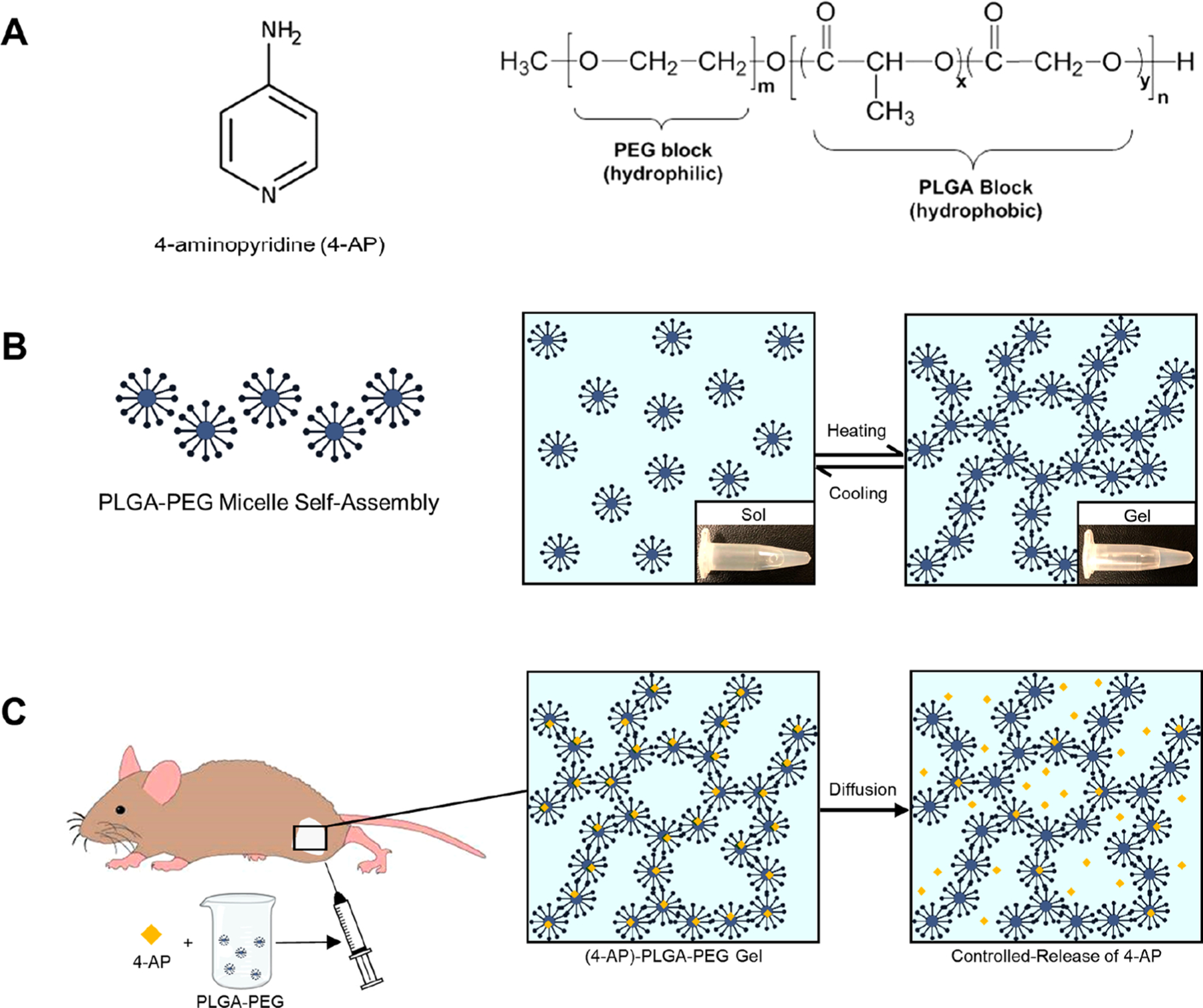
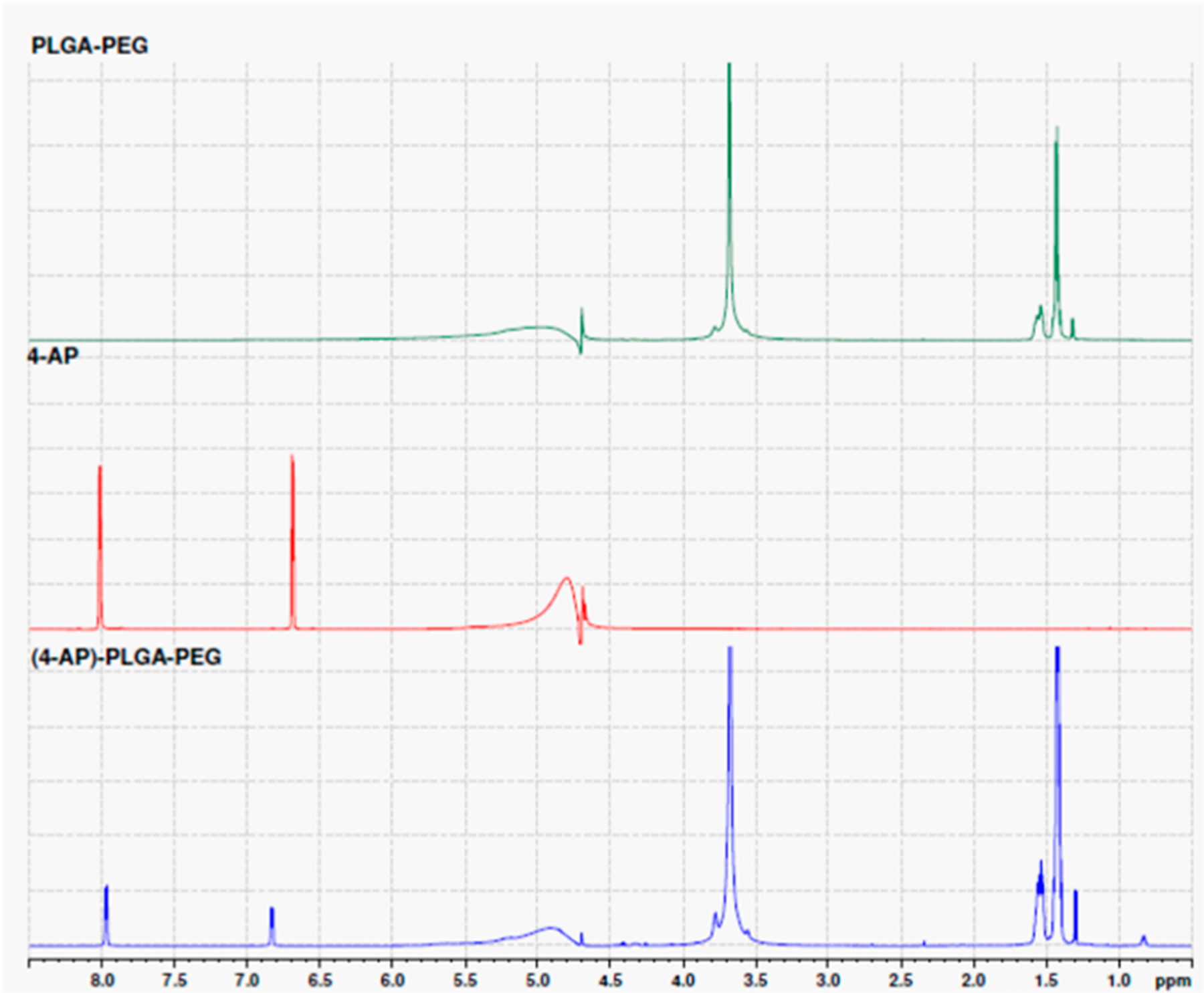
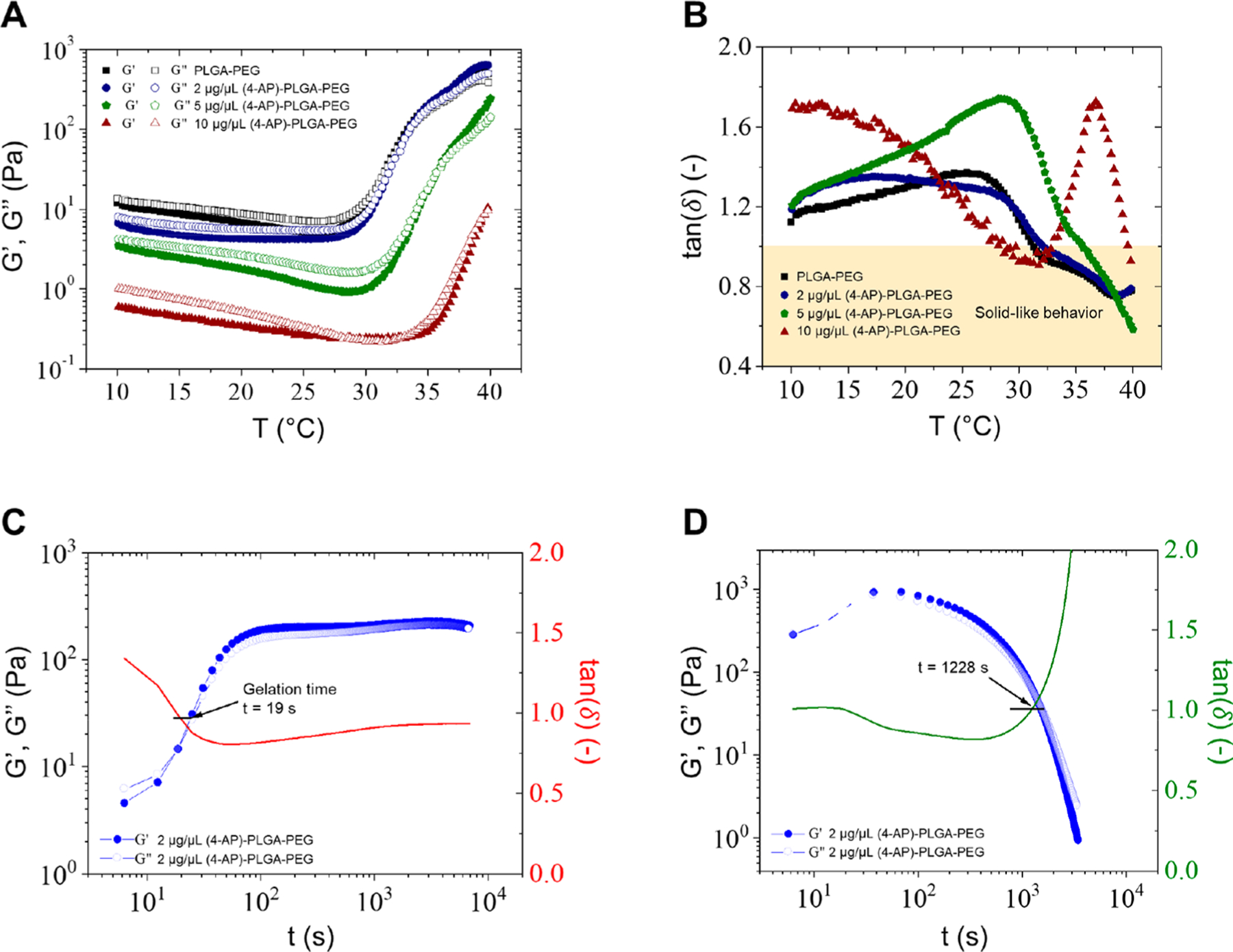
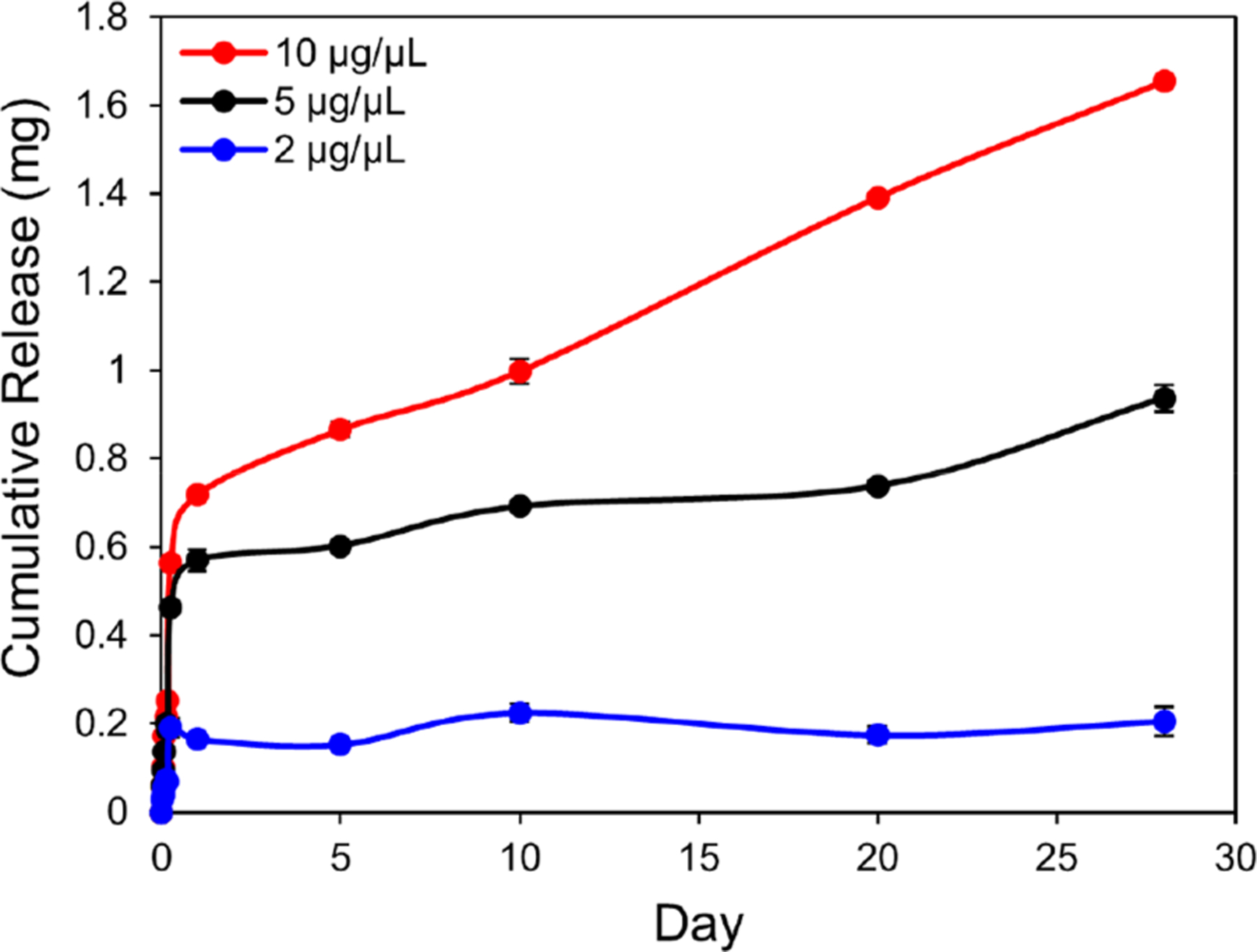
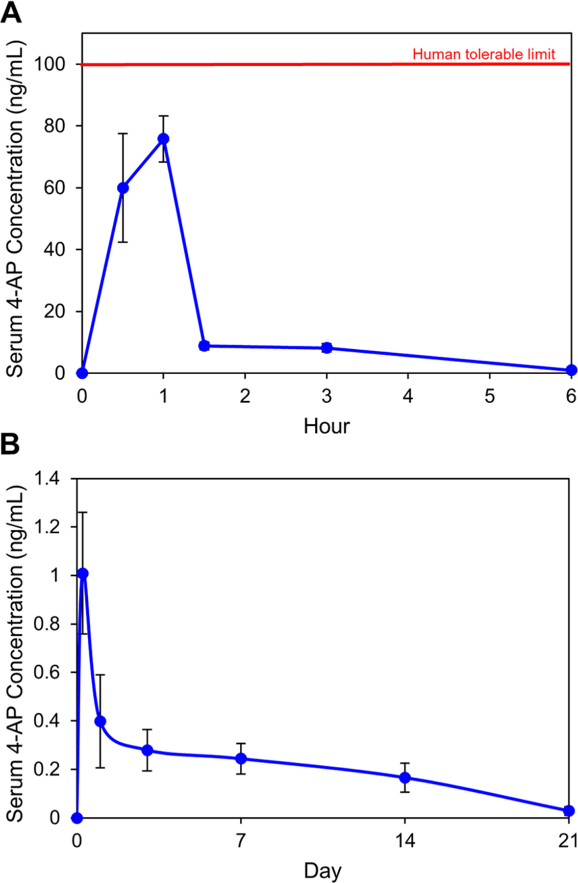
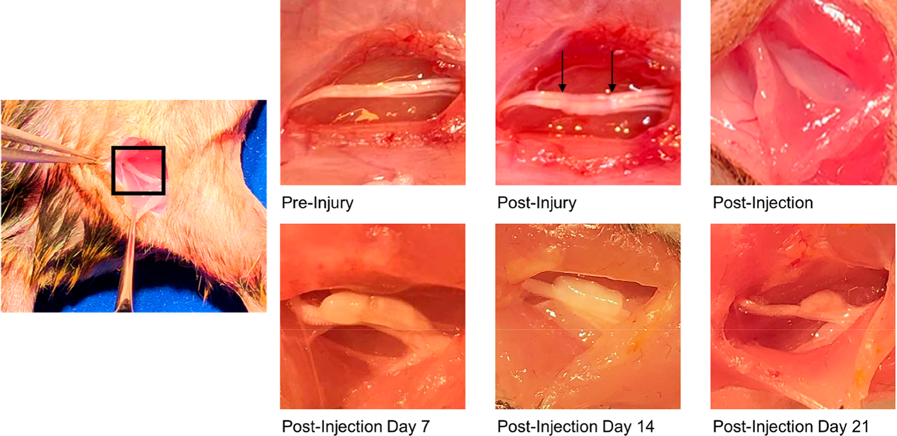
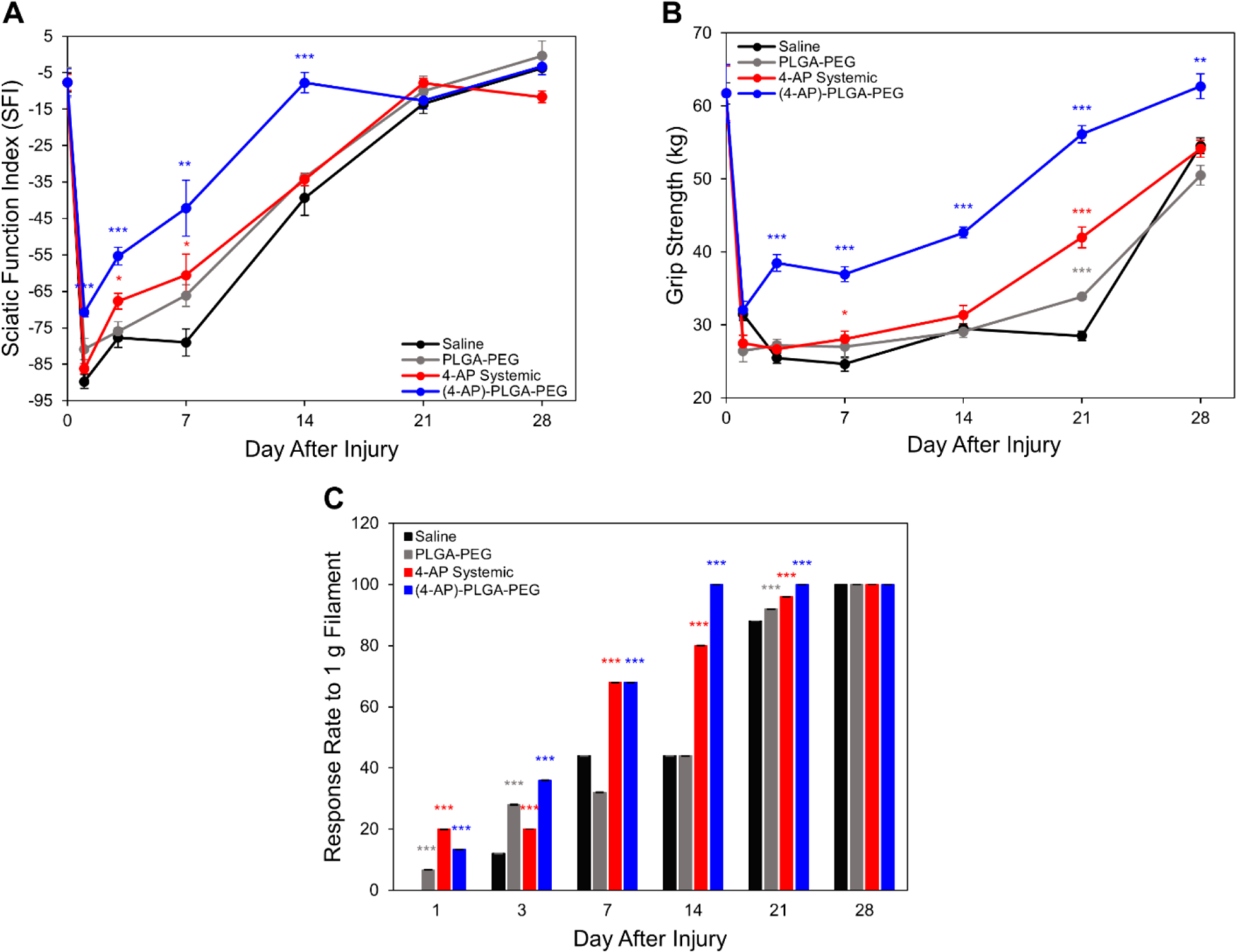
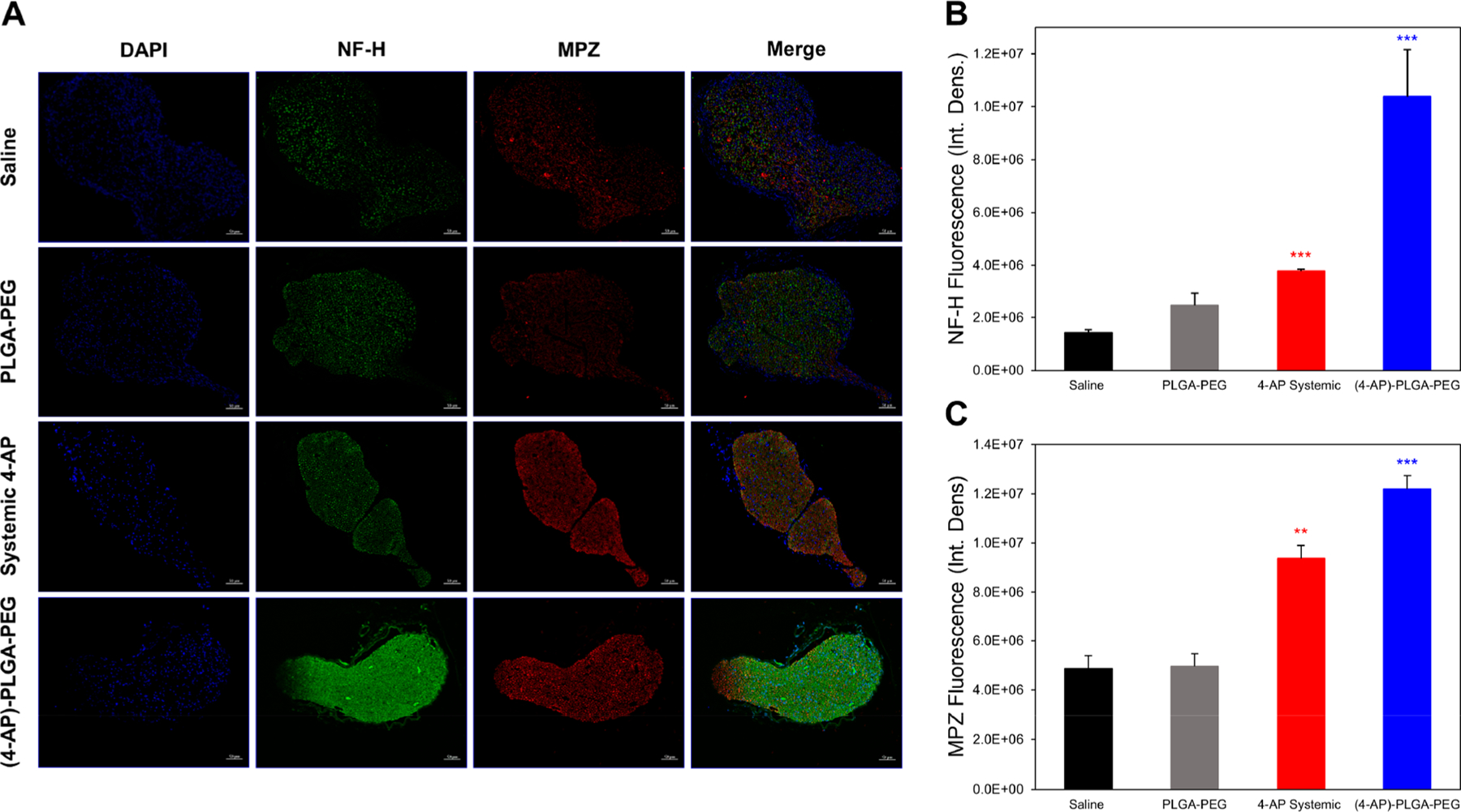
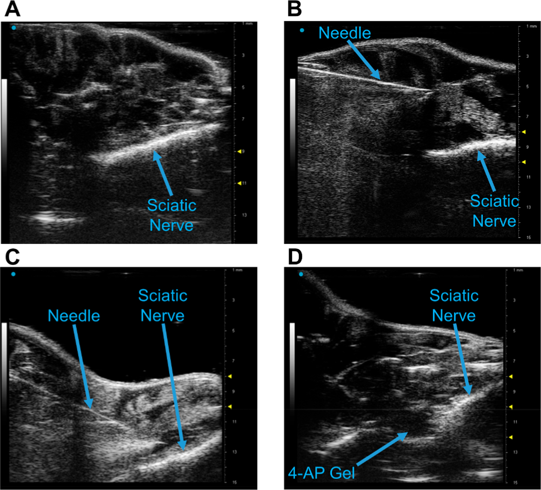
Traumatic peripheral nerve injury (TPNI) represents a major medical problem that results in loss of motor and sensory function, and in severe cases, limb paralysis and amputation. To date, there are no effective treatments beyond surgery in selective cases. In repurposing studies, we found that daily systemic administration of the FDA-approved drug 4-aminopyridine (4-AP) enhanced functional recovery after acute peripheral nerve injury. This study was aimed at constructing a novel local delivery system of 4-AP using thermogelling polymers. We optimized a thermosensitive (4-AP)-poly(lactide-co-glycolide)-b-poly(ethylene glycol)-b-poly(lactide-co-glycolide) (PLGA-PEG-PLGA) block copolymer formulation. (4-AP)-PLGA-PEG exhibited controlled release of 4-AP both in vitro and in vivo for approximately 3 weeks, with clinically relevant safe serum levels in animals. Rheological investigation showed that (4-AP)-PLGA-PEG underwent a solution to gel transition at 32 °C, a physiologically relevant temperature, allowing us to administer it to an injured limb while subsequently forming an in situ gel. A single local administration of (4-AP)-PLGA-PEG remarkably enhanced motor and sensory functional recovery on post-sciatic nerve crush injury days 1, 3, 7, 14, and 21. Moreover, immunohistochemical studies of injured nerves treated with (4-AP)-PLGA-PEG demonstrated an increased expression of neurofilament heavy chain (NF-H) and myelin protein zero (MPZ) proteins, two major markers of nerve regeneration. These findings demonstrate that (4-AP)-PLGA-PEG may be a promising long-acting local therapeutic agent in TPNI, for which no pharmacologic treatment exists.

Keywords: 4-aminopyridine; PEG; PLGA; block copolymer; crush injury; peripheral nerve; sciatic nerve; thermogel; traumatic nerve injury.


Conflict of interest statement

The authors declare the following competing financial interest(s): The senior author has an equity interest in and serves as an advisor to Peripheral Therapeutics Inc., a start-up company that may potentially benefit from the research results provided. The senior author’s ownership and role in the company have been disclosed and reviewed by The Pennsylvania State University in accordance with its conflict-of-interest policies.

Figures

Figure 1.
Figure 1.
Schematic representation of a (4-AP)-loaded thermogel as a local controlled-release delivery system. (A) Chemical structures of 4-AP and PLGA–PEG, respectively. (B) Amphiphilic polyester–PEG–polyester triblock copolymers self-assemble into micelles in aqueous solution at room temperature and form a solid gel via cross-links at higher temperature. (C) 4-AP can be incorporated into the triblock copolymer at room temperature and injected into an animal in liquid phase. Once the formulation reaches body temperature, it forms an in situ hydrogel for controlled release of 4-AP.
Figure 2.
Figure 2.
1H NMR spectra of PLGA–PEG, 4-AP, and (4-AP)–PLGA–PEG. The chemical shifts of the mixture showed significant change at both aromatic protons of 4-AP from δ8.02 to 7.94 and δ6.69 to 6.83, respectively, when mixed with PLGA–PEG. In addition, the PLGA–PEG aliphatic region was changed from δ1.31 to 1.29 after mixing.
Figure 3.
Figure 3.
Rheological investigation of copolymer aqueous solutions. (A) Gelation temperatures of PLGA–PEG and 2 μg/μL (4-AP)–PLGA–PEG were 31.3 and 32 °C, respectively. Gelation temperature increased with increasing 4-AP concentration. (B) Tan(δ) was less than 1, indicating solid-like behavior, for all concentrations at body temperature except 10 μg/μL, which exhibited a second solid to liquid crossover point at increasing temperature. (C) The solution to gel (sol–gel) transition of 2 μg/μL (4-AP)–PLGA–PEG occurred in 19 s and was followed by persistence of solid-like behavior over time. (D) The sol–gel transition of 2 μg/μL (4-AP)–PLGA–PEG was fully reversible and occurred in 1228 s.
Figure 4.
Figure 4.
Cumulative in vitro release of 4-AP from PLGA–PEG in PBS (pH 7.4) at 37 °C. PLGA–PEG carriers exhibited an overall burst biphasic profile with a burst release of 4-AP within 1 day followed by release of a low maintenance dose for approximately 28 days. Cumulative release was proportional to total loaded amount of 4-AP. Data are expressed as means ± SEM, n = 3/group.
Figure 5.
Figure 5.
Pharmacokinetic study of 4-AP serum levels in mice after 2 μg/μL (4-AP)–PLGA–PEG administration at a 4-AP dose of 1.4 mg/kg. (A) 4-AP serum levels peaked 1 h after administration and never exceeded the human tolerable limit of 100 ng/mL. (B) 4-AP serum levels were nearly undetectable by Day 21. Data are expressed as means ± SEM, n = 9/group.
Figure 6.
Figure 6.
In vivo biodegradation study of (4-AP)–PLGA–PEG. Pre-injury shows the mouse sciatic nerve. Post-injury shows the nerve after a moderate crush injury, as depicted by the arrows. Upon administration on the sciatic nerve post-injury, (4-AP)–PLGA–PEG turns opaque, indicating thermogelation. On post-injection days 7, 14, and 21, the gel remained directly on the nerve, although its mass decreased over time due to controlled polymeric degradation.
Figure 7.
Figure 7.
Effects of (4-AP)–PLGA–PEG on motor and sensory functional outcomes. (A) (4-AP)–PLGA–PEG significantly improved SFI on post-injury days 1, 3, 7, and 14 compared to saline, PLGA–PEG, and systemic 4-AP groups. (B) (4-AP)–PLGA–PEG significantly improved grip strength on post-injury days 3, 7, 14, 21, and 28 compared to all other treatment groups. (C) (4-AP)–PLGA–PEG significantly improved withdrawal reflex (percent response to filament) as compared to the saline group on post-injury days 1, 3, 7, 14, and 21. Data are expressed as means ± SEM, *p < 0.05, **p < 0.01, and ***p < 0.001 vs saline group, n = 5/group.
Figure 8.
Figure 8.
Effects of (4-AP)–PLGA–PEG on immunohistochemical markers of nerve regeneration. (A) Representative transverse sciatic nerve immunofluorescent images of nuclei (DAPI), NF-H, and MPZ on post-injury day 28. Each image represents nine images from three different mice. Scale bar: 50 μm; magnification: 20×. (B) Quantification of NF-H integrated density on post-injury day 28. (4-AP)–PLGA–PEG-treated nerves contained significantly more NF-H protein in the lesion area than nerves from saline-treated animals. (C) Quantification of MPZ integrated density on post-injury day 28. (4-AP)–PLGA–PEG-treated nerves contained significantly more MPZ in the lesion area than nerves from saline-treated animals. Data are expressed as means ± SEM, **p < 0.01 and ***p < 0.001 vs saline group, n = 3/group.
Figure 9.
Figure 9.
(4-AP)–PLGA–PEG injection on the mouse sciatic nerve using small animal ultrasonography. (A) Longitudinal visualization of the sciatic nerve using the Vevo 3100 40 MHz ultrasound probe. (B) Identification of the 20 G needle with the nerve. (C) Positioning the needle over the sciatic nerve pre-injection. (D) Visualization of (4-AP)–PLGA–PEG on the sciatic nerve post-injection.

Similar articles

Cited by

References

    1. Taylor CA; Braza D; Rice JB; Dillingham T The incidence of peripheral nerve injury in extremity trauma. Am. J. Phys. Med. Rehabil 2008, 87, 381–385. - PubMed
    1. Sakuma M; Minev IR; Gribi S; Singh B; Woolf CJ; Lacour SP Chronic electrical nerve stimulation as a therapeutic intervention for peripheral nerve repair. Bioelectron Med 2015, 2, 43–48.
    1. Asplund M; Nilsson M; Jacobsson A; von Holst H Incidence of traumatic peripheral nerve injuries and amputations in Sweden between 1998 and 2006. Neuroepidemiology 2009, 32, 217–228. - PubMed
    1. Campbell WW Evaluation and management of peripheral nerve injury. Clin. Neurophysiol 2008, 119, 1951–1965. - PubMed
    1. Niver GE; Ilyas AM Management of radial nerve palsy following fractures of the humerus. Orthop. Clin. North Am 2013, 44, 419–424. - PubMed

Publication types

MeSH terms

LinkOut - more resources