. 2021 Jun 2;2(2):100574.
doi: 10.1016/j.xpro.2021.100574. eCollection 2021 Jun 18.

Protocol for tissue slice cultures from human solid tumors to study therapeutic response

Affiliations

Protocol for tissue slice cultures from human solid tumors to study therapeutic response

Heidi L Kenerson et al. STAR Protoc. .

Abstract

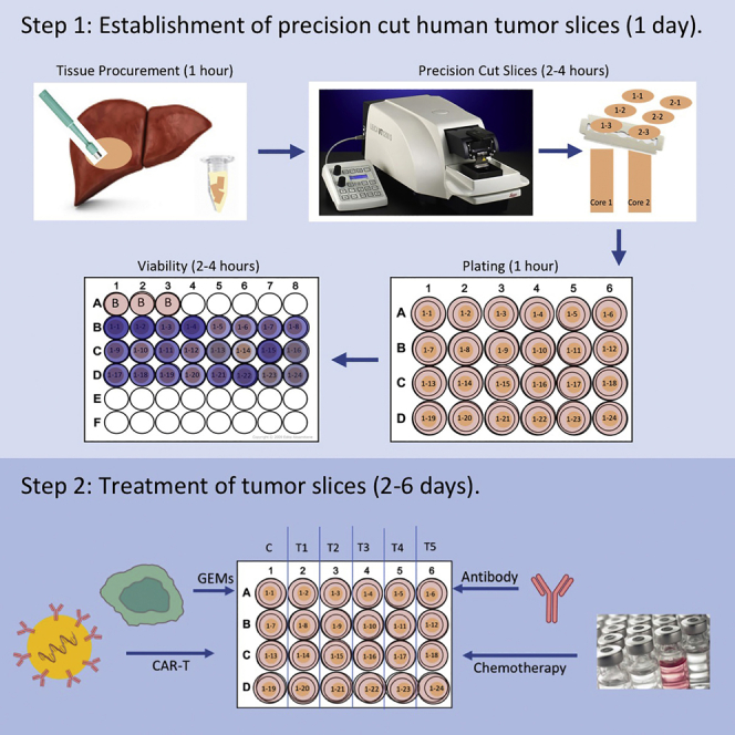
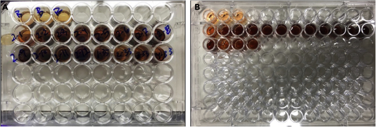
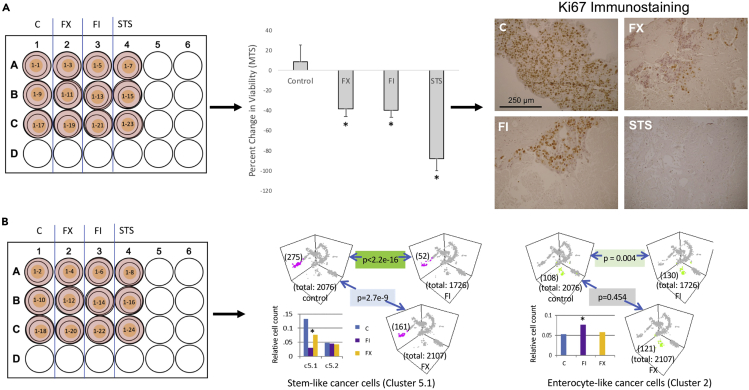
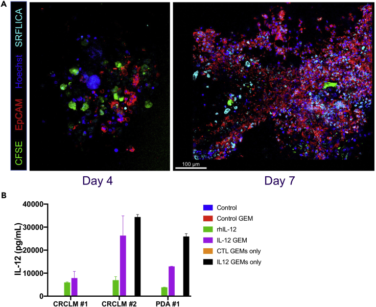
The impact of systemic therapy on the tumor microenvironment has been difficult to study in human solid tumors. Our protocol describes steps for establishing slice cultures to investigate response to chemotherapies, immunotherapies, or adoptive cell therapies. Endpoints include changes in viability, histology, live-cell imaging, and multi-omics analyses. The protocol has been applied to a broad array of gastrointestinal malignancies. Culture conditions and treatment parameters can be modified for specific experiments. The platform is highly flexible and easy to manipulate. For complete details on the use and execution of this protocol, please refer to Kenerson et al. (2020), Jabbari et al. (2020), Brempelis et al. (2020), and Jiang et al. (2017).

Keywords: Cancer; Cell culture; Cell-based Assays; High Throughput Screening; Immunology.


Conflict of interest statement

The authors declare no competing interests.

Figures

None
Graphical abstract
Figure 1
Figure 1
Sterile tumor tissue procurement and tumor core punches (A) Upon completion of liver resection, specimen is placed on a sterile field. With guidance of pathologist a 1 cm thick wedge of tissue is removed from specimen to access tumor tissue. Scale bar is equal to 5 cm. (B) A 6 mm biopsy punch is used to core tumor tissue for slice protocol. (C) Samples are taken from tumor periphery to avoid areas of central necrosis.
Figure 2
Figure 2
Tissue cores embedded in agarose (A) Cores are removed from transport solution and excess liquid removed. (B and C) (B) Cores are embedded in agarose individually in wells of a 24-well plate or (C) grouped in a well of a 6-well plate. (D) Agarose and tissue are adhered to specimen disc. (E) Specimen disc is placed in buffer tray with cutting solution on ice for slicing.
Figure 3
Figure 3
Plating tissue slices (A) Tumor slices are placed in wash buffer in a 48-well dish in order of core and slice number. (B) Slices are then transferred onto individual Millicell culture inserts placed in a 24-well plate with 450 μL of media. (C) One tumor slice plated on a 12 mm insert in a 24-well dish. (D) 4 tumor slices plated on a 30 mm insert in a 6-well dish.
Figure 4
Figure 4
MTS assay on tissue slices (A) Tissue slices are transferred to individual wells of a 48-well dish containing 400 μL of media. 80 μL of MTS reagent is added. Plate is incubated at 37°C and 5% CO2 for 3 h. (B) 200 μL of media is transferred to 96-well assay plate and absorbance read at 490 nm.
Figure 5
Figure 5
Tumor treatment scheme with multiple readouts Tumor slices from a CRLM were treated with vehicle control (0.2% DMSO), FX (1 μg/mL 5FU + 1 μg/mL Oxaliplatin), FI (1 μg/mL 5FU + 2 μg/mL Irinotecan, or STS (10 μm Staurosporine, to serve as a positive control) two times over 4 days. (A) One set of treated slices were used to calculate percent change in viability using pre-treatment and post-treatment MTS values and subsequently fixed for histology. Data are represented as mean ± standard deviation. Fixed slices were processed for immunohistochemistry staining with Ki67 (Dako, 1:200). Scale bar is equal to 250 μm. (B) Another set of treated slices were used to perform single-cell RNA sequencing. Figure reprinted with permission from Jabbari et al., 2020. Note how slices are dispersed over the treatment and endpoint assay.
Figure 6
Figure 6
GEMS infiltrate, persist, and function in tumor slice culture (A) Confocal fluorescent microscopic images of colorectal cancer liver metastasis slice four (left panel) and seven days (right panel) after addition of GEMs. Scale bar is equal to 100 μm. (B) IL-12 production quantified by ELISA in 3 slice culture experiments, 2 colorectal cancer liver metastasis (CRCLM) and 1 pancreatic ductal adenocarcinoma (PDA). Columns represent mean of 3 biologic replicates ± standard deviation.

References

    1. Brempelis K., Cowan C., Kreuser S., Labadie K., Prieskorn B., Lieberman N., Ene C., Moyes K., Chinn H., DeGolier K. Genetically engineered macrophages persist in solid tumors and locally deliver therapeutic proteins to activate immune responses. J. ImmunoTher. Cancer. 2020;8:e001356. - PMC - PubMed
    1. Jabbari N., Kenerson H., Lausted C., Yan X., Meng C., Sullivan K., Baloni P., Bergey D., Pillarisetty V., Hood L. Modulation of immune checkpoints by chemotherapy in human colorectal liver metastases. Cell Rep. Med. 2020;1:100160. - PMC - PubMed
    1. Jiang X., Seo Y., Chang J., Coveler A., Nigjeh E., Pan S., Jalikis F., Yeung R., Crispe I., Pillarisetty V. Long-lived pancreatic ductal adenocarcinoma slice cultures enable precise study of the immune microenvironment. OncoImmunology. 2017;6:e1333210. - PMC - PubMed
    1. Kenerson H., Sullivan K., Seo Y., Stadeli K., Ussakli C., Yan X., Lausted C., Pillarisetty V., Park J., Riehle K. Tumor slice culture as a biologic surrogate of human cancer. Ann. Transl. Med. 2020;8:114. - PMC - PubMed
    1. Leeman W.R., van de Gevel I.A., Rutten A.A. Cytotoxicity of retinoic acid, menadione and aflatoxin B(1) in rat liver slices using Netwell inserts as a new culture system. Toxicol. In Vitro. 1995;9:291–298. - PubMed

Publication types