. 2022 Jun 22;118(7):1742-1757.
doi: 10.1093/cvr/cvab207.

Chronically elevated branched chain amino acid levels are pro-arrhythmic

Affiliations

Chronically elevated branched chain amino acid levels are pro-arrhythmic

Vincent Portero et al. Cardiovasc Res. .

Abstract

Aims: Cardiac arrhythmias comprise a major health and economic burden and are associated with significant morbidity and mortality, including cardiac failure, stroke, and sudden cardiac death (SCD). Development of efficient preventive and therapeutic strategies is hampered by incomplete knowledge of disease mechanisms and pathways. Our aim is to identify novel mechanisms underlying cardiac arrhythmia and SCD using an unbiased approach.

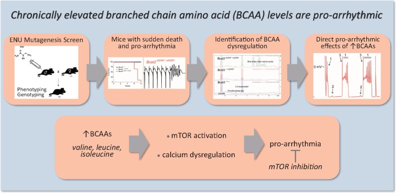
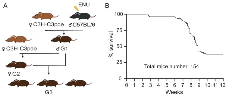
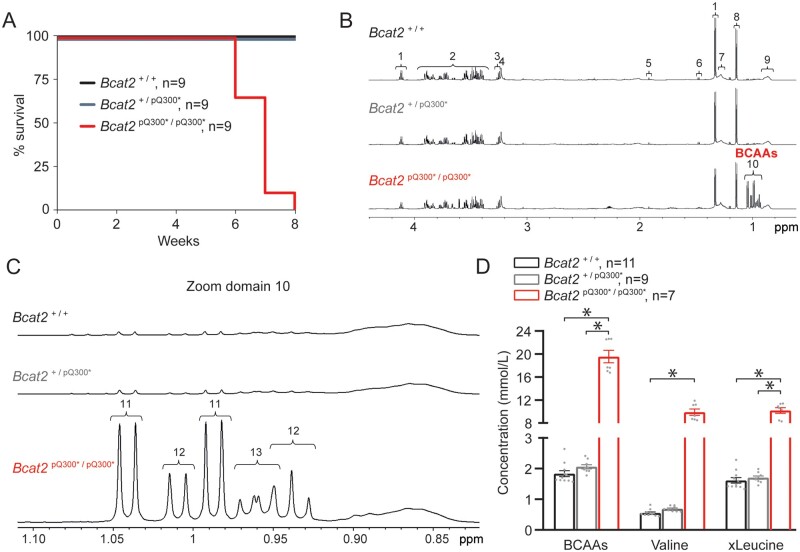
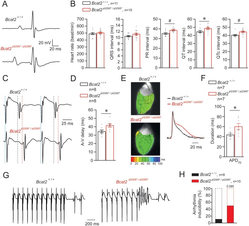
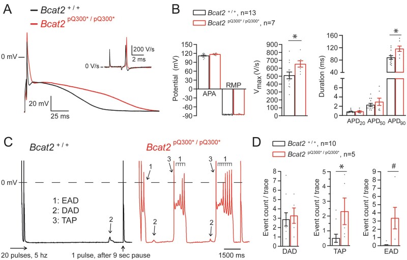
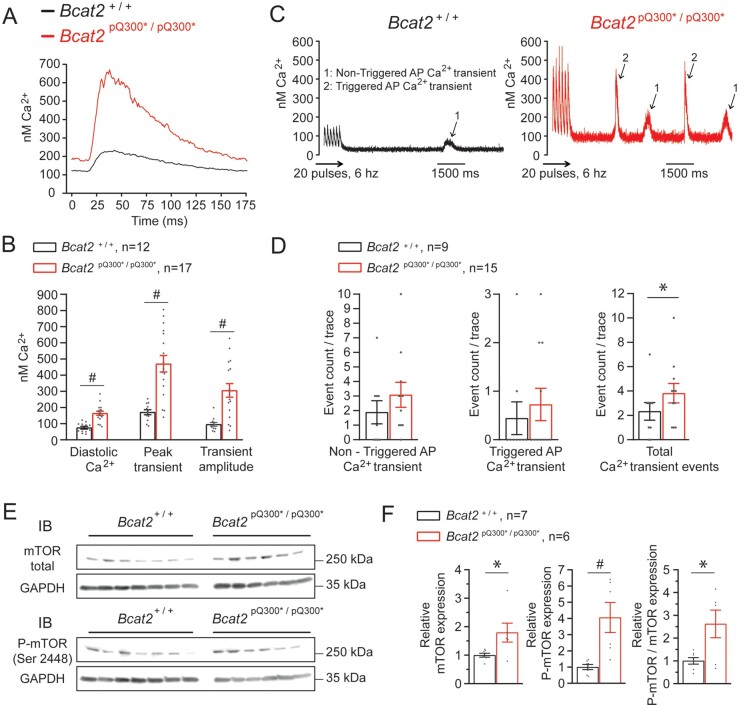
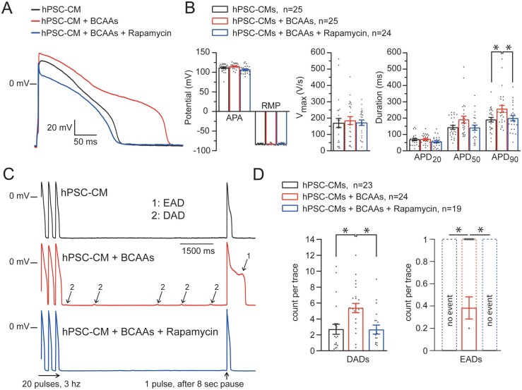
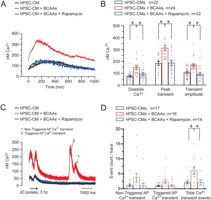
Methods and results: We employed a phenotype-driven N-ethyl-N-nitrosourea mutagenesis screen and identified a mouse line with a high incidence of sudden death at young age (6-9 weeks) in the absence of prior symptoms. Affected mice were found to be homozygous for the nonsense mutation Bcat2p.Q300*/p.Q300* in the Bcat2 gene encoding branched chain amino acid transaminase 2. At the age of 4-5 weeks, Bcat2p.Q300*/p.Q300* mice displayed drastic increase of plasma levels of branch chain amino acids (BCAAs-leucine, isoleucine, valine) due to the incomplete catabolism of BCAAs, in addition to inducible arrhythmias ex vivo as well as cardiac conduction and repolarization disturbances. In line with these findings, plasma BCAA levels were positively correlated to electrocardiogram indices of conduction and repolarization in the German community-based KORA F4 Study. Isolated cardiomyocytes from Bcat2p.Q300*/p.Q300* mice revealed action potential (AP) prolongation, pro-arrhythmic events (early and late afterdepolarizations, triggered APs), and dysregulated calcium homeostasis. Incubation of human pluripotent stem cell-derived cardiomyocytes with elevated concentration of BCAAs induced similar calcium dysregulation and pro-arrhythmic events which were prevented by rapamycin, demonstrating the crucial involvement of mTOR pathway activation.

Conclusions: Our findings identify for the first time a causative link between elevated BCAAs and arrhythmia, which has implications for arrhythmogenesis in conditions associated with BCAA metabolism dysregulation such as diabetes, metabolic syndrome, and heart failure.

Keywords: Arrhythmia; BCAA; Electrophysiology; Metabolism; Sudden death.


Figures

Graphical Abstract
Graphical Abstract
Figure 1
Figure 1
ENU mutagenesis-induced sudden death phenotype in young mice. (A) Breeding scheme used to produce G3 phenotyping cohorts. C57BL/6J males were mutagenized with ENU and crossed to C3H.Pde6b+ females. G1 males were then crossed again to C3H.Pde6b+ females, producing a G2 generation. Finally, G2 females were crossed to the G1 male parent to produce a G3 phenotyping cohort. (B) Mortality curve of the initial G3 phenotyping pedigrees (batch A; n = 154 mice) demonstrating a clear early death phenotype between 6 and 9 weeks of age.
Figure 2
Figure 2
Homozygous Bcat2p.Q300*/p.Q300* mice display sudden death and increased plasma BCAA levels at young age. (A) Mortality curve displaying death of all homozygous Bcat2p.Q300*/p.Q300* mice (batch B) by the age of 8 weeks. (B) Illustrative water suppressed spin-echo proton NMR spectra of free-fed plasma from Bcat2+/+, Bcat2+/p.Q300*, and Bcat2p.Q300*/p.Q300* mice at 5 weeks of age. The NMR spectra were plotted such that the αC1 proton of glucose at 5.23 ppm (not shown) was of similar intensity in each data set. The prominent metabolites shown in the expanded region are assigned to: (1) lactate, (2) glucose, (3) including β-glucose, (4) choline-containing compounds, (5) acetate, (6) alanine, (7) mobile lipid CH2, (8) 2,3-butanediol, (9) mobile lipid CH3, (10) branched chain amino acids (BCAAs). (C) NMR spectra from Bcat2+/+, Bcat2+/p.Q300*, Bcat2p.Q300*/p.Q300* further expanded (zoom domain 10) to illustrate the branched chain amino acid -CH3 region with metabolites assigned to (11) valine, (12) isoleucine, (13) leucine. (D) BCAA plasmatic quantification in Bcat2+/+ (n = 11), Bcat2+/p.Q300* (n = 9) and Bcat2p.Q300*/p.Q300* (n = 7) mice. *P < 0.05, one-way ANOVA on ranks with post hoc Dunn’s method.
Figure 3
Figure 3
Homozygous Bcat2p.Q300*/p.Q300* mice display ECG abnormalities in vivo and atrio-ventricular delay, prolonged repolarization and arrhythmia inducibility ex vivo. (A) Typical ECG traces obtained under isoflurane anaesthesia for Bcat2+/+ and Bcat2p.Q300*/p.Q300* mice. (B) Average values for heart rate, QRS interval, PR interval, QT and QTc (corrected) for both Bcat2+/+ (n = 11) and Bcat2p.Q300*/p.Q300* (n = 15) mice aged 4–5 weeks. (C) Typical examples of atrio-ventricular (AV) delay measurements in Langendorff-perfused isolated hearts (right atrial stimulation at a basic cycle length of 120 ms). (D) Average values for AV-delay in Bcat2+/+ and Bcat2p.Q300*/p.Q300* hearts. (E) Typical left ventricular repolarization maps obtained from optical mapping experiments on perfused explanted hearts during central stimulation (6.6 Hz). (F) Average APD70 (action potential duration at 70% of repolarization) values for Bcat2+/+ (n = 7) and Bcat2p.Q300*/p.Q300* (n = 7) mice. *P < 0.05, Student’s t-test. (G) Typical example of a short run of ventricular tachycardia induced in an isolated, Langendorff-perfused Bcat2p.Q300*/p.Q300* heart following a short-coupled paced beat. (H) Bar graph representing the percentage of arrhythmic events recorded on Bcat2+/+ and Bcatp.Q300*/p.Q300* mouse explanted hearts. *P < 0.05, Student’s t-test.
Figure 4
Figure 4
Abnormal repolarization and pro-arrhythmic events in Bcatp.Q300*/p.Q300* isolated cardiomyocytes. (A) Typical examples of action potentials (AP) and upstroke velocities (dV/dtmax; inset) elicited at a stimulation frequency of 2 Hz in left ventricular (LV) isolated cardiomyocytes from Bcat2+/+ and Bcat2p.Q300*/p.Q300* mice aged 4–5 weeks. (B) Average values for APA (action potential amplitude), RMP (resting membrane potential), Vmax (upstroke velocity), and APD (action potential duration) at 20%, 50%, and 90% repolarization (APD20, APD50, and APD90) of LV cardiomyocytes isolated from Bcat2+/+ (n = 15 cells from 6 independent cardiomyocyte dissociations) and Bcat2p.Q300*/p.Q300* (n = 9 cells from 5 independent cardiomyocyte dissociations) mice. (C) Typical examples of early afterdepolarizations (EADs: 1), delayed afterdepolarizations (DADs: 2), and triggered action potentials (TAPs: 3) recorded after a fast pacing stimulation protocol (20 pulses at 5 Hz followed by a 9 s pause and 1 pulse followed by a 500 ms pause) in Bcat2+/+ and Bcat2p.Q300*/p.Q300* LV cardiomyocytes. (D) Average count per trace for DADs, TAPs and EADs observed in Bcat2+/+ (n = 10 cardiomyocytes from 6 mice) and Bcat2p.Q300*/p.Q300* mice (n = 5 cardiomyocytes from 4 mice). Average numbers were calculated using five consecutive traces. (with Student’s t-test or Mann–Whitney Rank sum test).
Figure 5
Figure 5
Abnormal intracellular calcium and myocardial mTOR upregulation in Bcat2p.Q300*/p.Q300* mice. (A) Typical intracellular calcium transient recording performed at a pacing frequency of 6 Hz and (B) average values for diastolic Ca2+, peak transient and transient amplitude of LV cardiomyocytes from Bcat2+/+ (n = 12 cardiomyocytes from 5 independent cardiomyocyte dissociations) and Bcat2p.Q300*/p.Q300* (n = 17 cardiomyocytes from 4 independent cardiomyocyte dissociations) mice aged 4-5 weeks. (C) Typical example of calcium after-transients in isolated cardiomyocytes from Bcat2+/+ and Bcat2p.Q300*/p.Q300* cardiomyocytes following a fast pacing protocol using field stimulation and (D) quantification of non-triggered action potential (AP) Ca2+ transient, triggered AP Ca2+ transient and total Ca2+ transient events in Bcat2+/+ (n = 9 cardiomyocytes from 3 mice) and Bcat2p.Q300*/p.Q300* mice (n = 15 cardiomyocytes from 4 mice). (E) Representative immunoblots of total mTOR, phosphorylated mTOR (P-mTOR) at ser2448 and GAPDH from heart lysates (apex) from Bcat2+/+ and Bcat2p.Q300*/p.Q300* mice. (F) Average total mTOR and phosphorylated P-mTOR expression normalized to GAPDH, and ratio of normalized P-mTOR on normalized total mTOR from Bcat2+/+ (n = 7) and Bcat2p.Q300*/p.Q300* (n = 6) mouse heart lysates.*P < 0.05; #P ≤ 0.001 (with Student’s t-test or Mann–Whitney Rank sum test).
Figure 6
Figure 6
Increased levels of BCAAs recapitulate the pro-arrhythmic phenotype in hPSC derived cardiomyocytes (hPSC-CMs) which is reversible upon mTOR inhibition. (A) Typical example of APs elicited at a stimulation frequency of 1 Hz from hPSC-CMs incubated with control medium or with increased levels of BCAAs in the presence or absence of the mTOR inhibitor rapamycin (500 nmol/L). (B) Average values for APA (AP amplitude), RMP (resting membrane potential), Vmax (upstroke velocity), and APD (action potential duration) at 20%, 50%, and 90% repolarization (APD20, APD50, and APD90) of hPSC-CMs incubated with the control medium (n = 25 hPSC-CMs from 5 dissociations), with increased levels of BCAAs (n = 25 hPSC-CMs from 5 dissociations), and with increased BCAA levels and rapamycin (n = 24 hPSC-CMs from 5 dissociations). (C) Typical examples of EADs (1) and DADs (2) recorded after a fast-pacing stimulation protocol (20 pulses at 3 Hz followed by an 8 s pause and 1 pulse followed by a 1 s pause). (D) Average count per trace for DADs and EADs observed in hPSC-CMs incubated with the control medium (n = 23 hPSC-CMs from 5 dissociations), with increased levels of BCAAs (n = 24 hPSC-CMs from 5 dissociations), and with increased BCAA levels and rapamycin (n = 19 hPSC-CMs from 5 dissociations). Average numbers were calculated using five consecutive traces. *P < 0.05, one-way ANOVA (with Student–Newman–Keuls Method post hoc test) or one-way ANOVA on ranks (with Dunn’s method post hoc test).
Figure 7
Figure 7
Abnormal intracellular calcium regulation in human PSC derived cardiomyocytes (hPSC-CMs) with elevated BCAAs is reversible upon mTOR inhibition. (A) Typical examples of intracellular calcium transient recordings in hPSC-CMs performed at a pacing frequency of 1 Hz. (B) Average values for diastolic Ca2+, peak transient and transient amplitude with control medium (n = 22 hPSC-CMs from 3 dissociations), increased BCAA levels (n = 24 hPSC-CMs from 3 dissociations), and with increased BCAA levels and rapamycin at 500 nmol.L−1 (n = 22 hPSC-CMs from 3 dissociations). (C) Typical example of calcium after-transients in hPSC-CMs incubated with a control medium or with increased levels of BCAAs following a fast-pacing protocol using field stimulation and (D) quantification of non-triggered action potential (AP) Ca2+ transient, triggered AP Ca2+ transient and total Ca2+ transient events in hPSC-CMs incubated with the control medium (n = 17 hPSC-CMs from 2 dissociations), with increased levels of BCAAs (n = 16 hPSC-CMs from 2 dissociations), and with increased BCAA levels and rapamycin (n = 14 hPSC-CMs from 2 dissociations). *P < 0.05, one-way ANOVA on ranks (with Dunn’s method post hoc test).

References

    1. Deo R, Albert CM.. Epidemiology and genetics of sudden cardiac death. Circulation 2012;125:620–637. - PMC - PubMed
    1. Priori SG, Blomström-Lundqvist C, Mazzanti A, Blom N, Borggrefe M, Camm J, Elliott PM, Fitzsimons D, Hatala R, Hindricks G, Kirchhof P, Kjeldsen K, Kuck K-H, Hernandez-Madrid A, Nikolaou N, Norekvål TM, Spaulding C, Veldhuisen DV; ESC Scientific Document Group .2015 ESC Guidelines for the management of patients with ventricular arrhythmias and the prevention of sudden cardiac death: the Task Force for the Management of Patients with Ventricular Arrhythmias and the Prevention of Sudden Cardiac Death of the European Society of Cardiology (ESC). Endorsed by: association for European Paediatric and Congenital Cardiology (AEPC). Eur Heart J 2015;36:2793–2867. - PubMed
    1. Hoeijen DV, Blom MT, Bardai A, Souverein PC, Boer AD, Tan HL.. Reduced pre-hospital and in-hospital survival rates after out-of-hospital cardiac arrest of patients with type-2 diabetes mellitus: an observational prospective community-based study. Europace 2015;17:753–760. - PubMed
    1. Jouven X, Lemaître RN, Rea TD, Sotoodehnia N, Empana J-P, Siscovick DS.. Diabetes, glucose level, and risk of sudden cardiac death. Eur Heart J 2005;26:2142–2147. - PubMed
    1. Tomaselli GF, Zipes DP.. What causes sudden death in heart failure? Circ Res 2004;95:754–763. - PubMed

Publication types

LinkOut - more resources