. 2021 Oct;56(5):982-990.
doi: 10.1111/jre.12911. Epub 2021 Jun 17.

Pyrophosphate inhibits periodontal ligament stem cell differentiation and mineralization through MAPK signaling pathways

Affiliations

Pyrophosphate inhibits periodontal ligament stem cell differentiation and mineralization through MAPK signaling pathways

Yongxi Liang et al. J Periodontal Res. 2021 Oct.

Abstract

Background and objective: Periodontal ligament stem cells (PDLSCs) are the primary cell source for the regeneration and remodeling of periodontal ligament (PDL). It is crucial to prevent PDLSCs from mineralization when using the PDLSCs for PDL regeneration. At present, little is known about how to inhibit PDLSC mineralization. This study investigates the effects of pyrophosphate (PPi) on inhibiting PDLSC osteogenic differentiation and mineralization as well as the underlying mechanism.

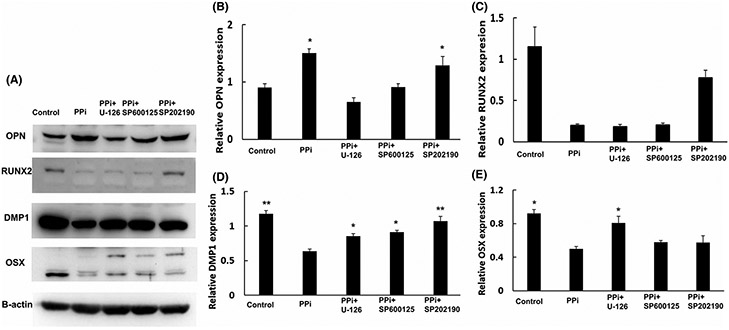
Materials and methods: Human PDLSCs were cultured in an osteogenic differentiation medium with different PPi concentrations (0, 10, or 100 μM). The effects of PPi on osteogenic differentiation were assessed by ALP activity and the expressions of osteogenic related proteins (OPN, RUNX2, OSX, and DMP1). The mineralization formation was detected by alizarin red staining. The activation of MAPK signaling pathways (ERK1/2, JNK, and p38) was determined by western blotting and pathway blockade assays. The gene expressions of PPi's regulators (Ank, Enpp1, and Alpl) were assessed by real-time PCR.

Results: Both low and high concentrations (10 μM and 100 μM) of PPi inhibited the mineralization of PDLSCs. The addition of PPi (10 μM or 100 μM) decreased the ALP activity of the PDLSCs to approximately two-thirds of the control group on day 3. PPi reduced the expressions of RUNX2, OSX, and DMP1 on days 7, 14, and 21, while it increased the expression of OPN at the three time points. PPi enhanced the phosphorylation of MAPK pathways, and the application of corresponding MAPK pathway inhibitors reversed the osteogenic inhibition effects of PPi.

Conclusion: PPi inhibits the osteogenic differentiation and mineralization of PDLSCs in vitro through activating ERK1/2, JNK, and p38 signaling pathways.

Keywords: inhibitor; mineralization; osteogenic differentiation; periodontal ligament stem cells; pyrophosphate.


Conflict of interest statement

CONFLICT OF INTERESTS

The authors declare no conflict of interests.

Figures

FIGURE 1
FIGURE 1
ALP activity of PDLSCs treated with PPi. (A) ALP staining of PDLSCs treated with osteogenic medium and different concentrations of PPi for 3 and 7 days. (B) Quantification of the ALP activity. On day 3, *p < 0.05 was compared to the 10 μM group. On day 7, *p < 0.05 was compared to the 100 μM group
FIGURE 2
FIGURE 2
Expressions of the osteogenic markers of PDLSCs treated with PPi. (A) Western blot images of OPN, RUNX2, DMP1, OSX, and β-actin. (B-E) The relative expressions of the osteogenic markers compared to β-actin. In (B),*p < 0.05 was compared to the control group in each time point. In (C), *p < 0.05 was compared to the 100 μM group on days 14 and 21. In (D) on day 7: *p < 0.05 was compared to the control group. On day 14 and 21, *p < 0.05 was compared to the 100 μM group. In (E), *p < 0.05 was compared to the 100 μM group on days 14 and 21
FIGURE 3
FIGURE 3
PPi inhibited mineralized tissue formation of PDLSCs. (A) Alizarin staining of PDLSCs treated with an osteogenic medium and different PPi concentrations for 14 and 21 days. (B) Quantification of the calcified nodules. *p < 0.05 was compared to the 100 μM group on day 14. **p < 0.05 was compared to 10 μM group on day 21. ***p < 0.05 was compared to the control group on day 14.
FIGURE 4
FIGURE 4
The protein expressions of phosphorylated/unphosphorylated ERK1/2, JNK, and p38 of PDLSCs treated with PPi. (A) Western blot images of phosphorylated/unphosphorylated ERK1/2 and β-actin, with quantification the relative expression of phosphorylated ERK1/2. 15 and 60 min: *p < 0.05 compare to 0 μM group. (B) Western blot images of phosphorylated/unphosphorylated JNK and β-actin, with quantification the relative expression of phosphorylated JNK. 7.5 min: *p < 0.05 compare to 0 μM group; 15 min: *p < 0.05 compare to 100 μM group. (C) Western blot images of phosphorylated/unphosphorylated p38 and β-actin, with quantification the relative expression of phosphorylated p38. *p < 0.05 compare to 0 μM group in each time points; ** p < 0.05 compare to 10 μM group in each time points
FIGURE 5
FIGURE 5
The protein expressions of osteogenic markers of PDLSC with PPi treatment after inhibiting ERK1/2, JNK, and p38 pathway for 21 days. (A) Western blot images of OPN, RUNX2, OSX, DMP1, and β-actin. (B-E) The relative expressions of OPN, RUNX2, DMP1, and OSX. In (B), *p < 0.05 was compared to ERK 1/2 inhibitor group. In (C), *p < 0.05 was compared to PPi only group. In (D), *p < 0.05 was compared to PPi only group. In (E), *p < 0.05 was compared to PPi only group; **p < 0.05 was compared to the ERK1/2 inhibitor group
FIGURE 6
FIGURE 6
The effects of PPi on the gene expressions of its regulators. (A) Ank expression after PPi treatment. *p < 0.05 was compared to the control group on day 3. (B) Enpp1 expression after PPi treatment. *p < 0.05 was compared to the control group and **p < 0.05 was compare to the 10 μM group on day 3. (C) Alpl expression after PPi treatment. *p < 0.05 was compared to the 10 μM group on day 3

Similar articles

Cited by

References

    1. Deas DE, Moritz AJ, Sagun RS Jr, Gruwell SF, Powell CA. Scaling and root planing vs. conservative surgery in the treatment of chronic periodontitis. Periodontology. 2000;2016(71):128–139. - PubMed
    1. Graziani F, Karapetsa D, Alonso B, Herrera D. Nonsurgical and surgical treatment of periodontitis: how many options for one disease? Periodontology. 2000;2017(75):152–188. - PubMed
    1. Smiley CJ, Tracy SL, Abt E, et al. Evidence-based clinical practice guideline on the nonsurgical treatment of chronic periodontitis by means of scaling and root planing with or without adjuncts. J Am Dent Assoc. 2015;146:525–535. - PubMed
    1. John MT, Michalowicz BS, Kotsakis GA, Chu H. Network meta-analysis of studies included in the Clinical Practice Guideline on the nonsurgical treatment of chronic periodontitis. J Clin Periodontol. 2017;44:603–611. - PMC - PubMed
    1. Liang Y, Luan X, Liu X. Recent advances in periodontal regeneration: a biomaterial perspective. Bioactive materials. 2020;5:297–308. - PMC - PubMed