. 2021 Jun 25;478(12):2247-2263.
doi: 10.1042/BCJ20210283.

Co-ordinated control of the Aurora B abscission checkpoint by PKCε complex assembly, midbody recruitment and retention

Affiliations

Co-ordinated control of the Aurora B abscission checkpoint by PKCε complex assembly, midbody recruitment and retention

Lisa Watson et al. Biochem J. .

Abstract

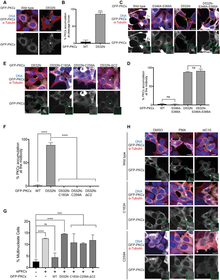
A requirement for PKCε in exiting from the Aurora B dependent abscission checkpoint is associated with events at the midbody, however, the recruitment, retention and action of PKCε in this compartment are poorly understood. Here, the prerequisite for 14-3-3 complex assembly in this pathway is directly linked to the phosphorylation of Aurora B S227 at the midbody. However, while essential for PKCε control of Aurora B, 14-3-3 association is shown to be unnecessary for the activity-dependent enrichment of PKCε at the midbody. This localisation is demonstrated to be an autonomous property of the inactive PKCε D532N mutant, consistent with activity-dependent dissociation. The C1A and C1B domains are necessary for this localisation, while the C2 domain and inter-C1 domain (IC1D) are necessary for retention at the midbody. Furthermore, it is shown that while the IC1D mutant retains 14-3-3 complex proficiency, it does not support Aurora B phosphorylation, nor rescues division failure observed with knockdown of endogenous PKCε. It is concluded that the concerted action of multiple independent events facilitates PKCε phosphorylation of Aurora B at the midbody to control exit from the abscission checkpoint.

Keywords: 14-3-3; abscission checkpoint; aurora kinases; midbody; protein kinase C.


Conflict of interest statement

The authors declare that there are no competing interests associated with the manuscript.

Figures

Figure 1.
Figure 1.. A S350 dephosphorylated PKCε-14-3-3 complex is required for Aurora B S227 phosphorylation at the midbody.
(A) Schematic representing the organisation of PKCε, highlighting the three serine residues of the V3 domain required for 14-3-3 binding denoted in red alongside the kinases responsible for their phosphorylation. (B) Quantitation of multinucleate cells upon treatment of cells with non-targeting siRNA (control) or PKCε siRNA, with expression of siRNA-resistant mouse GFP-PKCε, GFP-PKCε-D532N, GFP-PKCε-S346A-S368A or GFP-PKCε-S350A. Values plotted are means for n = 3 experiments, with error bars denoting SD. Two-hundred cells were counted per condition per experiment. One-way analysis of variance: **** = P ≤ 0.0001; *** = P ≤ 0.001; ns (not significant) = P > 0.05. (C) Representative immunofluorescence images of DLD1 FRT-TREx cells treated with non-targeting siRNA (control) or PKCε siRNA, with expression of either siRNA-resistant mouse GFP-PKCε, GFP-PKCε-D532N or GFP-PKCε-S346A-S368A induced with doxycycline. Cells stained for DNA (blue), tubulin (red) and Aurora B Ser227 phosphorylation (green), with GFP-PKCε in white if expressed. The Aurora B S227 phosphorylation is highlighted in the lower greyscale panels with the midbody region indicated expanded for each image. Scale bar = 20µm. (D) Quantitation of cells with Aurora B pSer227 signal at the midbody. DLD1 FRT-TREx cells with inducible expression of GFP-PKCε, GFP-PKCε-D532N or GFP-PKCε-S346A-S368A were treated with either non-targeting siRNA (control), PKCε siRNA or PKCε siRNA and doxycycline to induce expression of siRNA-resistant mouse GFP-PKCε constructs, and the Aurora B pSer227 signal at the midbody quantified. Values plotted are means for n = 3 experiments, with error bars denoting SD. Twenty cells with midbodies were counted per condition per experiment. One-way analysis of variance: **** = P ≤ 0.0001; ns (not significant) = P > 0.05. (E) Schematic of incorporation of the diazirine amino acid AbK (red star) into the V3 domain (N371AbK) of PKCε. (FG) Western blots probed with (F) PKCε antibody and (G) phosphoSer346 antibody, showing cross-linking in HEK293T cells expressing GFP-PKCε-N371AbK, GFP-PKCε-N371AbK-S346A or GFP-PKCε-N371AbK-S350A upon exposure to 365 nm UV for 10 min. The upper and lower arrowheads indicate cross-linked (X-link) PKCε and uncross-linked PKCε, respectively. Western blot images are a representative example of three independent experiments. (H) Table showing the number of PKCε peptide spectrum matches (#PSM) identified in the mass spectrometry analysis in samples containing uncross-linked PKCε (PKCε) vs samples containing PKCε cross-linked to 14-3-3 (PKCε-14-3-3). (I) The percentage of the different types of V3 domain PKCε derived phosphopeptides identified in the uncross-linked and 14-3-3 cross-linked sampled.
Figure 2.
Figure 2.. C1 and C2 domains are required for retention and/or recruitment of PKCε to the midbody region.
(A) Representative immunofluorescence images of HEK293T cells expressing GFP-PKCε and GFP-PKCε-D532N. Cells stained for DNA (blue) and tubulin (red), with PKCε or PKCε-D532N shown in grey. Scale bar = 20 µm. (B) Quantitation of dividing cells with GFP-PKCε and GFP-PKCε-D532N accumulation at the midbody. Values plotted are means for n = 3 experiments, with error bars denoting SD. Thirty cells with midbodies were observed per condition per experiment. (C) Representative immunofluorescence images of dividing HEK293T cells expressing GFP-PKCε, GFP-PKCε-S346A-S368A, GFP-PKCε-D532N or GFP-PKCε-D532N-S346A-S368A. Cells stained for DNA (blue) and tubulin (red), with GFP-PKCε shown in grey; the midbody region highlighted is expanded for each image. Scale bar = 20 µm. (D) Quantitation of dividing cells with GFP-PKCε, GFP-PKCε-S346A-S368A, GFP-PKCε-D532N or GFP-PKCε-D532N-S346A-S368A accumulation at the midbody. Values plotted are means for n = 3 experiments, with error bars denoting SD. Thirty cells with midbodies were observed per condition per experiment. (E) Representative immunofluorescence images of dividing HEK293T cells expressing GFP-PKCε-D532N, GFP-PKCε-D532N-C183A, GFP-PKCε-D532N-C259A or GFP-PKCε-D532N-ΔC2. Cells stained for DNA (blue) and tubulin (red), with GFP-PKCε shown in grey; the midbody region highlighted is expanded for each image. Scale bar = 20 µm. (F) Quantitation of dividing cells with GFP-PKCε-D532N, GFP-PKCε-D532N-C183A, GFP-PKCε-D532N-C259A or GFP-PKCε-D532N-ΔC2 accumulation at the midbody. Values plotted are means for n = 3 experiments, with error bars denoting SD. Thirty cells with midbodies were observed per condition per experiment. (G) Quantitation of multinucleate cells upon treatment with non-targeting siRNA (control) or PKCε siRNA, with expression of siRNA-resistant mouse GFP-PKCε, GFP-PKCε-D532N, GFP-PKCε-C183A, GFP-PKCε-C259A or GFP-PKCε-ΔC2. Values plotted are means for n = 3 experiments, with error bars denoting SD. Two-hundred cells were counted per condition per experiment. (H) Representative immunofluorescence images of HEK293T cells expressing GFP-PKCε, GFP-PKCε-C183A or GFP-PKCε-C259A and treated with DMSO, PMA or diC10. Cells were treated for 20 min with 1 µM PMA, or 3 min with 10 µM diC10. Cells stained for DNA (blue) and tubulin (red), with GFP-PKCε shown in grey; the midbody region highlighted is expanded for each image. Twenty cells with midbodies were observed per condition across three experiments. Scale bar = 20 µm.
Figure 3.
Figure 3.. The IC1D domain is necessary for retention but not recruitment to the midbody region.
(A) Schematic representing the organisation of PKCε, with the residues of the Inter-C1 Domain (IC1D) noted with the three residues that make up the actin binding motif in red, and the two phosphorylation sites in blue and green. (B) Representative immunofluorescence images of dividing HEK293T cells expressing GFP-PKCε, GFP-PKCε-D532N, GFP-PKCε-D532N-L223A-K224A-E227A, GFP-PKCε-D532N-T228A or GFP-PKCε-D532N-S234A. Cells stained for DNA (blue) and tubulin (red), with GFP-PKCε shown in grey; the midbody region highlighted is expanded for each image. Scale bar = 20 µm. (C) Quantitation of dividing cells with GFP-PKCε, GFP-PKCε-D532N, GFP-PKCε-D532N-L223A-K224A-E227A, GFP-PKCε-D532N-T228A or GFP-PKCε-D532N-S234A accumulation at the midbody. Values plotted are means for n = 3 experiments, with error bars denoting SD. Thirty cells with midbodies were observed per condition per experiment. One-way analysis of variance: **** = P ≤ 0.0001; *** = P ≤ 0.001; ns (not significant) = P > 0.05. (D) Representative immunofluorescence images of HEK293T cells expressing GFP-PKCε, GFP-PKCε-D532N or GFP-PKCε-L223A-K224A-E227A and treated with DMSO, PMA or diC10 as indicated. Cells were treated for 20 min with 1 µM PMA, or 3 min with 10 µM diC10. Cells stained for DNA (blue) and tubulin (red), with GFP-PKCε shown in grey; the midbody region highlighted is expanded for each image. Twenty cells with midbodies were observed per condition across three experiments. Scale bar = 20 µm.
Figure 4.
Figure 4.. Retention at the midbody region is required for Aurora B S227 phosphorylation.
(A) Representative immunofluorescence images of DLD1 FRT-TREx cells treated with non-targeting siRNA (control) or PKCε siRNA, with expression of either siRNA-resistant mouse GFP-PKCε, GFP-PKCε-D532N or GFP-PKCε-L223-K224-E227A induced with doxycycline. Cells stained for DNA (blue), tubulin (red) and Aurora B pSer227 (green), with GFP-PKCε in white if expressed. The Aurora B S227 phosphorylation is highlighted in the lower greyscale panels with the midbody region indicated expanded for each image. Scale bar = 20 µm. (B) Quantitation of cells with Aurora B pSer227 signal at the midbody. DLD1 FRT-TREx cells with inducible expression of GFP-PKCε, GFP-PKCε-D532N or GFP-PKCε-L223-K224-E227A were treated with either non-targeting siRNA (control), PKCε siRNA or PKCε siRNA and doxycycline to induce expression of siRNA-resistant mouse GFP-PKCε constructs, and the Aurora B pSer227 signal at the midbody quantified. Values plotted are means for n = 3 experiments, with error bars denoting SD. Twenty cells with midbodies were counted per condition per experiment. One-way analysis of variance: **** = P ≤ 0.0001; ns (not significant) = P > 0.05. (C) Representative immunofluorescence images of HEK293T cells treated with non-targeting siRNA (control) or PKCε siRNA, with expression of siRNA-resistant mouse GFP-PKCε, GFP-PKCε-D532N or GFP-PKCε-L223-K224-E227A. Cells stained for DNA (blue) and tubulin (grey), with GFP-PKCε in green. Scale bar = 10 µm; red asterisks indicate multinucleate cells. (D) Quantitation of multinucleate cells upon treatment with non-targeting siRNA (control) or PKCε siRNA, with expression of siRNA-resistant mouse GFP-PKCε, GFP-PKCε-D532N or GFP-PKCε-L223-K224-E227A. Values plotted are means for n = 3 experiments, with error bars denoting SD. Two-hundred cells were counted per condition per experiment. One-way analysis of variance: *** = P ≤ 0.001; ** = P ≤ 0.01; ns (not significant) = P > 0.05. (E) Working model of PKCε turnover at midbody and mediation of exit from the abscission checkpoint. The different states of PKCε are indicated alongside the involved upstream regulators. Predicted compartments are indicated by coloured boxes. The endpoint of the partner bound, membrane associated, 14-3-3 complex is the form of PKCε required for Aurora B S227 phosphorylation at the midbody and exit from the abscission checkpoint.

Similar articles

Cited by

References

    1. Christ, L., Raiborg, C., Wenzel, E. M., Campsteijn, C. and Stenmark, H. (2017) Cellular functions and molecular mechanisms of the ESCRT membrane-scission machinery. Trends Biochem. Sci. 42, 42–56 10.1016/j.tibs.2016.08.016 - DOI - PubMed
    1. Petsalaki, E. and Zachos, G. (2019) Building bridges between chromosomes: novel insights into the abscission checkpoint. Cell Mol. Life Sci. 76, 4291–4307 10.1007/s00018-019-03224-z - DOI - PMC - PubMed
    1. Norden, C., Mendoza, M., Dobbelaere, J., Kotwaliwale, C. V., Biggins, S. and Barral, Y. (2006) The NoCut pathway links completion of cytokinesis to spindle midzone function to prevent chromosome breakage. Cell 125, 85–98 10.1016/j.cell.2006.01.045 - DOI - PubMed
    1. Barr, F. A. and Gruneberg, U. (2007) Cytokinesis: placing and making the final cut. Cell 131, 847–860 10.1016/j.cell.2007.11.011 - DOI - PubMed
    1. Carlton, J. G., Caballe, A., Agromayor, M., Kloc, M. and Martin-Serrano, J. (2012) ESCRT-III governs the Aurora B-mediated abscission checkpoint through CHMP4C. Science 336, 220–225 10.1126/science.1217180 - DOI - PMC - PubMed

Publication types