. 2021 Jul 13;16(7):1818-1831.
doi: 10.1016/j.stemcr.2021.05.016. Epub 2021 Jun 17.

Auxin-degron system identifies immediate mechanisms of OCT4

Affiliations

Auxin-degron system identifies immediate mechanisms of OCT4

Lawrence E Bates et al. Stem Cell Reports. .

Abstract

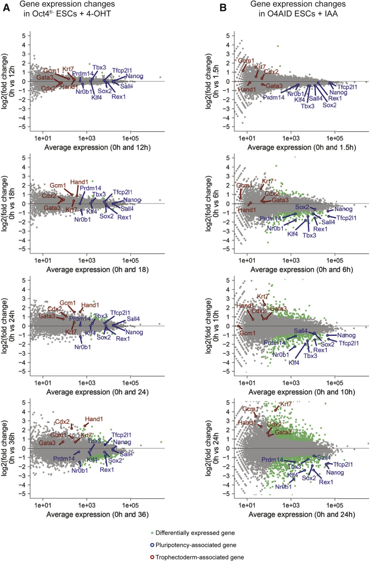
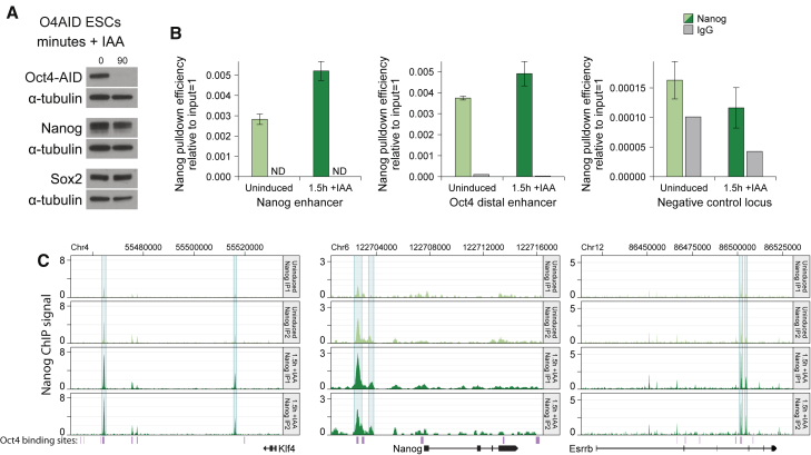
The pluripotency factor OCT4 is essential for the maintenance of naive pluripotent stem cells in vitro and in vivo. However, the specific role of OCT4 in this process remains unknown. Here, we developed a rapid protein-level OCT4 depletion system that demonstrates that the immediate downstream response to loss of OCT4 is reduced expression of key pluripotency factors. Our data show a requirement for OCT4 for the efficient transcription of several key pluripotency factors and suggest that expression of trophectoderm markers is a subsequent event. In addition, we find that NANOG is able to bind to the genome in the absence of OCT4, and this binding is in fact enhanced. Globally, however, the active enhancer-associated histone mark H3K27ac is depleted. Our work establishes that, while OCT4 is required for the maintenance of the naive transcription factor network, at a normal embryonic stem cell levels it antagonizes this network through inhibition of NANOG binding.

Keywords: Nanog; Oct4; Pou5f1; auxin-inducible degron; embryonic stem cells; mouse nPSCs.


Figures

None
Graphical abstract
Figure 1
Figure 1
Auxin-degron-tagged OCT4 sustains nPSC self-renewal and permits rapid loss of OCT4 (A and B) The kinetics of OCT4 depletion in conventional Oct4Fl/− ESCs were examined. (A) Oct4 expression level (qRT-PCR) following addition of 4-OHT and medium change to SL. (B) OCT4 protein level (western blot) following addition of 4-OHT and medium change to SL. α-TUBULIN shown as a loading control. (C) Schematic showing the generation and use of O4AID ESCs. (D) Expression profiling (qRT-PCR) of pluripotency markers. (E) Oct4-AID fusion and wild-type OCT4 protein level (western blot) following addition of IAA. α-Tubulin was used as a loading control. (F) Schematic showing the generation and use of O4AID iPSCs. rOKM, retroviral Oct4, Klf4, and cMyc. (G) Expression profiling (qRT-PCR) of retroviruses and pluripotency factor Nanog in partially and fully reprogrammed cells. NSC, neural stem cell; r.Oct4, retroviral Oct4; r.cMyc, retroviral cMyc; r.Klf4, retroviral Klf4. (H) Oct4-AID fusion and wild-type protein level (western blot) following addition of IAA. qRT-PCR data represent the mean ± SD of three technical replicates. Dox, doxycycline; WT, wild type. See also Figure S1.
Figure 2
Figure 2
OCT4 is required for the expression of key pluripotency factors (A and B) MA plots showing gene expression (RNA-sequencing) changes following (A) deletion of Pou5f1 in conventional Oct4F/− CreER ESCs by addition of 4-OHT or (B) degradation of OCT4 protein in O4AID ESCs by addition of IAA. Differentially expressed genes are highlighted in green (q > 0.9, NOISeq-sim). Selected pluripotency- (blue) and differentiation- (red) associated genes are indicated. (A) Top to bottom: 0 versus 12, 0 versus 18, 0 versus 24, and 0 versus 36 h. (B) Top to bottom: 0 versus 1.5, 0 versus 6, 0 versus 10, and 0 versus 24 h. Note that for Oct4F/− CreER ESCs two replicate datasets were merged, and for O4AID ESCs a single replicate was used. A non-parametric algorithm designed for use on data lacking replicates was used for the analysis (see experimental procedures for more details). Key findings were corroborated by qRT-PCR analysis in two independent cell lines; see Figure S2.
Figure 3
Figure 3
OCT4 is required for enhancer activity at key pluripotency loci and for maintaining global H3K27ac (A) Mapped RNA-sequencing reads (single replicate) of enhancer RNAs at the Prdm14 distal enhancer (top) and the Klf4 distal enhancer (bottom) in O4AID ESCs before and 1.5 h after addition of IAA. (B) Visualization of H3K27ac ChIP-seq signal (signal per kilobase per million mapped reads) at the Prdm14 distal enhancer (top) and the Klf4 distal enhancer (bottom) in O4AID ESCs before and 1.5 h after addition of IAA. Genomic coordinates refer to the GRCm38/mm10 genome assembly, and gene intron/exon annotations are taken from Ensembl. OCT4 binding sites generated from ChIP-seq data from Marson et al. (2008) are indicated in purple. (C) Violin and box plot showing log2-fold change in H3K27ac signal between uninduced and 1.5 h IAA-treated O4AID ESCs. Data were generated by merging mapped ChIP-seq reads from three independent immunoprecipitations. Note that the key findings were corroborated by ChIP-qPCR in two independent cell lines (Figure S3B). Boxes show the median value and extend to the 25th and 75th quartiles, and whiskers extend to 1.5 times the interquartile range. All H3K27ac peaks (n = 26,457) above a (background) threshold and the complementary subsets of peaks within 1 kb of an OCT4 binding site (n = 4,662) and not within 1 kb of an OCT4 binding site (n = 21,795) are plotted. For each set a paired t test of H3K27ac signal before and after treatment showed highly significant change, p < 10−10. See also Figure S3.
Figure 4
Figure 4
OCT4 is dispensable for NANOG binding to pluripotent regulatory sequences (A) Protein level of OCT4 and NANOG (western blot) in O4AID ESCs before and 1.5 h after addition of IAA, with α-tubulin as a loading control. (B) ChIP qPCR following pull-down of NANOG or using normal immunoglobulin G (IgG) negative control at NANOG binding sites or a negative control locus in O4AID ESCs. ChIP qPCR data for NANOG pull-down represent the mean ± SD of three IPs; IgG pull-down represents the mean of three technical replicates of a single IP. ND, not detected. Note that this is corroborated by ChIP qPCR in an independent cell line (Figure S4). (C) Visualization of NANOG ChIP-seq signal (signal per kilobase per million mapped reads) across indicated loci before and 1.5 h after addition of IAA, shown for two IPs. Genomic coordinates refer to the GRCm38/mm10 genome assembly, and gene intron/exon annotations are taken from Ensembl. OCT4 binding sites generated from ChIP-seq data from Marson et al. (2008) are indicated in purple. See also Figure S4.
Figure 5
Figure 5
NANOG binding is increased globally, independent of OCT4 co-binding (A) Summary distribution (top) and heatmap (bottom) of NANOG signal (sum of mapped reads from two independent IPs, normalized to library size) centered at the summit of all NANOG binding sites across the genome, before and 1.5 h after addition of IAA. (B) Violin and box plot of log2 of the fold change in the average normalized NANOG signal at each NANOG peak in the genome (n = 16,608) before and after addition of IAA, further broken down into loci within 1 kb of (n = 3,930) or farther away than 1 kb from (n = 12,678) the OCT4 binding sites generated from ChIP-seq data from Marson et al. (2008). Mapped reads from two independent IPs each, before and after treatment, were merged to generate NANOG ChIP-seq data. Boxes show the median value and extend to the 25th and 75th quartiles, and whiskers extend to 1.5 times the interquartile range. (C) Euler plot showing the number of NANOG peaks assigned to various chromatin environments and the overlap between assignations. Numbers indicate the number of peaks uniquely in that section of the diagram. (D) Violin and box plot of log2 of the fold change in the average normalized NANOG signal (data as in [B]) at each NANOG peak in the genome before and 1.5 h after addition of IAA, further broken down into non-exclusive chromatin environments, as indicated in (C). Boxes show the median value and extend to the 25th and 75th quartiles, and whiskers extend to 1.5 times the interquartile range. (E) Model indicating the proposed relationship between the quantity of OCT4 protein and the capacity for NANOG to bind to the genome and consequently the ability of cells to maintain a naive identity. See also Figure S5.

References

    1. Abranches E., Bekman E., Henrique D. Generation and characterization of a novel mouse embryonic stem cell line with a dynamic reporter of nanog expression. PLoS One. 2013;8:e59928. - PMC - PubMed
    1. Alvarez-Dominguez J.R., Knoll M., Gromatzky A.A., Lodish H.F. The super-enhancer-derived alncRNA-EC7/bloodlinc potentiates red blood cell development in trans. Cell Rep. 2017;19:2503–2514. - PMC - PubMed
    1. Baker O., Gupta A., Obst M., Zhang Y., Anastassiadis K., Fu J., Stewart A.F. RAC-tagging: recombineering and Cas9-assisted targeting for protein tagging and conditional analyses. Sci. Rep. 2016;6:25529. - PMC - PubMed
    1. Le Bin G.C., Muñoz-Descalzo S., Kurowski A., Leitch H., Lou X., Mansfield W., Etienne-Dumeau C., Grabole N., Mulas C., Niwa H. Oct4 is required for lineage priming in the developing inner cell mass of the mouse blastocyst. Development. 2014;141:1001–1010. - PMC - PubMed
    1. Chambers I., Silva J., Colby D., Nichols J., Nijmeijer B., Robertson M., Vrana J., Jones K., Grotewold L., Smith A. Nanog safeguards pluripotency and mediates germline development. Nature. 2007;450:1230–1234. - PubMed

Publication types