

Integration of quantitative imaging biomarkers in clinical trials for MR-guided radiotherapy: Conceptual guidance for multicentre studies from the MR-Linac Consortium Imaging Biomarker Working Group
- PMID: 34144436
- PMCID: PMC8340311
- DOI: 10.1016/j.ejca.2021.04.041
Integration of quantitative imaging biomarkers in clinical trials for MR-guided radiotherapy: Conceptual guidance for multicentre studies from the MR-Linac Consortium Imaging Biomarker Working Group
Abstract
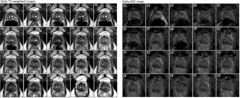
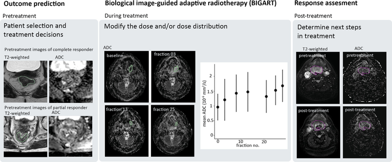
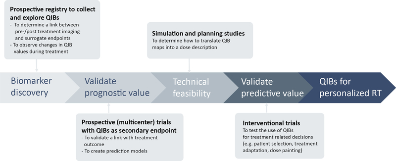
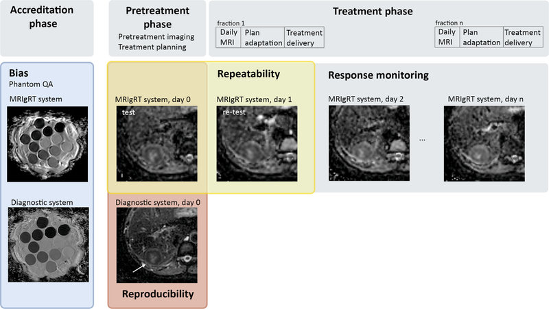
Quantitative imaging biomarkers (QIBs) derived from MRI techniques have the potential to be used for the personalised treatment of cancer patients. However, large-scale data are missing to validate their added value in clinical practice. Integrated MRI-guided radiotherapy (MRIgRT) systems, such as hybrid MRI-linear accelerators, have the unique advantage that MR images can be acquired during every treatment session. This means that high-frequency imaging of QIBs becomes feasible with reduced patient burden, logistical challenges, and costs compared to extra scan sessions. A wealth of valuable data will be collected before and during treatment, creating new opportunities to advance QIB research at large. The aim of this paper is to present a roadmap towards the clinical use of QIBs on MRIgRT systems. The most important need is to gather and understand how the QIBs collected during MRIgRT correlate with clinical outcomes. As the integrated MRI scanner differs from traditional MRI scanners, technical validation is an important aspect of this roadmap. We propose to integrate technical validation with clinical trials by the addition of a quality assurance procedure at the start of a trial, the acquisition of in vivo test-retest data to assess the repeatability, as well as a comparison between QIBs from MRIgRT systems and diagnostic MRI systems to assess the reproducibility. These data can be collected with limited extra time for the patient. With integration of technical validation in clinical trials, the results of these trials derived on MRIgRT systems will also be applicable for measurements on other MRI systems.
Keywords: Biomarkers; Image-guided; Magnetic resonance imaging; Multicentre study; Radiotherapy.
Copyright © 2021 Elsevier Ltd. All rights reserved.
Conflict of interest statement
Conflict of interest statement The authors declare the following financial interests/personal relationships which may be considered as potential competing interests: Dr. van der Heide receives industry grant support from Philips Healthcare and Elekta AB and ITEA project (‘Starlit’). Dr. Fuller received/receives funding and salary support related to this project from: the National Institutes of Health (NIH) National Institute of Biomedical Imaging and Bioengineering (NIBIB) Research Education Programs for Residents and Clinical Fellows Grant (R25EB025787-01); the National Institute for Dental and Craniofacial Research Establishing Outcome Measures Award (1R01DE025248/R56DE025248) and Academic Industrial Partnership Grant (R01DE028290); National Cancer Institute (NCI) Early Phase Clinical Trials in Imaging and Image-Guided Interventions Program (1R01CA218148); an NIH/NCI Cancer Center Support Grant (CCSG) Pilot Research Program Award from the UT MD Anderson CCSG Radiation Oncology and Cancer Imaging Program (P30CA016672); and an NCI-NSF Smart Connected Health Program (R01 CA257814). Direct infrastructure support is provided to Dr. Fuller by the multidisciplinary Stiefel Oropharyngeal Research Fund of the University of Texas MD Anderson Cancer Center Charles and Daneen Stiefel Center for Head and Neck Cancer and the Cancer Center Support Grant (P30CA016672) and the MD Anderson Program in Image-guided Cancer Therapy. Dr. Fuller has received direct industry grant support, in-kind support, honoraria, and travel funding from Elekta AB related to this project. Ms. McDonald receives salary and funding support from the NIH NIDCR Ruth L. Kirschstein National Research Service Award (NRSA) Individual Predoctoral Fellowship (F31DE029093) and a Dr. John J. Kopchick Fellowship through the UT MD Anderson UTHealth Graduate School of Biomedical Sciences. All remaining authors have declared no conflicts of interest.
Figures
Similar articles
-
Quantitative MRI on MR-Linacs: Towards Biological Image-Guided Adaptive Radiotherapy.Semin Radiat Oncol. 2024 Jan;34(1):107-119. doi: 10.1016/j.semradonc.2023.10.010. Semin Radiat Oncol. 2024. PMID: 38105085 Review.
-
Use of magnetic resonance image-guided radiotherapy for breast cancer: a scoping review.J Med Radiat Sci. 2022 Mar;69(1):122-133. doi: 10.1002/jmrs.545. Epub 2021 Sep 15. J Med Radiat Sci. 2022. PMID: 34523823 Free PMC article.
-
Magnetic Resonance Image-Guided Radiotherapy (MRIgRT): A 4.5-Year Clinical Experience.Clin Oncol (R Coll Radiol). 2018 Nov;30(11):720-727. doi: 10.1016/j.clon.2018.08.010. Epub 2018 Sep 7. Clin Oncol (R Coll Radiol). 2018. PMID: 30197095 Free PMC article.
-
News in magnetic resonance imaging use for radiation oncology.Cancer Radiother. 2022 Oct;26(6-7):784-788. doi: 10.1016/j.canrad.2022.06.028. Epub 2022 Aug 26. Cancer Radiother. 2022. PMID: 36031496 Review.
-
Does the presence of Magtrace preclude adaptive breast radiotherapy on an MR-Linac?J Med Imaging Radiat Sci. 2024 Dec;55(4):101716. doi: 10.1016/j.jmir.2024.101716. Epub 2024 Jul 20. J Med Imaging Radiat Sci. 2024. PMID: 39032239
Cited by
-
Machine learning identifies multi-parametric functional PET/MR imaging cluster to predict radiation resistance in preclinical head and neck cancer models.Eur J Nucl Med Mol Imaging. 2023 Aug;50(10):3084-3096. doi: 10.1007/s00259-023-06254-9. Epub 2023 May 6. Eur J Nucl Med Mol Imaging. 2023. PMID: 37148296 Free PMC article.
-
Dataset of weekly intra-treatment diffusion weighted imaging in head and neck cancer patients treated with MR-Linac.Sci Data. 2024 May 11;11(1):487. doi: 10.1038/s41597-024-03217-z. Sci Data. 2024. PMID: 38734679 Free PMC article.
-
Longitudinal Correlations Between Intravoxel Incoherent Motion (IVIM) and Dynamic Contrast-Enhanced (DCE) MRI During Radiotherapy in Prostate Cancer Patients.Front Oncol. 2022 Jun 7;12:897130. doi: 10.3389/fonc.2022.897130. eCollection 2022. Front Oncol. 2022. PMID: 35747819 Free PMC article.
-
Optimising the MR-Linac as a standard treatment modality.J Med Radiat Sci. 2023 Dec;70(4):491-497. doi: 10.1002/jmrs.712. Epub 2023 Aug 4. J Med Radiat Sci. 2023. PMID: 37540059 Free PMC article.
-
Diffusion-weighted magnetic resonance imaging as an early prognostic marker of chemoradiotherapy response in squamous cell carcinoma of the anus: An individual patient data meta-analysis.Phys Imaging Radiat Oncol. 2024 Jul 31;31:100618. doi: 10.1016/j.phro.2024.100618. eCollection 2024 Jul. Phys Imaging Radiat Oncol. 2024. PMID: 39188809 Free PMC article.
References
-
- Hockings P, Saeed N, Simms R, Smith N, Hall MG, Waterton JC, et al. MRI biomarkers. 2020. 10.1016/B978-0-12-817057-1.00002-0. liii–lxxxvi. - DOI
Publication types
MeSH terms
Substances
Grants and funding
LinkOut - more resources
Full Text Sources
Medical
Research Materials
