

The effects of different accumulated pressure-time integral stimuli on plantar blood flow in people with diabetes mellitus
- PMID: 34144680
- PMCID: PMC8214278
- DOI: 10.1186/s12891-021-04437-9
The effects of different accumulated pressure-time integral stimuli on plantar blood flow in people with diabetes mellitus
Abstract
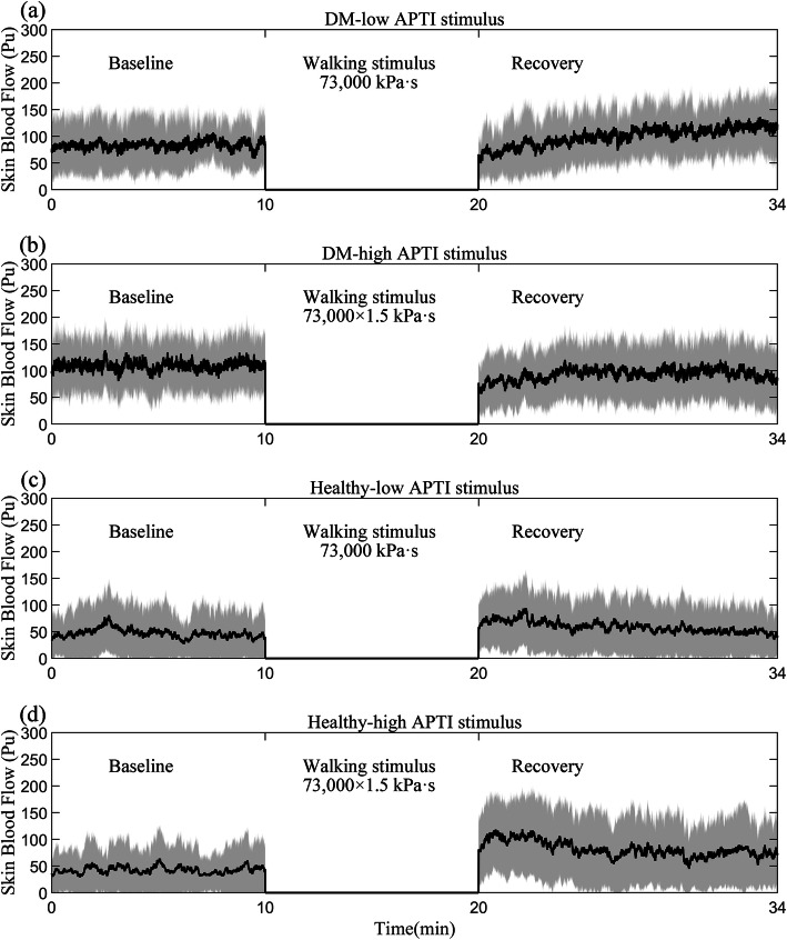
Background: Exercise, especially weight-bearing exercise (e.g. walking), may affect plantar tissue viability due to prolonged repetitive high vertical and high shear pressure stimulus on the plantar tissue, and further induce development of diabetic foot ulcers (DFUs). This study aimed to investigate the effects of different accumulated pressure-time integral (APTI) stimuli induced by walking on plantar skin blood flow (SBF) responses in people with diabetes mellitus (DM).
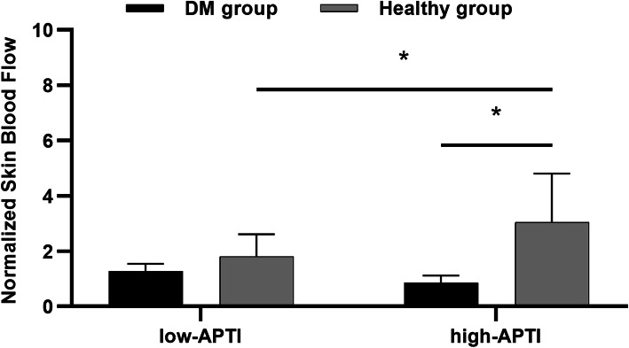
Methods: A repeated measures design was used in this study. Two walking protocols (low APTI (73,000 kPa·s) and high APTI (73,000 × 1.5 kPa·s)) were randomly assigned to ten people with DM and twenty people without DM. The ratio of SBF measured by laser Doppler flowmetry after walking to that before (normalized SBF) was used to express the SBF responses.
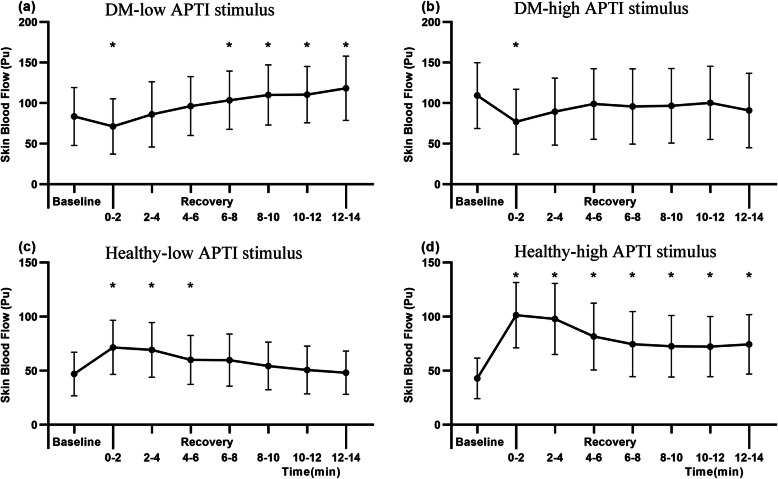
Results: After low APTI, plantar SBF of people with DM showed a similar response to people without DM (P = 0.91). However, after high APTI, people with DM had a significantly lower plantar SBF compared to people without DM (P < 0.05). In people with DM, plantar SBF in the first 2 min after both APTI stimuli significantly decreased compared to plantar SBF before walking (P < 0.05).
Conclusions: People with DM had a normal SBF response after low APTI walking but had an impaired SBF response after high APTI walking, which suggests that they should avoid weight-bearing physical activity with intensity more than 73,000 kPa·s and should rest for more than 2 min after weight-bearing physical activity to allow a full vasodilatory response to reduce risk of DFUs.
Keywords: Accumulated pressure-time integral; Diabetic foot ulcers; Microcirculation; Plantar skin blood flow; Weight-bearing exercise.
Conflict of interest statement
The authors declare that they have no competing interests.
Figures
Similar articles
-
Differences in skin blood flow oscillations between the plantar and dorsal foot in people with diabetes mellitus and peripheral neuropathy.Microvasc Res. 2019 Mar;122:45-51. doi: 10.1016/j.mvr.2018.11.002. Epub 2018 Nov 9. Microvasc Res. 2019. PMID: 30414870
-
Effects of Preconditioning Local Vibrations on Subsequent Plantar Skin Blood Flow Response to Walking.Int J Low Extrem Wounds. 2021 Jun;20(2):143-149. doi: 10.1177/1534734620905744. Epub 2020 Feb 25. Int J Low Extrem Wounds. 2021. PMID: 32098542 Clinical Trial.
-
Dynamic Microcirculation Characteristics of Plantar Skin Under Metatarsal Head of Human Foot in Response to Life-Like Pressure Stimulus.Microcirculation. 2024 Jul;31(5):e12860. doi: 10.1111/micc.12860. Epub 2024 Jun 5. Microcirculation. 2024. PMID: 38837938 Clinical Trial.
-
Emerging technologies for the prevention and management of diabetic foot ulcers.J Tissue Viability. 2020 May;29(2):61-68. doi: 10.1016/j.jtv.2020.03.003. Epub 2020 Mar 17. J Tissue Viability. 2020. PMID: 32197948 Review.
-
Should weight-bearing activity be reduced during healing of plantar diabetic foot ulcers, even when using appropriate offloading devices?Diabetes Res Clin Pract. 2021 May;175:108733. doi: 10.1016/j.diabres.2021.108733. Epub 2021 Mar 10. Diabetes Res Clin Pract. 2021. PMID: 33713722 Review.
Cited by
-
Measurement of plantar pressure differences in the contralateral limb when using offloading modalities for diabetic foot ulcerations.J Foot Ankle Res. 2025 Mar;18(1):e70028. doi: 10.1002/jfa2.70028. J Foot Ankle Res. 2025. PMID: 39797703 Free PMC article. Clinical Trial.
-
In-shoe plantar temperature, normal and shear stress relationships during gait and rest periods for people living with and without diabetes.Sci Rep. 2025 Mar 14;15(1):8804. doi: 10.1038/s41598-025-91934-9. Sci Rep. 2025. PMID: 40087292 Free PMC article.
-
Relationship Between Plantar Tissue Hardness and Plantar Pressure Distributions in People With Diabetic Peripheral Neuropathy.Front Bioeng Biotechnol. 2022 Apr 4;10:836018. doi: 10.3389/fbioe.2022.836018. eCollection 2022. Front Bioeng Biotechnol. 2022. PMID: 35445007 Free PMC article.
-
Plantar pressure gradient and pressure gradient angle are affected by inner pressure of air insole.Front Bioeng Biotechnol. 2024 Mar 11;12:1353888. doi: 10.3389/fbioe.2024.1353888. eCollection 2024. Front Bioeng Biotechnol. 2024. PMID: 38529404 Free PMC article.
-
Targeting oxidative damage in diabetic foot ulcers: integrative strategies involving antioxidant drugs and nanotechnologies.Burns Trauma. 2025 Mar 10;13:tkaf020. doi: 10.1093/burnst/tkaf020. eCollection 2025. Burns Trauma. 2025. PMID: 40718700 Free PMC article. Review.
References
-
- International Diabetes Federation. IDF Diabetes Atlas. 9th ed. Brussels, Belgium; 2019. https://www.diabetesatlas.org
-
- Lazzarini PA, Crews RT, van Netten JJ, Bus SA, Fernando ME, Chadwick PJ, Najafi B. Measuring plantar tissue stress in people with diabetic peripheral neuropathy: a critical concept in diabetic foot management. J Diabetes Sci Technol. 2019;13(5):869–880. doi: 10.1177/1932296819849092. - DOI - PMC - PubMed
Publication types
MeSH terms
LinkOut - more resources
Full Text Sources
Medical
