

Control of cardiomyocyte differentiation timing by intercellular signaling pathways
- PMID: 34144893
- PMCID: PMC8968240
- DOI: 10.1016/j.semcdb.2021.06.002
Control of cardiomyocyte differentiation timing by intercellular signaling pathways
Abstract
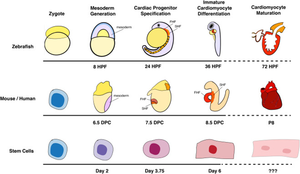
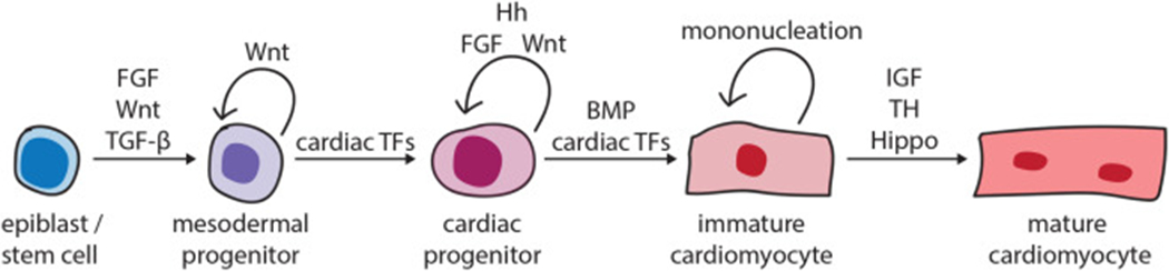
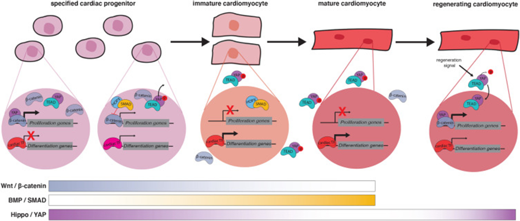
Congenital Heart Disease (CHD), malformations of the heart present at birth, is the most common class of life-threatening birth defect (Hoffman (1995) [1], Gelb (2004) [2], Gelb (2014) [3]). A major research challenge is to elucidate the genetic determinants of CHD and mechanistically link CHD ontogeny to a molecular understanding of heart development. Although the embryonic origins of CHD are unclear in most cases, dysregulation of cardiovascular lineage specification, patterning, proliferation, migration or differentiation have been described (Olson (2004) [4], Olson (2006) [5], Srivastava (2006) [6], Dunwoodie (2007) [7], Bruneau (2008) [8]). Cardiac differentiation is the process whereby cells become progressively more dedicated in a trajectory through the cardiac lineage towards mature cardiomyocytes. Defects in cardiac differentiation have been linked to CHD, although how the complex control of cardiac differentiation prevents CHD is just beginning to be understood. The stages of cardiac differentiation are highly stereotyped and have been well-characterized (Kattman et al. (2011) [9], Wamstad et al. (2012) [10], Luna-Zurita et al. (2016) [11], Loh et al. (2016) [12], DeLaughter et al. (2016) [13]); however, the developmental and molecular mechanisms that promote or delay the transition of a cell through these stages have not been as deeply investigated. Tight temporal control of progenitor differentiation is critically important for normal organ size, spatial organization, and cellular physiology and homeostasis of all organ systems (Raff et al. (1985) [14], Amthor et al. (1998) [15], Kopan et al. (2014) [16]). This review will focus on the action of signaling pathways in the control of cardiomyocyte differentiation timing. Numerous signaling pathways, including the Wnt, Fibroblast Growth Factor, Hedgehog, Bone Morphogenetic Protein, Insulin-like Growth Factor, Thyroid Hormone and Hippo pathways, have all been implicated in promoting or inhibiting transitions along the cardiac differentiation trajectory. Gaining a deeper understanding of the mechanisms controlling cardiac differentiation timing promises to yield insights into the etiology of CHD and to inform approaches to restore function to damaged hearts.
Keywords: Bone morphogenic protein; Cardiac progenitor; Cardiac regeneration; Cardiomyocyte; Developmental timing; Differentiation; Fibroblast growth factor; Hedgehog; Hippo; Insulin-like growth factor; Signaling; Thyroid hormone; Wnt.
Copyright © 2021. Published by Elsevier Ltd.
Figures
Similar articles
-
Signaling Pathways Governing Cardiomyocyte Differentiation.Genes (Basel). 2024 Jun 18;15(6):798. doi: 10.3390/genes15060798. Genes (Basel). 2024. PMID: 38927734 Free PMC article. Review.
-
GATA6 regulates WNT and BMP programs to pattern precardiac mesoderm during the earliest stages of human cardiogenesis.Elife. 2025 Mar 13;13:RP100797. doi: 10.7554/eLife.100797. Elife. 2025. PMID: 40080060 Free PMC article.
-
Harnessing developmental cues for cardiomyocyte production.Development. 2023 Aug 1;150(15):dev201483. doi: 10.1242/dev.201483. Epub 2023 Aug 10. Development. 2023. PMID: 37560977 Free PMC article. Review.
-
Enhanced Canonical Wnt Signaling During Early Zebrafish Development Perturbs the Interaction of Cardiac Mesoderm and Pharyngeal Endoderm and Causes Thyroid Specification Defects.Thyroid. 2021 Mar;31(3):420-438. doi: 10.1089/thy.2019.0828. Epub 2020 Sep 16. Thyroid. 2021. PMID: 32777984
-
Revisiting the Role of Autophagy in Cardiac Differentiation: A Comprehensive Review of Interplay with Other Signaling Pathways.Genes (Basel). 2023 Jun 24;14(7):1328. doi: 10.3390/genes14071328. Genes (Basel). 2023. PMID: 37510233 Free PMC article. Review.
Cited by
-
The homeodomain transcription factor Ventx2 regulates respiratory progenitor cell number and differentiation timing during Xenopus lung development.Dev Growth Differ. 2022 Sep;64(7):347-361. doi: 10.1111/dgd.12807. Epub 2022 Sep 16. Dev Growth Differ. 2022. PMID: 36053777 Free PMC article.
-
Signaling Pathways Governing Cardiomyocyte Differentiation.Genes (Basel). 2024 Jun 18;15(6):798. doi: 10.3390/genes15060798. Genes (Basel). 2024. PMID: 38927734 Free PMC article. Review.
-
Heart generation and regeneration.Semin Cell Dev Biol. 2021 Oct;118:92-93. doi: 10.1016/j.semcdb.2021.07.014. Epub 2021 Jul 22. Semin Cell Dev Biol. 2021. PMID: 34304994 Free PMC article. No abstract available.
-
Sonic Hedgehog signaling regulates the optimal differentiation pace from early-stage mesoderm to cardiogenic mesoderm in mice.Dev Growth Differ. 2025 Feb;67(2):75-84. doi: 10.1111/dgd.12955. Epub 2025 Jan 9. Dev Growth Differ. 2025. PMID: 39783159 Free PMC article.
-
Dissecting the Complexity of Early Heart Progenitor Cells.J Cardiovasc Dev Dis. 2021 Dec 26;9(1):5. doi: 10.3390/jcdd9010005. J Cardiovasc Dev Dis. 2021. PMID: 35050215 Free PMC article. Review.
References
-
- Hoffman JI, Incidence of congenital heart disease: II. Prenatal incidence, Pedia Cardiol. 16 (4) (1995) 155–165. - PubMed
-
- Gelb BD, Genetic basis of congenital heart disease, Curr. Opin. Cardiol 19 (2) (2004) 110–115. - PubMed
-
- Olson EN, A decade of discoveries in cardiac biology, Nat. Med 10 (5) (2004) 467–474. - PubMed
Publication types
MeSH terms
Grants and funding
LinkOut - more resources
Full Text Sources
