. 2021 Jun 18;12(1):3764.
doi: 10.1038/s41467-021-24104-w.

Preventing post-surgical cardiac adhesions with a catechol-functionalized oxime hydrogel

Affiliations

Preventing post-surgical cardiac adhesions with a catechol-functionalized oxime hydrogel

Masaki Fujita et al. Nat Commun. .

Abstract

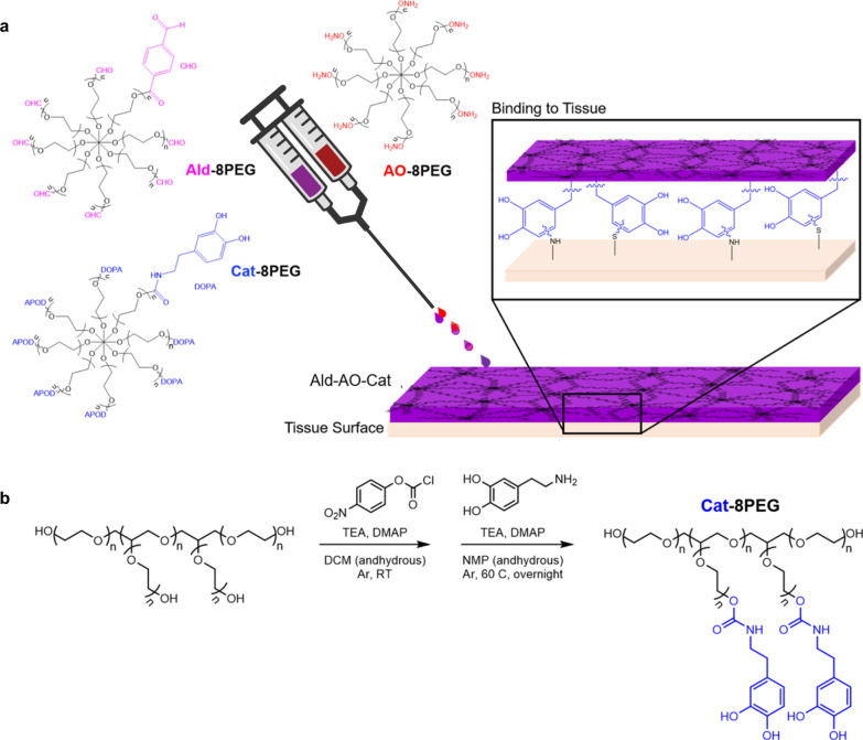
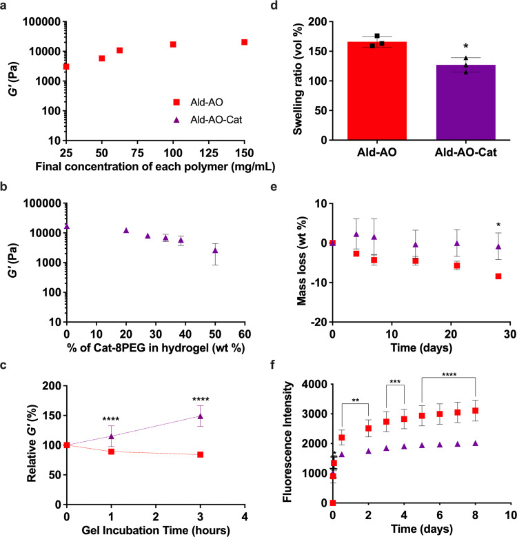
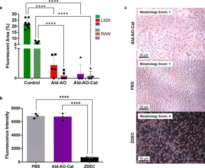
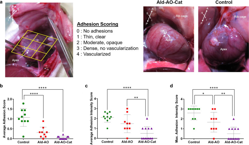
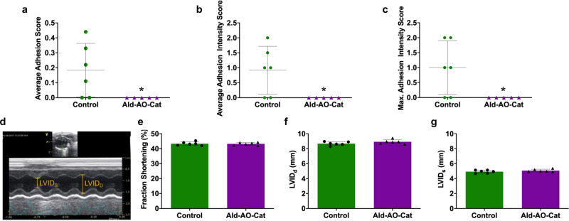
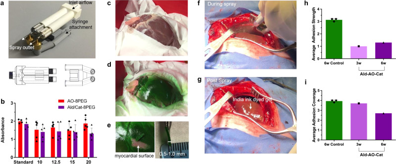
Post-surgical cardiac adhesions represent a significant problem during routine cardiothoracic procedures. This fibrous tissue can impair heart function and inhibit surgical access in reoperation procedures. Here, we propose a hydrogel barrier composed of oxime crosslinked poly(ethylene glycol) (PEG) with the inclusion of a catechol (Cat) group to improve retention on the heart for pericardial adhesion prevention. This three component system is comprised of aldehyde (Ald), aminooxy (AO), and Cat functionalized PEG mixed to form the final gel (Ald-AO-Cat). Ald-AO-Cat has favorable mechanical properties, degradation kinetics, and minimal swelling, as well as superior tissue retention compared to an initial Ald-AO gel formulation. We show that the material is cytocompatible, resists cell adhesion, and led to a reduction in the severity of adhesions in an in vivo rat model. We further show feasibility in a pilot porcine study. The Ald-AO-Cat hydrogel barrier may therefore serve as a promising solution for preventing post-surgical cardiac adhesions.


Conflict of interest statement

K.L.C. is co-founder, consultant, and holds equity interest and M.M.M. is a consultant for Karios Technologies, Inc. M.F., M.M.M., and K.L.C. are inventors on patent/patent applications (US10611880B2, WO2018183284A1) related to this work. The other authors declare no competing interests.

Figures

Fig. 1
Fig. 1. Fabrication of catechol-functionalized oxime hydrogel.
a Three distinct 8-arm star PEG polymers were used to form the oxime hydrogel. The Ald-8PEG/Cat-8PEG solution is mixed with the AO-8PEG solution in a 1:1 ratio and sprayed onto the tissue surface using a FibriJet gas-assisted applicator head from Nordson Micromedics. Oxime bonds rapidly form between Ald-8PEG and AO-8PEG. The catechol forms covalent bonds with the primary amines present on the tissue surface to promote hydrogel-tissue attachment. b Synthesis of Cat-8PEG required a two-step process by activating PEG–OH with NPC and then deprotecting with dopamine hydrochloride. Ald-8PEG and AO-8PEG were synthesized as previously reported. Poly(ethylene glycol) PEG, aldehyde Ald, catechol Cat, aminooxy AO, triethylamine TEA, 4-(dimethylamino) pyridine DMAP, dichloromethane DCM, N-methyl-2-pyrrolidone NMP, 4-nitrophenyl chloroformate NPC.
Fig. 2
Fig. 2. Characterization of rapidly gelling oxime hydrogels.
a Storage moduli (G′) were measured as a function of final polymer mass concentration in Ald–AO gels using a frequency sweep from 10−2 to 102 Hz at 37°C; G′ at 1Hz is shown. b Ald–AO–Cat gels with varying Cat-8PEG content were gelled and G′ was measured under the same conditions. Increasing Cat-8PEG content of the gels resulted in decreasing G′. c G′ for Ald–AO–Cat and Ald–AO were measured up to 3h post gelation and relative G′ was calculated. Over the 3h incubation period, G′ significantly increased for Ald–AO–Cat indicating the formation of secondary crosslinks between Cat and AO. d Low swelling ratios were observed for both Ald–AO–Cat and Ald–AO gels, with significantly lower swelling ratio observed for Ald–AO–Cat. e In vitro degradation of the oxime hydrogels revealed significantly greater mass loss after 28 days for Ald–AO gels compared to Ald–AO–Cat gels. f Ex vivo retention of Ald-AO-Cat shows significantly greater retention on the tissue surface compared to Ald-AO over 8 days. Note that some error bars are too small to be visible in ac, e, f. Mean ± SD and analyzed with a two-way ANOVA with a Tukey’s post-hoc test in c, e, f and an unpaired two-sided t-test in d (*p < 0.05, **p < 0.005, ***p < 0.001, ****p < 0.0001 between hydrogels). n = 21 individual frequencies were used to determine the means in ac. Measurements in a were repeatedly independently with similar results for 100mg/mL (n = 3) and 62.5mg/mL (n = 3), and measurements in b were repeatedly independently with similar results for 0% (n = 3) and all other concentrations (n = 2). n = 3 independent gel samples for each condition in df. Source data are provided as a Source Data file.
Fig. 3
Fig. 3. In vitro cell adhesion and cytocompatibility testing of Ald–AO–Cat.
a Inflammatory cell adhesion was studied on oxime hydrogels and tissue culture plastic as a control. L929 fibroblasts and RAW macrophages were seeded on each surface and imaged after 24 h of incubation for cell adhesion, as indicated by the fluorescent area. <1% fluorescent area was observed for both gels for L929 and RAW cells. Conversely, 22% and 6.6% fluorescence area were observed for L929 and RAW in control wells, indicating oxime hydrogels resist inflammatory cell adhesion. n = 10, 5, and 5 independent wells with L929 cells for control, Ald–AO, and Ald–AO–Cat, respectively. n = 6, 5, and 5 independent wells with RAW cells for control, Ald–AO, and Ald–AO–Cat, respectively. b Cytocompatibility of Ald–AO–Cat gels was assessed after 24 h of incubation in direct contact with the L929 fibroblast cell monolayer. Phosphate-buffered saline (PBS) and zinc diethyldithiocarbamate (ZDEC) was doped into the media for positive and negative controls, respectively. Ald–AO–Cat did not significantly affect metabolic activity compared to the positive control. n = 3 independent samples for each condition. c Representative brightfield images of the fibroblasts in each condition (n = 9 independent wells per group) reveal that no effect on cell morphology and spreading was observed for Ald–AO–Cat compared to the positive control. A cell morphology score was assigned to each brightfield image for quantification of cell morphology. Morphology scores suggest that fibroblasts are well spread and that no apparent cell lysis occurs when in direct contact with Ald–AO–Cat. Data are reported as mean ± SD and analyzed with a one-way ANOVA with a Tukey’s posthoc test (****p < 0.0001). Source data are provided as a Source Data file.
Fig. 4
Fig. 4. Reduced adhesion formation 2 weeks after application in rat surgical model.
Adhesion formation was  assessed for administration of Ald–AO–Cat, Ald–AO, and the untreated control after 2 weeks. a Adhesion scores were assigned to 9 discrete segments of the exposed heart surface when the chest was re-entered during euthanasia and harvest. Representative images of adhesions upon re-entry for Ald–AO–Cat and control groups are shown. Anatomical directions are shown to provide a point of reference: A—anterior, P—posterior, S—superior, I—inferior. b The individual scores for the 9 segments were averaged to obtain the adhesion score for each heart. At 2 weeks there was a significant reduction in adhesion score for Ald–AO–Cat and Ald–AO compared to the untreated group. c The average intensity score was calculated over regions of adhesion formation. At 2 weeks, Ald–AO–Cat show significantly reduced adhesion intensity compared to Ald–AO and the untreated control. d The maximum adhesion intensity score was also reported for each animal. Significantly higher maximum intensity scores were assigned to adhesions in rats treated with Ald–AO and the untreated group, compared to Ald–AO–Cat. Together these data suggest reduced adhesion formation and intensity when Ald–AO–Cat is applied following abrasion. Data are reported as mean ± SD and analyzed with a one-way ANOVA with a Tukey’s posthoc test (*p < 0.05, **p < 0.005, ****p < 0.0001) n = 12, 9, and 10 independent rats for Ald–AO–Cat, Ald–AO, and Control groups, respectively. Source data for bd are provided as a Source Data file.
Fig. 5
Fig. 5. Reduced adhesion formation and maintained cardiac function 4 weeks after application in rat surgical model.
Adhesion formation and heart function were assessed for administration of untreated control and Ald–AO–Cat. ac At 4 weeks, all parameters for comparing adhesion formation and intensity showed significantly lower values for Ald–AO–Cat when compared to the untreated control. It is also worth noting that all Ald–AO–Cat-treated animals received an average adhesion and intensity score of 0, indicating the complete reduction of adhesion formation. dg Cardiac function was assessed 3 ± 1 days post material application using M-mode echocardiography. Fraction shortening was calculated from measured left ventricle internal diameter systole (LVIDs) and left ventricle internal diameter diastole (LVIDD), and regardless of treatment, animals showed identical cardiac function, indicating no adverse effects from the application of Ald-AO-Cat gels. Data are reported as mean ± SD and analyzed with an unpaired two-sided Mann–Whitney t-test (*p < 0.05). n = 5 and 6 independent animals for Ald–AO–Cat and Control groups, respectively, in ag. Source data for ac and eg are provided as a Source Data file.
Fig. 6
Fig. 6. Histology of Ald–AO–Cat in a rat model.
H&E staining was performed for Ald–AO–Cat tissue samples (n = 6 independent animals) to verify the biocompatibility of the oxime hydrogel systems. At 4 weeks, thin capsule formation around some remaining gel (black arrows) was visible, however, there was no indication of chronic toxicity as a result of application or degradation of the hydrogel system. Furthermore, no cell infiltration into the Ald–AO–Cat gel coincides with our expectations, due to the non-cell adhesive nature of PEG polymers.
Fig. 7
Fig. 7. Spray device design, testing, and application in a pilot porcine study.
a A specialized device was developed to deliver each component together in a 1:1 ratio in the form of an atomized spray for application in a large animal model. This device has two separate parts, one of which allows the flow of air from an air compressor through two separate pathways, while a second piece supports the addition of two syringes containing sterile solutions of the component polymers injected into the path of air through two pathways. b The mixing ratio of the sprayed solutions at various working distances was tested using mock Ald–Cat-8PEG and AO-8PEG solutions dyed blue and yellow, respectively. The standard was prepared by pipetting equal volumes of both solutions as a control (n = 4 independent samples). Equal mixing of both solutions was observed at all spray distances (n = 5 independent samples for each working distance and polymer). c The dual spray device was tested on an ex vivo porcine heart to determine an effective working distance and the amount of spray necessary for complete coverage of the anterior surface with a gel ~0.5–1.0 mm thick. The heart was set in a shallow insulated ice bucket surrounded by Kim wipes to simulate the exposure of the epicardium during a sternotomy. d The anterior surface of the heart was sprayed from a working distance of ~10–12 cm with a total of 8 mL (4 mL AO-8PEG (dyed blue) and 4 mL Ald–Cat-8EPG (dyed yellow)) of hydrogel solution. Equal mixing was visually determined by the green gel that formed on the surface of the heart. Minimal overspray is seen on the Kim wipes surrounding the heart surface, indicating effective targeting of the spray. e For clinical translation, in the case of an emergency, it would be advantageous that the gel could be manually removed by the surgeon. The gel thickness was measured after removal, showing the targeted 0.5–1.0 mm gel thickness has been obtained with the 8 mL of solution. f, g The Ald–AO–Cat gel applied using the dual spray device was tested in a pilot porcine study against a non-treated control. The solutions were prepared with a 1:100 dilution of India Ink for visualization of the gel once sprayed. h and i 3 and 6 weeks post-sternotomy and abrasion, resternotomy and adhesion scoring was performed based on the defined adhesion scoring system (Table 3). Assessment of adhesions revealed a decrease in adhesion strength in Ald–AO–Cat-treated animals (n = 1 pig at 3 weeks, n = 1 pig at 6 weeks), compared to controls (n = 3 pigs at 6 weeks). The treated animals required only simple blunt dissection of the adhesions to access the anterior surface of the heart, while the control required sharp dissection, resulting in tissue damage with adhesion removal. Average adhesion coverage was similar for all animals with a trending decrease in adhesion coverage at 6 weeks for the Ald–AO–Cat-treated animal. This observation agrees with clinical observations of reduced cardiac adhesion coverage at longer time points post sternotomy and surgical trauma. Data are reported as mean ± SD. Source data for b, h, i are provided as a Source Data file.

Similar articles

Cited by

References

    1. Alizzi AM, et al. Reduction of post-surgical pericardial adhesions using a pig model. Heart Lung Circ. 2012;21:22–29. doi: 10.1016/j.hlc.2011.10.002. - DOI - PubMed
    1. Konertz WF, et al. Reducing the incidence and severity of pericardial adhesions with a sprayable polymeric matrix. Ann. Thorac. Surg. 2003;76:1270–1274. doi: 10.1016/S0003-4975(03)00733-1. - DOI - PubMed
    1. Nkere UU, et al. Pericardial trauma and adhesions in relation to reoperative cardiac surgery TT—Perikardiales Trauma und Verwachsungen im Rahmen von Re-Eingriffen in der Herzchirurgie. Thorac. Cardiovasc. Surg. 1995;43:338–346. doi: 10.1055/s-2007-1013806. - DOI - PubMed
    1. Cannata A, et al. Postsurgical intrapericardial adhesions: mechanisms of formation and prevention. Ann. Thorac. Surg. 2013;95:1818–1826. doi: 10.1016/j.athoracsur.2012.11.020. - DOI - PubMed
    1. Cliff WJ, Grobety J, Ryan GB. Postoperative pericardial adhesions. The role of mild serosal injury and spilled blood. J. Thorac. Cardiovasc. Surg. 1973;65:744–750. doi: 10.1016/S0022-5223(19)40710-1. - DOI - PubMed

Publication types