. 2021 Sep;23(9):1636-1647.
doi: 10.1038/s41436-021-01204-y. Epub 2021 Jun 18.

Inherited duplications of PPP2R3B predispose to nevi and melanoma via a C21orf91-driven proliferative phenotype

Affiliations

Inherited duplications of PPP2R3B predispose to nevi and melanoma via a C21orf91-driven proliferative phenotype

Satyamaanasa Polubothu et al. Genet Med. 2021 Sep.

Abstract

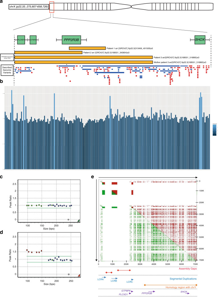
Purpose: Much of the heredity of melanoma remains unexplained. We sought predisposing germline copy-number variants using a rare disease approach.

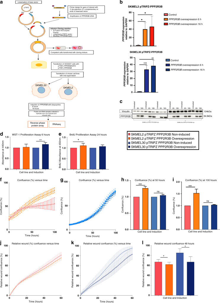
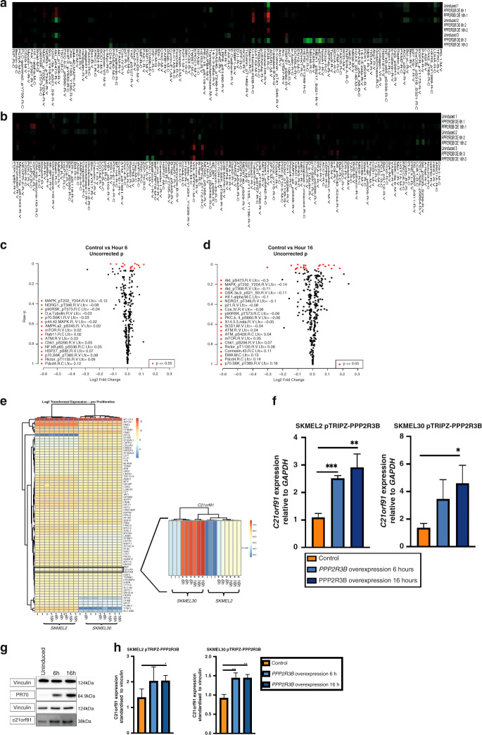
Methods: Whole-genome copy-number findings in patients with melanoma predisposition syndrome congenital melanocytic nevus were extrapolated to a sporadic melanoma cohort. Functional effects of duplications in PPP2R3B were investigated using immunohistochemistry, transcriptomics, and stable inducible cellular models, themselves characterized using RNAseq, quantitative real-time polymerase chain reaction (qRT-PCR), reverse phase protein arrays, immunoblotting, RNA interference, immunocytochemistry, proliferation, and migration assays.

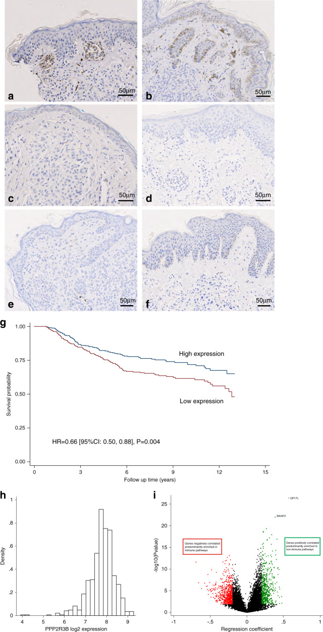
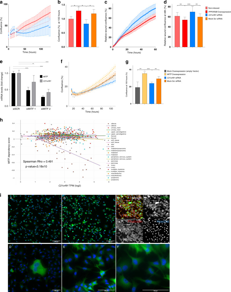
Results: We identify here a previously unreported genetic susceptibility to melanoma and melanocytic nevi, familial duplications of gene PPP2R3B. This encodes PR70, a regulatory unit of critical phosphatase PP2A. Duplications increase expression of PR70 in human nevus, and increased expression in melanoma tissue correlates with survival via a nonimmunological mechanism. PPP2R3B overexpression induces pigment cell switching toward proliferation and away from migration. Importantly, this is independent of the known microphthalmia-associated transcription factor (MITF)-controlled switch, instead driven by C21orf91. Finally, C21orf91 is demonstrated to be downstream of MITF as well as PR70.

Conclusion: This work confirms the power of a rare disease approach, identifying a previously unreported copy-number change predisposing to melanocytic neoplasia, and discovers C21orf91 as a potentially targetable hub in the control of phenotype switching.


Conflict of interest statement

The authors declare no competing interests.

Figures

Fig. 1
Fig. 1. Germline duplications involving PPP2R3B are found at increased frequency in individuals with melanocytic neoplasia.
(a) Schematic of Xp22.33 demonstrating the location of three novel duplications (yellow) found in 24 congenital melanocytic nevi (CMN) patients using whole-genome array comparative genomic hybridization (CGH) of leukocyte DNA, with one identical parental duplication demonstrating inheritance. Previously described copy-number variants in that region are shown below, duplications in blue, deletions in red, with each bar representing a single publication. The publication representing a duplication involving PPP2R3B described a single variant in a cohort of approximately 36,000 (asterisk; see text for details), confirming that the CMN duplications are rare in the normal population. (b) PPP2R3B duplications in a UK nonsyndromic melanoma cohort (4 duplications in nonselected cohort n = 168), and CMN cohort (3 duplications in known preselected cohort n = 5) shown by targeted next-generation sequencing (NGS) of PPP2R3B, in addition to the two telomeric genes (GTPBP6 and PLCXD1) and the next centromeric gene (SHOX). Data represent the ratio of corrected read depth (see text for details) across the whole of PPP2R3B with respect to the ratio across the whole of SHOX. Each bar represents an individual patient. PPP2R3B duplications called are shown in light blue: validation of the array CGH findings in the three CMN patients are clustered to the right of the figure, and new duplications in the melanoma cohort in the rest of the figure (n = 4, 2.4%). Validation of PPP2R3B duplications detected by array CGH. Custom-designed multiplex ligation-dependent probe amplification (MLPA) ratio plots validating copy-number measurement of PPP2R3B (4 probes) and the two telomeric genes GTPBP6 and PLCXD1 (one probe each), to the left of each figure and less than 150 bp in length; control probes of greater than 160 kb targeting genes of known normal copy number across the genome are shown to the right at greater than 150 bp size. A representative example of normal copy number for all genes (c), and of a duplication of PPP2R3B and GTPBP6 and PLCXD1 in a CMN patient (red dots) (d). While this method was able to validate the array CGH findings, it was not as robust as the targeted NGS panel for novel discovery of copy-number changes, likely due to the repetitive, GC-rich, and polymorphic nature of the region studied. Low-copy repeats at Xp22.33. (e) The upper panel depicts a regional similarity search across Xp22.33 with YASS software (http://bioinfo.cristal.univ-lille.fr/yass/index.php) both forward (green) and backward (red) revealing three segmental duplications (LCRA, LCRB, and LCRC) 5’ of PPP2R3B and a high density of SINE and LINE repeats. No segmental duplications are detected 3’ to PPP2R3B before SHOX. The assembly gaps (red), local genes (purple), and the homology region (orange) with the Y chromosome are indicated.
Fig. 2
Fig. 2. Germline duplications of PPP2R3B lead to increased expression of protein product PR70 in congenital melanocytic nevi (CMN) tissue, compared to that of normal copy-number controls.
(a, b) Immunohistochemical staining of formalin fixed paraffin embedded (FFPE) CMN tissue demonstrates moderate intensity PR70 staining throughout the cytoplasm of nevus cells in two patients where tissue was available with a confirmed germline PPP2R3B duplication and (cf). Negative PR70 staining in four patients with confirmed normal copy number of PPP2R3B. Stained sections were assessed by two independent blinded assessors and assigned a score of 1–3, based on the intensity of staining observed and scores averaged. Scores were as follows: a = 3, b = 2.5, c = 0, d = 0, e = 0, and f = 0. Increased PPP2R3B expression in melanoma tissue is correlated with improved melanoma specific survival. (g) Kaplan–Meier curve generated from transcriptomic data from 703 FFPE melanoma tumors from the Leeds Melanoma Cohort, hazard ratio (HR) = 0.66, (95% confidence interval [CI] 0.50–0.88), p = 0.004. The effect remains significant after adjusting for age, sex, American Joint Committee for Cancer (AJCC) stage, vascular invasion, site, BRAF/NRAS pathogen variant status, and tumor invading lymphocytes (TILs). (h) Log intensity distribution of PPP2R3B DASL probe (ILMN_1689720) is close to a normal distribution. Improved melanoma specific survival observed with increased expression of PPP2R3B appears not to be immune mediated. Tumor expression of PPP2R3B correlates with expression of a large number of other genes in the genome: 596 positively correlated at FDR < 0.05 with regression coefficient >0.20; 731 negatively correlated at FDR < 0.05 with a regression coefficient < −0.2. (i) The genes positively correlated with PPP2R3B are predominantly enriched in nonimmune pathways, consistent with the lack of association between PPP2R3B expression and TILs or any specific immune cell score. The genes negatively correlated with PPP2R3B expression are predominantly enriched in immune pathways (Table S1, 2).
Fig. 3
Fig. 3. Generation of stable inducible overexpression model for PPP2R3B in melanoma cell lines SKMEL2 and SKMEL30.
(a) Diagrammatic representation of stable inducible PPP2R3B overexpression system (SKMEL2-pTRIPZ-PPP2R3B and SKMEL30-pTRIPZ-PPP2R3B), and generation of samples for reverse phase protein arrays (RPPA) and RNA sequencing. Validation of PPP2R3B overexpression in samples for reverse phase protein array and RNAseq: (b) quantitative real-time polymerase chain reaction (qRT-PCR) demonstrating increased PPP2R3B messenger RNA (mRNA) in both induced cell lines at 6 hours and 16 hours (relative fold change in PPP2R3B expression, standardized to GAPDH, mean + SD of samples in quadruplicate), and (c) western blot confirming PR70 overexpression in induced cell lines at 6 hours and 16 hours with vinculin loading control. Statistical significance was determined using a Student’s t-test (Prism v7.0, Graphpad). Statistically significant values are indicated by a single asterisk (p < 0.05), a double asterisk (p < 0.01), a triple asterisk (p < 0.001), or a quadruple asterisk (p < 0.0001). Overexpression of PPP2R3B increases proliferation in melanoma cell lines. (d) Increased proliferation following PPP2R3B overexpression using WST1 proliferation assay in SKMEL2-pTRIPZ-PPP2R3B at 6 hours, (e) in SKMEL30-pTRIPZ-PPP2R3B by BrdU assay at 24 hours (mean absorbance of colormetric assay of eight replicates shown with standard deviation), and (f, g) by IncuCyte® cell count proliferation assay in SKMEL2-pTRIPZ-PPP2R3B and SKMEL30-pTRIPZPPP2R3B respectively over 100 hours, measuring confluence (%) versus time (hours) (mean confluence of eight replicates with standard deviation). (h, i) Mean confluence in each cell line is shown at timepoints of 50 hours and 100 hours respectively (mean of eight replicates standardized to noninduced cell line shown with standard error). Statistical analysis was performed and depicted as described above in figure legend. Overexpression of PPP2R3B decreases cellular migration in melanoma cell lines. (j, k) Scratch wound assay in SKMEL2-pTRIPZ-PPP2R3B and SKMEL30-pTRIPZ-PPP2R3B respectively leads to decreased relative wound confluence (%) versus time (hours) compared to noninduced controls. (l) Mean relative wound confluence in both cell lines shown at 48 hours (mean of eight replicates shown with error bars). Statistical analysis was performed and depicted as described above in figure legend.
Fig. 4
Fig. 4. PPP2R3B overexpression affects mTOR/p70S6K1 and HIF-1 signaling pathways.
(a, b) Heat map of protein expression observed by reverse phase protein arrays (RPPA) following overexpression of PPP2R3B in SKMEL2-pTRIPZ-PPP2R3B and SKMEL30-pTRIPZ-PPP2R3B respectively, demonstrating low background activity as expected from a controlled cellular model. (c, d) Volcano plots of log fold change in protein expression versus p value for differentially expressed proteins common to both cell lines following PPP2R3B overexpression at 6 hours and 16 hours respectively. Unadjusted p values < 0.05 are shown in red. Raw data is available in the Supplementary material (Table S4). PPP2R3B overexpression leads to significant and sustained rise in expression of gene C21orf91. C21orf91 was the most differentially expressed gene on PPP2R3B induction common to both cell lines and at both time points by RNA sequencing (Table S5), other than PPP2R3B itself. (e) Heat map from pathway signature analysis of RNAseq data at 6 hours and 16 hours, focusing on pro-proliferative anti-invasive melanoma signature genes38, demonstrating increased expression of C21orf91 following PPP2R3B overexpression observed in both cell lines, at 6 and 16 hours. Validation of significantly increased C21orf91 expression following PPP2R3B overexpression at 6 hours and 16 hours in both cell lines, shown by (f) quantitative real-time polymerase chain reaction (qRT-PCR) relative fold change in C21orf91 mRNA levels, samples standardized to GAPDH (mean + SD of samples in quadruplicate) and (g, h) representative western blot with quantification of fold change of C21orf91—samples standardized to vinculin (mean shown with standard deviation of samples in triplicate). Statistical significance was determined using a Student’s t-test (Prism v7.0, Graphpad). Statistically significant values are indicated by a single asterisk (p < 0.05), a double asterisk (p < 0.01), a triple asterisk (p < 0.001), or a quadruple asterisk (p < 0.0001).
Fig. 5
Fig. 5. Knockdown of C21orf91 rescues increased proliferation associated with PPP2R3B overexpression.
IncuCyte® proliferation assay confluence (%) versus time (hours) for PPP2R3B-induced SKMEL2-pTRIPZ-PPP2R3B, with and without small interfering RNA (siRNA) knockdown of C21orf91 (a), with means taken at 100 hours (b) (mean confluence at given time point of eight replicates shown with standard deviation) shows a significant increase in proliferation following PPP2R3B induction, rescued by C21orf91 knockdown and significantly different from knockdown with Scr siRNA. Statistical significance was determined using a Student’s t-test (Prism v7.0, Graphpad). Statistically significant values are indicated by a single asterisk (p < 0.05), a double asterisk (p < 0.01), a triple asterisk (p < 0.001), or a quadruple asterisk (p < 0.0001). Knockdown of C21orf91 rescues decreased migration associated with PPP2R3B overexpression. IncuCyte® scratch wound assay relative wound confluence (%) versus time (hours) for PPP2R3B-induced SKMEL2-pTRIPZ-PPP2R3B, with and without siRNA knockdown of C21orf91 (c), with mean taken at 48 hours (d) (mean relative wound confluence at each time point of twelve replicates shown with standard deviation) shows a significant decrease in migration following PPP2R3B induction, rescued by C21orf91 knockdown and significantly different from knockdown with Scr siRNA. Statistical significance was determined using a Student’s t-test (Prism v7.0, Graphpad). Statistically significant values are indicated by a single asterisk (p < 0.05), a double asterisk (p < 0.01), a triple asterisk (p < 0.001), or a quadruple asterisk (p < 0.0001). Knockdown of MITF leads to decreased expression of C21orf91. Quantitative real-time polymerase chain reaction (qRT-PCR) demonstrating decrease in both MITF and C21orf91 messenger RNA (mRNA) following transfection by two different siRNAs targeting MITF transcript (siMITF 1 and siMITF 2) in SKMEL2 cell line (e). Relative fold change in gene expression compared to control cells transfected by nontarget siRNA (siSCRA), standardized to GAPDH, mean + SD of 3 independent experiments. Knockdown of C21orf91 in cells rescues MITF-driven increase in proliferation. IncuCyte® proliferation assay confluence (%) versus time (hours) following overexpression of MITF in SKMEL2, with and without siRNA knockdown of C21orf91 (f), with means taken at 100 hours (g) (mean confluence at given time point of four replicates shown with standard deviation). MITF overexpression drives proliferation as expected, which is rescued by knockdown of C21orf91. Controls for both transfection with the MITF overexpression vector and the C21orf91 siRNA are included. Statistical significance was determined using a Student’s t-test (Prism v7.0, Graphpad). Statistically significant values are indicated by a single asterisk (p < 0.05), a double asterisk (p < 0.01), a triple asterisk (p < 0.001), or a quadruple asterisk (p < 0.0001). Increased expression of C21orf91 is associated with genetic dependency on MITF in melanoma cell lines. (h) CRISPR-Cas9 genome-scale knockout of MITF in melanoma cell lines (n = 30) reveals increased expression of C21orf91 in cells with greater MITF dependency, suggesting that C21orf91 is downstream of MITF. Dependency score as described in https://depmap.org. PR70 and C21orf91 are expressed throughout the cytoplasm with increased expression of C21orf91 in dividing cells. (i) Immunocytochemistry of SKMEL30-pTRIPZ-PPP2R3B in uninduced cells at 10× (A) and induced cells at 10× (B) and 20× (C) confirms increased PR70 and C21orf91 expression throughout the cytoplasm following induction of PPP2R3B. PR70 is stained with Alex Fluor® 488 (green) secondary antibody, and nuclei stained with Hoescht (blue) and in (C) Phalloidin (actin) are visualized with a conjugated Alex Fluor® 647 (far red) antibody. C21orf91 expression is increased in cells with two nuclei, which could be due to various causes including cell division or a cytokinesis defect 10× (D), 20× (E), 40× (F), stained with Alexa Fluor® 488 (green) secondary antibody, and Hoescht nuclear stain (blue). Scale bars represent 100 microns. RE: Inherited duplications of PPP2R3B promote nevi and melanoma via a novel C21orf91-driven proliferative phenotype.

References

    1. Harland M, et al. Prevalence and predictors of germline CDKN2A mutations for melanoma cases from Australia, Spain and the United Kingdom. Hered. Cancer Clin. Pract. 2014;12:20. doi: 10.1186/1897-4287-12-20. - DOI - PMC - PubMed
    1. Kinsler VA, et al. Multiple congenital melanocytic nevi and neurocutaneous melanosis are caused by postzygotic mutations in codon 61 of NRAS. J. Invest. Dermatol. 2013;133:2229–2236. doi: 10.1038/jid.2013.70. - DOI - PMC - PubMed
    1. Polubothu, S. et al. Genotype-phenotype-outcome cohort study of congenital melanocytic nevi – the relevance of genotype to clinical management. Br. J. Dermatol.182, 434–443 (2020). - PMC - PubMed
    1. Etchevers, H. C. et al. Giant congenital melanocytic nevus with vascular malformation and epidermal cysts associated with a somatic activating mutation in BRAF. Pigment Cell. Melanoma Res.31, 437–441 (2018). - PubMed
    1. Kinsler VA, Birley J, Atherton DJ. Great Ormond Street Hospital for Children Registry for congenital melanocytic nevi: prospective study 1988-2007. Part 1-epidemiology, phenotype and outcomes. Br. J. Dermatol. 2009;160:143–150. doi: 10.1111/j.1365-2133.2008.08849.x. - DOI - PubMed

Publication types