Dermal Nanoemulsion Treatment Reduces Burn Wound Conversion and Improves Skin Healing in a Porcine Model of Thermal Burn Injury
- PMID: 34145458
- PMCID: PMC8633125
- DOI: 10.1093/jbcr/irab118
Dermal Nanoemulsion Treatment Reduces Burn Wound Conversion and Improves Skin Healing in a Porcine Model of Thermal Burn Injury
Abstract
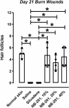
Burn wound progression is an inflammation-driven process where an initial partial-thickness thermal burn wound can evolve over time to a full-thickness injury. We have developed an oil-in-water nanoemulsion formulation (NB-201) containing benzalkonium chloride for use in burn wounds that is antimicrobial and potentially inhibits burn wound progression. We used a porcine burn injury model to evaluate the effect of topical nanoemulsion treatment on burn wound conversion and healing. Anesthetized swine received thermal burn wounds using a 25-cm2 surface area copper bar heated to 80°C. Three different concentrations of NB-201 (10, 20, or 40% nanoemulsion), silver sulfadiazine cream, or saline were applied to burned skin immediately after injury and on days 1, 2, 4, 7, 10, 14, and 18 postinjury. Digital images and skin biopsies were taken at each dressing change. Skin biopsy samples were stained for histological evaluation and graded. Skin tissue samples were also assayed for mediators of inflammation. Dermal treatment with NB-201 diminished thermal burn wound conversion to a full-thickness injury as determined by both histological and visual evaluation. Comparison of epithelial restoration on day 21 showed that 77.8% of the nanoemulsion-treated wounds had an epidermal injury score of 0 compared to 16.7% of the silver sulfadiazine-treated burns (P = .01). Silver sulfadiazine cream- and saline-treated wounds (controls) converted to full-thickness burns by day 4. Histological evaluation revealed reduced inflammation and evidence of skin injury in NB-201-treated sites compared to control wounds. The nanoemulsion-treated wounds often healed with complete regrowth of epithelium and no loss of hair follicles (NB-201: 4.8 ± 2.1, saline: 0 ± 0, silver sulfadiazine: 0 ± 0 hair follicles per 4-mm biopsy section, P < .05). Production of inflammatory mediators and sequestration of neutrophils were also inhibited by NB-201. Topically applied NB-201 prevented the progression of a partial-thickness burn wound to full-thickness injury and was associated with a concurrent decrease in dermal inflammation.
© The Author(s) 2021. Published by Oxford University Press on behalf of the American Burn Association. All rights reserved. For permissions, please e-mail: journals.permissions@oup.com.
Figures


References
-
- http://www.cdc.gov/masstrauma/factsheets/public/burns.pdf; accessed 5/25/21.
-
- D’Avignon LC, Chung KK, Saffle JR, Renz EM, Cancio LC; Prevention of Combat-Related Infections Guidelines Panel . Prevention of infections associated with combat-related burn injuries. J Trauma 2011;71:S282–9. - PubMed
-
- University of Michigan Burn Registry.
-
- Clark RAF, Fenner J, Sasson A, McClain SA, Singer AJ, Tonnesen MG. Blood vessel occlusion with erythrocyte aggregates causes burn injury progression-microvasculature dilation as a possible therapy. Exp Dermatol 2018;27:625–9. - PubMed
Publication types
MeSH terms
Substances
Grants and funding
LinkOut - more resources
Full Text Sources
Other Literature Sources
Medical

