

Imaging modalities for endoleak surveillance
- PMID: 34145780
- PMCID: PMC8655756
- DOI: 10.1002/jmrs.522
Imaging modalities for endoleak surveillance
Abstract
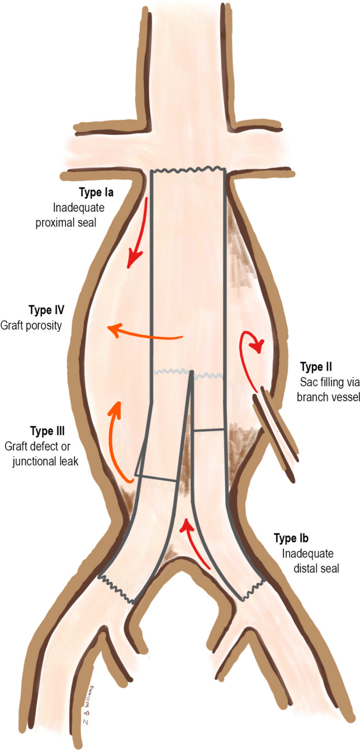
As the global population ages, the issue of abdominal aortic aneurysm continues to grow. With the evolution of new devices and refined operative technique, aneurysm treatment via endovascular aortic repair is becoming increasingly favourable. This, however, is not without drawbacks, where regular surveillance is paramount to long-term success and detection of post-procedure complications. Of these complications, endoleak is the most notable and poses the greatest risk of potential future aortic rupture. The purpose of this review paper is to discuss the armada of imaging modalities used in the detection and evaluation of endoleak and their varying usefulness. Plain abdominal X-ray is a cost-effective tool in detecting gross graft abnormalities such as stent migration or deformity (kinking or fracture). Though it may raise suspicion for endoleak, X-ray does not allow accurate classification of endoleak type when used alone. Duplex ultrasonography quantifies both aortic anatomy and real time flow dynamics. Most screening programmes are conducted using two-dimensional ultrasound. Unfortunately, observer and equipment variability may lead to surveillance discrepancies-but reduced when utilising a dedicated vascular sonography laboratory. Contrast enhanced ultrasonography is a promising alternative to computed tomography, though still is emerging. Computed tomography angiography certainly has disadvantages (ionising radiation, contrast-nephropathy, limited differentiation of endoleak type)-however, it provides near-real surgical dimensions and highlights graft complications and concomitant disease (such as neighbouring infection). With widespread availability and short scan time, it certainly remains valuable in surveillance. Magnetic resonance angiography has a similar sensitivity to computed tomography (minus the radiation), however is plagued by movement and metal artefact. Other novel modalities in endoleak surveillance include four-dimensional ultrasound, multiplanar intra-operative probes, nuclear medicine and wall stress analysis.
Keywords: Aortic aneurysm; diagnostic imaging; endoleak; endovascular procedures; vascular grafting.
© 2021 The Authors. Journal of Medical Radiation Sciences published by John Wiley & Sons Australia, Ltd on behalf of Australian Society of Medical Imaging and Radiation Therapy and New Zealand Institute of Medical Radiation Technology.
Figures
References
-
- Derubertis BG, Trocciola SM, Ryer EJ, et al. Abdominal aortic aneurysm in women: prevalence, risk factors, and implications of screening. J Vasc Surg 2007; 46: 630–5. - PubMed
-
- Cornuz J, Pinto CS, Tevaearai H, Egger M. Risk factors for asymptomatic abdominal aortic aneurysm: systematic review and meta‐analysis of population‐based screening studies. Eur J Public Health 2004; 14: 343–9. - PubMed
-
- Towne JB. Endovascular treatment of abdominal aortic aneurysms. Am J Surg 2005; 189: 140–9. - PubMed
Publication types
MeSH terms
LinkOut - more resources
Full Text Sources
Research Materials
