

Dual role for CXCR3 and CCR5 in asthmatic type 1 inflammation
- PMID: 34146578
- PMCID: PMC8674372
- DOI: 10.1016/j.jaci.2021.05.044
Dual role for CXCR3 and CCR5 in asthmatic type 1 inflammation
Abstract
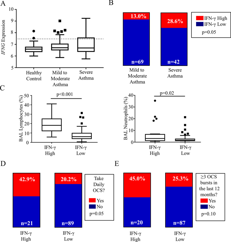
Background: Many patients with severe asthma (SA) fail to respond to type 2 inflammation-targeted therapies. We previously identified a cohort of subjects with SA expressing type 1 inflammation manifesting with IFN-γ expression and variable type 2 responses.
Objective: We investigated the role of the chemotactic receptors C-X-C chemokine receptor 3 (CXCR3) and C-C chemokine receptor 5 (CCR5) in establishing type 1 inflammation in SA.
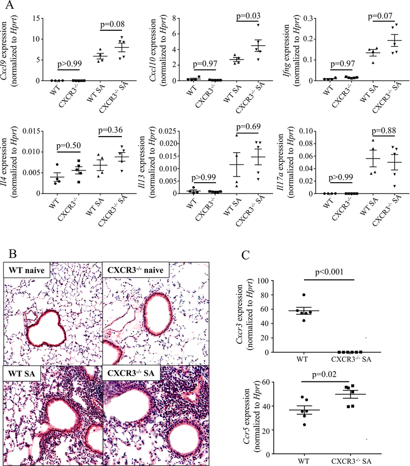
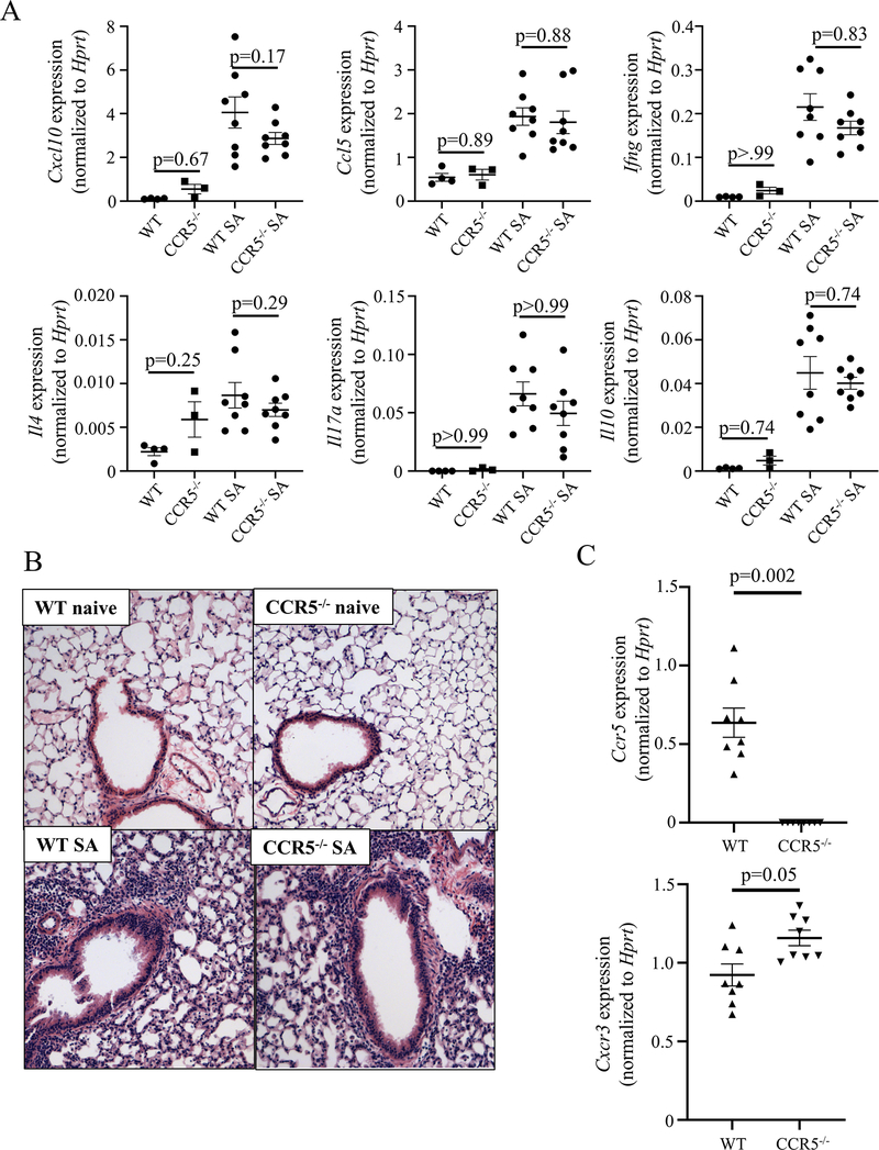
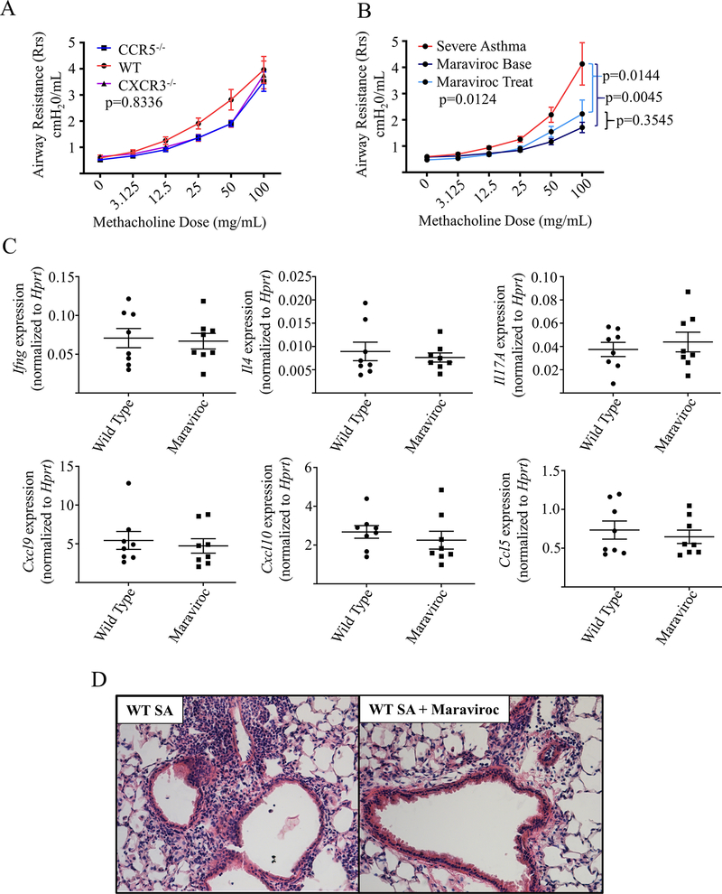
Methods: Bronchoalveolar lavage microarray data from the Severe Asthma Research Program I/II were analyzed for pathway expression and paired with clinical parameters. Wild-type, Cxcr3-/-, and Ccr5-/- mice were exposed to a type 1-high SA model with analysis of whole lung gene expression and histology. Wild-type and Cxcr3-/- mice were treated with a US Food and Drug Administration-approved CCR5 inhibitor (maraviroc) with assessment of airway resistance, inflammatory cell recruitment by flow cytometry, whole lung gene expression, and histology.
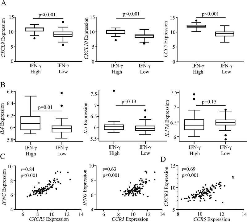
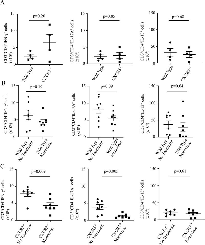
Results: A cohort of subjects with increased IFN-γ expression showed higher asthma severity. IFN-γ expression was correlated with CXCR3 and CCR5 expression, but in Cxcr3-/- and Ccr5-/- mice type 1 inflammation was preserved in a murine SA model, most likely owing to compensation by the other pathway. Incorporation of maraviroc into the experimental model blunted airway hyperreactivity despite only mild effects on lung inflammation.
Conclusions: IFNG expression in asthmatic airways was strongly correlated with expression of both the chemokine receptors CXCR3 and CCR5. Although these pathways provide redundancy for establishing type 1 lung inflammation, inhibition of the CCL5/CCR5 pathway with maraviroc provided unique benefits in reducing airway hyperreactivity. Targeting this pathway may be a novel approach for improving lung function in individuals with type 1-high asthma.
Keywords: CCL5; CCR5; CXCL10; CXCL9; CXCR3; IFN-γ; maraviroc; severe asthma.
Copyright © 2021 American Academy of Allergy, Asthma & Immunology. Published by Elsevier Inc. All rights reserved.
Conflict of interest statement
The Authors have no potential conflicts of interest to disclose.
Figures
Similar articles
-
A small-molecule compound targeting CCR5 and CXCR3 prevents airway hyperresponsiveness and inflammation.Eur Respir J. 2008 Apr;31(4):783-9. doi: 10.1183/09031936.00111507. Epub 2007 Dec 19. Eur Respir J. 2008. PMID: 18094012
-
Characteristics of Allergic Pulmonary Inflammation in CXCR3Knockout Mice Sensitized and Challenged with House Dust Mite Protein.PLoS One. 2016 Oct 11;11(10):e0162905. doi: 10.1371/journal.pone.0162905. eCollection 2016. PLoS One. 2016. PMID: 27727269 Free PMC article.
-
Liver inflammation in a mouse model of Th1 hepatitis despite the absence of invariant NKT cells or the Th1 chemokine receptors CXCR3 and CCR5.Lab Invest. 2012 Oct;92(10):1461-71. doi: 10.1038/labinvest.2012.104. Epub 2012 Aug 20. Lab Invest. 2012. PMID: 22906987 Free PMC article.
-
Hypersensitivity pneumonitis and alpha-chemokines.Clin Ter. 2017 Mar-Apr;168(2):e140-e145. doi: 10.7417/CT.2017.1996. Clin Ter. 2017. PMID: 28383627 Review.
-
Role of C-C chemokine receptor type 5 in pathogenesis of malaria and its severe forms.Int J Immunogenet. 2024 Dec;51(6):369-379. doi: 10.1111/iji.12700. Epub 2024 Oct 25. Int J Immunogenet. 2024. PMID: 39449652 Review.
Cited by
-
Cytotoxic CD4+ tissue-resident memory T cells are associated with asthma severity.Med. 2023 Dec 8;4(12):875-897.e8. doi: 10.1016/j.medj.2023.09.003. Epub 2023 Oct 20. Med. 2023. PMID: 37865091 Free PMC article.
-
T1-T2 Interplay in the Complex Immune Landscape of Severe Asthma.Immunol Rev. 2025 Mar;330(1):e70011. doi: 10.1111/imr.70011. Immunol Rev. 2025. PMID: 39991821 Free PMC article. Review.
-
Functional immunophenotyping of children with critical status asthmaticus identifies differential gene expression responses in neutrophils exposed to a poly(I:C) stimulus.Sci Rep. 2022 Nov 16;12(1):19644. doi: 10.1038/s41598-022-24261-y. Sci Rep. 2022. PMID: 36385161 Free PMC article.
-
CCL5 is a potential bridge between type 1 and type 2 inflammation in asthma.J Allergy Clin Immunol. 2023 Jul;152(1):94-106.e12. doi: 10.1016/j.jaci.2023.02.028. Epub 2023 Mar 8. J Allergy Clin Immunol. 2023. PMID: 36893862 Free PMC article.
-
Parkin Promotes Airway Inflammatory Response to Interferon Gamma.Biomedicines. 2023 Oct 20;11(10):2850. doi: 10.3390/biomedicines11102850. Biomedicines. 2023. PMID: 37893223 Free PMC article.
References
-
- Akinbami LJ, Moorman JE, Liu X. Asthma Prevalence, Health Care Use, and Mortality: United States, 2005–2009. In: Services UDoHaH, ed. National Health Statistics Reports. Hyattsville, MD: National Center for Health Statistics, 2011:1–15. - PubMed
-
- Nurmagambetov T, Kuwahara R, Garbe P. The Economic Burden of Asthma in the United States, 2008–2013. Ann Am Thorac Soc 2018; 15:348–56. - PubMed
Publication types
MeSH terms
Substances
Grants and funding
LinkOut - more resources
Full Text Sources
Medical
Research Materials
